The DPPH Assay: A Comprehensive Guide for Evaluating Antioxidant Activity in Medicinal Plants for Drug Development
This article provides a definitive, up-to-date guide to the DPPH (2,2-diphenyl-1-picrylhydrazyl) free radical scavenging assay tailored for researchers, scientists, and drug development professionals.
The DPPH Assay: A Comprehensive Guide for Evaluating Antioxidant Activity in Medicinal Plants for Drug Development
Abstract
This article provides a definitive, up-to-date guide to the DPPH (2,2-diphenyl-1-picrylhydrazyl) free radical scavenging assay tailored for researchers, scientists, and drug development professionals. It explores the fundamental chemistry and significance of antioxidants in medicinal plants, delivers a detailed, step-by-step methodological protocol for reliable application, addresses common troubleshooting and optimization challenges to enhance data precision, and critically examines validation strategies and comparative analyses with other antioxidant assays. The goal is to equip professionals with the knowledge to generate robust, reproducible, and biologically relevant data for pre-clinical phytochemical screening and natural product development.
Understanding DPPH: The Chemistry, Principle, and Role in Screening Medicinal Plant Antioxidants
The Oxidative Stress Challenge in Biomedicine and the Quest for Natural Antioxidants
1. Introduction: Oxidative Stress in Disease Pathogenesis Oxidative stress arises from an imbalance between the production of reactive oxygen species (ROS) and the biological system's ability to detoxify them. This imbalance is a critical pathological mechanism in numerous chronic diseases.
Table 1: Key Diseases Linked to Oxidative Stress and Associated Biomarkers
| Disease Category | Specific Conditions | Key ROS/RNS Involved | Common Biomarkers |
|---|---|---|---|
| Neurodegenerative | Alzheimer's, Parkinson's | •OH, ONOO-, H₂O₂ | 4-HNE, 8-OHdG, protein carbonyls |
| Cardiovascular | Atherosclerosis, Hypertension | O₂•⁻, LOOH, ONOO- | ox-LDL, F₂-isoprostanes |
| Metabolic | Type 2 Diabetes, NAFLD | O₂•⁻, H₂O₂ | AGEs, MDA, HbA1c |
| Cancer | Various solid & hematologic tumors | H₂O₂, O₂•⁻, •OH | 8-OHdG, nitrotyrosine |
2. The DPPH Radical Scavenging Assay: A Cornerstone in Antioxidant Research The 2,2-diphenyl-1-picrylhydrazyl (DPPH) assay is a stable, rapid, and widely used colorimetric method to evaluate the free radical scavenging capacity of natural plant extracts, providing a primary screen for antioxidant potential.
3. Application Notes & Protocols
Application Note AN-001: Standardization of Plant Extract Preparation for DPPH Assay Objective: Ensure reproducible extraction of antioxidant compounds from medicinal plant material. Background: Extraction efficiency directly impacts measured activity. Key parameters include solvent polarity, temperature, and time. Table 2: Effect of Solvent Polarity on Antioxidant Yield from *Ocimum sanctum (Leaf)*
| Solvent System (v/v) | Total Phenolic Content (mg GAE/g) | DPPH IC₅₀ (μg/mL) | Key Compound Class Extracted |
|---|---|---|---|
| 80% Methanol-Water | 45.2 ± 3.1 | 18.5 ± 1.2 | Phenolic acids, Flavonoids |
| 70% Ethanol-Water | 42.8 ± 2.7 | 20.1 ± 1.5 | Flavonoids, Rosmarinic acid |
| 100% Acetone | 28.4 ± 2.3 | 35.7 ± 2.8 | Terpenoids, Less polar flavonoids |
| Water | 15.6 ± 1.8 | 58.9 ± 3.5 | Polar glycosides, Tannins |
Protocol P-01: Detailed DPPH Radical Scavenging Assay Principle: The purple-colored DPPH• radical (λmax ~517 nm) is reduced to a yellow-colored diphenylpicrylhydrazine upon reaction with an antioxidant, causing decolorization proportional to antioxidant strength.
Materials:
- 0.1 mM DPPH solution in methanol (freshly prepared, kept in dark)
- Plant extract samples (dissolved in methanol or DMSO <1% final)
- Standard antioxidant (e.g., Trolox, Ascorbic acid)
- 96-well microplate (clear, flat-bottom)
- Microplate reader capable of measuring absorbance at 515-517 nm
- Methanol (HPLC grade)
Procedure:
- Sample Preparation: Prepare serial dilutions of the test extract and standard in methanol.
- Reaction Setup: In each well, mix 100 μL of DPPH solution with 100 μL of sample/standard/methanol control. Run in triplicate.
- Incubation: Cover plate and incubate in dark at room temperature for 30 minutes.
- Measurement: Measure absorbance at 517 nm against a methanol blank.
- Calculation:
Scavenging Activity (%) = [(A_control - A_sample) / A_control] × 100- Generate dose-response curve. Calculate IC₅₀ (concentration scavenging 50% of radicals) using linear regression.
Validation & Troubleshooting:
- Positive Control: Trolox IC₅₀ typically 5-10 μg/mL. Re-calibrate if deviated.
- Negative Control: Ensure DPPH + solvent shows no significant decay.
- Interference: Colored samples require a sample-only background correction well.
- Kinetics: For slow-reacting antioxidants, monitor absorbance up to 60-90 min.
Application Note AN-002: Integrating DPPH Screening with Cellular Oxidative Stress Models Objective: Bridge chemical antioxidant activity with relevant biological activity. Workflow: DPPH-positive extracts are advanced to cell-based assays (e.g., H₂O₂-induced stress in HepG2 or SH-SY5Y cells) measuring intracellular ROS (DCFH-DA probe), glutathione levels, and cell viability (MTT assay).
4. Visualization of Concepts & Workflows
Diagram Title: Oxidative Stress Balance & Antioxidant Action Pathway
Diagram Title: DPPH Screening Workflow for Plant Extracts
5. The Scientist's Toolkit: Key Research Reagent Solutions
Table 3: Essential Reagents & Kits for Antioxidant Research
| Item Name/Type | Primary Function in Research | Key Considerations |
|---|---|---|
| DPPH (Free Radical) | Core reagent for primary antioxidant screening. Provides a stable radical source. | Purchase high-purity crystalline form. Prepare methanolic solution fresh daily. Store solid in desiccator at -20°C. |
| Trolox (6-hydroxy-2,5,7,8-tetramethylchroman-2-carboxylic acid) | Water-soluble vitamin E analog. Standard for quantifying antioxidant capacity (TEAC). | Primary standard for calibration curves. Prepare stock in methanol or buffer. |
| DCFH-DA Probe (2',7'-Dichlorodihydrofluorescein diacetate) | Cell-permeable probe for measuring intracellular ROS. Becomes fluorescent upon oxidation. | Requires de-esterification in cells. Sensitive to light; can auto-oxidize. Use with positive control (e.g., H₂O₂). |
| Total Antioxidant Capacity Assay Kits (e.g., ABTS, FRAP, ORAC) | Validated, ready-to-use kits for complementary antioxidant mechanism profiling. | ABTS for hydrophilic/lipophilic antioxidants. FRAP for reducing power. ORAC for peroxyl radical scavenging. |
| MTT (3-(4,5-Dimethylthiazol-2-yl)-2,5-Diphenyltetrazolium Bromide) | Yellow tetrazolium salt reduced to purple formazan by metabolically active cells. Assesses cell viability post-oxidative insult. | Formazan crystals require solubilization. Not suitable for highly colored plant extracts without stringent controls. |
| Glutathione Assay Kit (GSH/GSSG) | Quantifies reduced (GSH) and oxidized (GSSG) glutathione, a major endogenous antioxidant. | Critical for assessing redox state. Requires rapid cell quenching to prevent GSH auto-oxidation. |
| Hydrogen Peroxide (H₂O₂) | Used as a direct, stable inducer of exogenous oxidative stress in cell culture models. | Dose and time-critical. High variability between cell lines. Always freshly diluted from stock. |
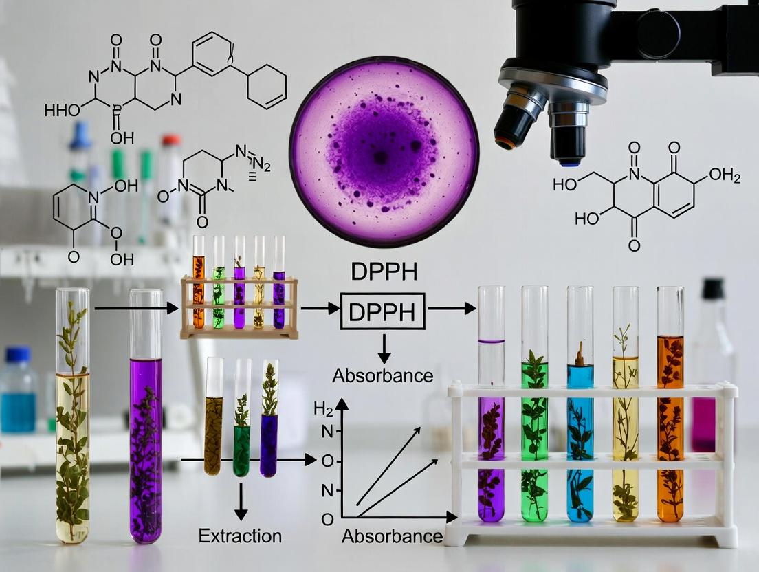
Within the context of a thesis investigating the antioxidant potential of medicinal plant extracts, understanding the Diphenylpicrylhydrazyl (DPPH•) radical is fundamental. DPPH• is a stable, nitrogen-centered free radical characterized by its deep violet color, with an absorption maximum typically around 517 nm. The assay principle is based on a colorimetric redox reaction: when an antioxidant molecule donates a hydrogen atom to the DPPH• radical, it is reduced to its non-radical form, diphenylpicrylhydrazine, resulting in a color change from violet to pale yellow. The degree of discoloration, measured spectrophotometrically, correlates directly with the radical-scavenging activity of the sample.
Table 1: Fundamental Properties of the DPPH Radical
| Property | Specification / Value | Notes |
|---|---|---|
| Chemical Name | 2,2-Diphenyl-1-picrylhydrazyl | Also known as 1,1-Diphenyl-2-picrylhydrazyl |
| State | Dark violet crystalline powder | Stable in solid form at room temperature. |
| Molecular Weight | 394.32 g/mol | - |
| Solubility | Soluble in methanol, ethanol, acetone | Not soluble in water. Methanol is the preferred solvent to avoid interference. |
| λ_max (Absorption) | 515 - 520 nm | Exact peak should be confirmed for the specific solvent and instrument used. |
| Molar Absorptivity (ε) | ~10,000 - 12,000 L·mol⁻¹·cm⁻¹ | Must be determined experimentally for precise quantitative work. |
Table 2: Standard Calibration Data for DPPH Solution (Example)
| DPPH Concentration (µM) | Expected Absorbance at 517 nm (ε=12,000) | Visual Color Description |
|---|---|---|
| 100 | ~1.2 (requires dilution) | Deep Violet |
| 50 | ~0.6 | Violet |
| 25 | ~0.3 | Light Purple |
| 10 | ~0.12 | Very Pale Purple |
| 0 (Blank) | 0.0 | Colorless (Solvent) |
Core Protocol: DPPH Radical Scavenging Activity Assay
Principle: Measurement of the decrease in absorbance of the DPPH radical solution at 517 nm after reaction with an antioxidant-containing sample.
Materials & Reagents:
- DPPH powder (high purity, ≥95%)
- Absolute methanol or ethanol (HPLC grade)
- Antioxidant standard (e.g., Trolox, Ascorbic acid)
- Plant extract samples (dissolved in same solvent as DPPH)
- Microplate reader or UV-Vis spectrophotometer
- 96-well microplates or cuvettes
- Piperettes and micropipettes
- Amber vials and volumetric flasks (light-sensitive)
Detailed Protocol:
A. Preparation of Reagents:
- DPPH Stock Solution (2 mM): Accurately weigh 1.576 mg of DPPH powder. Transfer to a 2 mL amber volumetric flask and dilute to the mark with methanol. Sonicate briefly to ensure complete dissolution. Prepare fresh daily.
- DPPH Working Solution (100 µM): Dilute the 2 mM stock solution 1:20 with methanol (e.g., 500 µL stock + 9.5 mL methanol). The absorbance should be ~1.0 ± 0.02 at 517 nm.
- Sample & Standard Solutions: Prepare serial dilutions of the plant extract or standard antioxidant in methanol. A typical range is 1-100 µg/mL for crude extracts.
B. Assay Procedure (Microplate Method):
- Control (Blank): Add 100 µL of methanol to 100 µL of DPPH working solution in a well. (Corrects for solvent).
- Sample Test: Add 100 µL of sample solution to 100 µL of DPPH working solution.
- Negative Control (DPPH Baseline): Add 100 µL of methanol to 100 µL of DPPH working solution in a separate well. This is used to determine the initial absorbance of DPPH (A_control).
- Mix thoroughly and incubate the plate in the dark at room temperature for 30 minutes.
- Measure the absorbance at 515-517 nm using a microplate reader.
C. Data Analysis:
Calculate the percentage of DPPH Radical Scavenging Activity (% RSA):
% RSA = [(A_control - A_sample) / A_control] × 100
where Acontrol is the absorbance of the negative control and Asample is the absorbance of the test sample/standard.
Generate a dose-response curve and calculate the IC₅₀ value (concentration required to scavenge 50% of DPPH radicals).
The Scientist's Toolkit: Essential Research Reagents & Materials
Table 3: Key Research Reagent Solutions
| Item | Function & Specification | Notes for Thesis Research |
|---|---|---|
| DPPH Crystalline Reagent | Source of the stable free radical. Purity ≥95% is critical for reproducibility. | Store desiccated at -20°C in the dark. Weigh accurately. |
| HPLC-Grade Methanol | Primary solvent for DPPH. Minimizes solvent-related absorbance artifacts. | Use the same solvent batch for all experiments in a series. |
| Trolox (6-hydroxy-2,5,7,8-tetramethylchroman-2-carboxylic acid) | Water-soluble vitamin E analog used as a standard reference antioxidant. | Enables expression of results as "Trolox Equivalents (TEAC)". |
| Ascorbic Acid (Vitamin C) | Common natural antioxidant standard for method validation. | Check stability in solution; prepare fresh. |
| Gallic Acid / Quercetin | Phenolic compound standards relevant to plant extract analysis. | Useful for standardizing assays focused on polyphenolic antioxidants. |
| 96-Well Flat-Bottom Microplates | High-throughput reaction vessel for spectrophotometry. | Use clear plates for absorbance reading. Ensure compatibility with reader. |
| Absorbent Plate Sealer / Aluminum Foil | Prevents solvent evaporation and protects light-sensitive DPPH during incubation. | Critical for consistent results during the 30-min incubation. |
Visualizing the DPPH Assay Workflow and Chemistry
Title: DPPH Assay Experimental Workflow
Title: DPPH Radical Scavenging Reaction Mechanism
Within the broader thesis on utilizing the DPPH (2,2-diphenyl-1-picrylhydrazyl) assay for evaluating antioxidant activity in medicinal plants, understanding the core reaction mechanisms is paramount. The DPPH• stable free radical is reduced to its corresponding hydrazine (DPPH-H) upon reaction with an antioxidant (AH). This reduction can proceed via two primary pathways: Hydrogen Atom Transfer (HAT) and Single Electron Transfer (SET). The dominant mechanism depends on the antioxidant's structure, solvent system, and pH, influencing the interpretation of results for natural product drug discovery.
Mechanism Pathways
Hydrogen Atom Transfer (HAT) Mechanism
In the HAT mechanism, the antioxidant (AH) directly donates a hydrogen atom to the DPPH• radical in a single step. This is a concerted process where the bond between the antioxidant's hydrogen and its parent atom breaks, and a new bond forms with the nitrogen radical of DPPH•.
Reaction: DPPH• + AH → DPPH-H + A•
Single Electron Transfer (SET) Mechanism
The SET mechanism involves two steps. First, the antioxidant donates a single electron to DPPH•, forming a radical cation (AH•+) and the reduced DPPH anion (DPPH-). This is often followed by a subsequent proton transfer or disproportionation.
Reactions: Step 1: DPPH• + AH → DPPH- + AH•+ Step 2: AH•+ → H+ + A• (may occur) Follow-up: DPPH- + H+ → DPPH-H
Comparative Analysis of HAT vs. SET Mechanisms
Table 1: Key Characteristics of HAT and SET Pathways in the DPPH Assay
| Parameter | Hydrogen Atom Transfer (HAT) | Single Electron Transfer (SET) |
|---|---|---|
| Primary Step | Direct H-atom donation | Electron donation followed by proton transfer |
| Solvent Dependence | Favored in non-polar solvents (e.g., toluene) | Favored in polar/protic solvents (e.g., methanol, ethanol) |
| pH Influence | Less sensitive to pH | Highly sensitive; basic pH favors SET |
| Antioxidant Type | Preferred by phenols with low ionization potential, O-H bond strength critical | Preferred by compounds easily oxidized (low reduction potential), e.g., flavonoids, ascorbate |
| Kinetics | Typically faster, diffusion-controlled | Can be slower, depends on solvent stabilization of ions |
| Role in Plant Extracts | Major pathway for simple phenolic acids (e.g., gallic acid) | Likely for complex flavonoids and ascorbic acid |
Table 2: Experimental Conditions Favoring Each Mechanism
| Condition | Favors HAT Mechanism | Favors SET Mechanism |
|---|---|---|
| Solvent | Hydrocarbons (toluene, hexane) | Alcohols (MeOH, EtOH), aqueous mixtures, acetonitrile |
| pH | Acidic to neutral | Neutral to basic |
| Antioxidant Structure | Non-ionizable phenols, thiols | Ionizable phenols, anions (e.g., ascorbate, flavonoid anions) |
| Additives | None (pure system) | Presence of metal ions or bases |
Detailed Experimental Protocols
Protocol 1: Standard DPPH Radical Scavenging Assay
Objective: To determine the percentage inhibition and IC50 of a medicinal plant extract.
- Reagent Prep: Prepare a 0.1 mM DPPH solution in methanol (or ethanol). Protect from light using aluminum foil.
- Sample Prep: Prepare serial dilutions of the plant extract (or pure compound) in the same solvent.
- Reaction: Mix 2.0 mL of DPPH solution with 2.0 mL of sample solution. For control, mix 2.0 mL DPPH with 2.0 mL pure solvent.
- Incubation: Incubate the mixture in the dark at room temperature for 30 minutes.
- Measurement: Measure absorbance at 517 nm against a blank of pure solvent.
- Calculation: % Inhibition = [(Acontrol - Asample) / A_control] × 100 Plot % Inhibition vs. concentration to calculate IC50.
Protocol 2: Kinetic Assay to Probe Mechanism Dominance
Objective: To gather evidence for HAT or SET dominance via reaction kinetics.
- Setup: Use a UV-Vis spectrophotometer with kinetics capability.
- Initial Rate: Prepare a reaction mixture with final [DPPH] = 0.05 mM and antioxidant at a concentration giving 50-80% final inhibition. Rapidly mix and initiate scanning at 517 nm immediately.
- Data Collection: Record absorbance every 5-10 seconds for the first 2-5 minutes.
- Analysis: Plot Absorbance vs. Time. A rapid, exponential decay suggests HAT dominance. A slower, more linear decay suggests SET or mixed mechanisms. Calculate initial reaction rates (d[DPPH]/dt).
Protocol 3: Solvent Polarity Experiment
Objective: To infer mechanism by testing antioxidant activity in solvents of different polarities.
- Solvent Selection: Prepare DPPH solutions (0.1 mM) in toluene (non-polar) and methanol (polar protic).
- Sample Prep: Dissolve the same antioxidant (e.g., a purified plant flavonoid) in both solvents.
- Assay: Perform the standard assay (Protocol 1) in both solvents.
- Interpretation: A significantly higher activity in toluene suggests a HAT pathway. Similar or higher activity in methanol suggests SET contribution.
Visualizing the Mechanisms and Workflow
Title: DPPH Assay: HAT vs SET Reaction Pathways
Title: DPPH Assay Workflow for Medicinal Plant Research
The Scientist's Toolkit: Research Reagent Solutions
Table 3: Essential Materials for DPPH Assay Research
| Item | Function & Rationale |
|---|---|
| DPPH (2,2-diphenyl-1-picrylhydrazyl) | Stable free radical source. Its deep purple color (λ_max ~517 nm) bleaches upon reduction, enabling spectrophotometric quantification. |
| Methanol (HPLC/UV grade) | Common solvent for DPPH. Polar and protic, favors SET mechanisms. Must be free of stabilizers that can act as antioxidants. |
| Ethanol (Absolute) | Alternative to methanol. Less toxic, suitable for extractions. Also favors SET pathways. |
| Toluene (Anhydrous) | Non-polar solvent used to probe HAT mechanisms, as it suppresses ionization. |
| Gallic Acid / Ascorbic Acid | Reference standard antioxidants. Gallic acid often acts via HAT/mixed, ascorbic acid via SET. Used for calibration and IC50 comparison. |
| UV-Vis Spectrophotometer | Essential instrument for measuring absorbance change at 517 nm. Requires micro-cuvettes or plate reader capability for high-throughput. |
| pH Meter & Buffers | For preparing extracts or adjusting reaction pH to study SET mechanism sensitivity (e.g., phosphate buffer pH 6-8). |
| Microplate Reader (96-well) | Enables high-throughput screening of multiple plant extracts or fractions simultaneously, significantly increasing efficiency. |
| Reaction Vials (Amber) | Protect light-sensitive DPPH solutions and reactions from photodegradation during incubation. |
Within the thesis research on the DPPH assay for evaluating the antioxidant activity of medicinal plants, the IC50 value emerges as the fundamental, quantitative endpoint. It is the concentration of an antioxidant required to scavenge 50% of the initial DPPH free radicals. A lower IC50 indicates higher antioxidant potency, allowing for direct comparison between complex plant extracts and pure compounds, guiding the isolation of bioactive constituents and establishing structure-activity relationships.
Key Quantitative Data
Table 1: Comparative IC50 Values of Standard Antioxidants & Plant Extracts
| Substance / Extract | Reported IC50 (µg/mL) | Class / Source | Key Implication |
|---|---|---|---|
| Ascorbic Acid | 1.2 - 2.5 | Standard Reference | Benchmark for strong, pure antioxidants. |
| Trolox | 4.8 - 6.0 | Standard Reference (Vitamin E analog) | Common standard for hydrophilic antioxidants. |
| Quercetin | 7.5 - 12.0 | Pure Flavonoid | High-potency plant compound benchmark. |
| Ginkgo biloba leaf extract | 25.0 - 40.0 | Standardized Plant Extract | Represents a potent commercial extract. |
| Curcuma longa (Turmeric) rhizome extract | 45.0 - 65.0 | Medicinal Plant Extract | Moderate potency, varies with curcuminoid content. |
| Olea europaea (Olive) leaf extract | 10.0 - 20.0 | Medicinal Plant Extract | High potency linked to oleuropein. |
| Green Tea Catechins Extract | 8.0 - 15.0 | Plant Extract | Very high potency due to epigallocatechin gallate. |
Table 2: Interpretation of IC50 Values in Research Context
| IC50 Range (µg/mL) | Potency Rating | Research Utility & Next Steps |
|---|---|---|
| < 10 | Very High | Prioritize for bioassay-guided fractionation, compound identification, and in vivo studies. |
| 10 - 50 | High | Strong candidate for further purification, synergy studies, and standardization. |
| 50 - 100 | Moderate | May be significant in complex mixtures; investigate synergies or use as supporting data. |
| > 100 | Low | Likely not a primary source of potent antioxidants via DPPH mechanism. |
Experimental Protocols
Protocol 1: Standard DPPH Assay for IC50 Determination
- Objective: To determine the IC50 value of a plant extract or pure compound.
- Principle: The purple DPPH• radical is reduced to yellow diphenylpicrylhydrazine by an antioxidant, with color change measurable at 517 nm.
- Reagents: 0.1 mM DPPH in methanol (freshly prepared), sample solutions at varying concentrations, methanol (blank), ascorbic acid/Trolox (positive control).
- Procedure:
- Prepare serial dilutions of the test sample (e.g., 1, 5, 10, 25, 50, 100 µg/mL in methanol).
- Add 2 mL of DPPH solution to 2 mL of each sample concentration in test tubes. For control, add 2 mL methanol to 2 mL DPPH.
- Vortex mixtures and incubate in the dark at room temperature for 30 minutes.
- Measure absorbance at 517 nm against a methanol blank.
- Calculate % Inhibition:
[(A_control - A_sample) / A_control] * 100. - Plot % Inhibition vs. sample concentration. Use non-linear regression (log inhibitor vs. response) to calculate the IC50 value.
Protocol 2: Validation and Quality Control for IC50 Measurement
- Objective: To ensure assay reproducibility and accuracy.
- Procedure:
- Linear Range Verification: Test a range of standard (e.g., Trolox) concentrations to ensure the response is dose-dependent.
- Precision (Repeatability): Perform the assay on the same sample in triplicate on the same day.
- Intermediate Precision: Perform the assay on three different days.
- Accuracy/Recovery: Spike a known concentration of standard into a sample matrix and measure recovery (should be 90-110%).
- Positive Control: Include a standard antioxidant in every assay plate/run. The IC50 must fall within the expected historical range.
The Scientist's Toolkit: Research Reagent Solutions
| Item | Function & Significance |
|---|---|
| DPPH (2,2-Diphenyl-1-picrylhydrazyl) | Stable free radical compound; the core reagent that provides the spectrophotometric signal. |
| Trolox (6-hydroxy-2,5,7,8-tetramethylchroman-2-carboxylic acid) | Water-soluble vitamin E analog; the gold-standard calibrator for reporting TEAC (Trolox Equivalent Antioxidant Capacity). |
| Ascorbic Acid | Primary reference standard; validates assay performance for strong, rapid antioxidants. |
| Spectrophotometer/Microplate Reader | Essential for high-throughput measurement of absorbance change at 517 nm. |
| Methanol (HPLC Grade) | Preferred solvent for DPPH; minimizes interference and stabilizes the radical solution. |
Visualizations
Within the broader thesis of evaluating antioxidant activity for drug discovery from medicinal plants, the 2,2-diphenyl-1-picrylhydrazyl (DPPH) assay remains a cornerstone preliminary screening tool. Its role is pivotal in the rapid identification and ranking of antioxidant candidates from complex phytochemical matrices before advancing to more physiologically relevant, but resource-intensive, cellular and in vivo models. This document outlines its definitive advantages, critical limitations, and provides detailed protocols for robust implementation.
Core Principles and Data Interpretation
The DPPH assay measures the free radical scavenging capacity of a compound or extract. The stable, violet-colored DPPH radical (λ_max ~517 nm) is reduced to a yellow-colored diphenylpicrylhydrazine, causing a measurable decrease in absorbance. Results are typically expressed as IC50 (concentration required to scavenge 50% of radicals), percent inhibition, or Trolox Equivalents (TEAC).
Table 1: Standard Quantitative Benchmarks for DPPH Assay Interpretation
| Parameter | Typical Range/Value | Interpretation in Phytochemical Screening |
|---|---|---|
| DPPH Working Solution Concentration | 100-200 µM in methanol/ethanol | Ensures linearity and avoids self-quenching. |
| Reaction Time | 30 min - 1 hour (stable endpoint) | Plant polyphenols require varying times; must be standardized. |
| Control Absorbance (A_control) | 0.6 - 1.2 AU | Optimizes spectrophotometric accuracy. |
| IC50 Value (Strong Antioxidant) | < 50 µg/mL | Extract/compound considered highly active. |
| IC50 Value (Moderate Antioxidant) | 50 - 200 µg/mL | Warrant further fractionation. |
| IC50 Value (Weak Antioxidant) | > 200 µg/mL | May be deprioritized. |
| Linear Range for Calibration (Trolox) | 10 - 100 µM | For accurate TEAC calculation. |
Table 2: Key Advantages of the DPPH Assay for Phytochemical Research
| Advantage | Practical Implication for Researchers |
|---|---|
| Rapid & High-Throughput | Enables screening of hundreds of plant extracts/fractions in a single day. |
| Technical Simplicity | No complex instrumentation; requires only a UV-Vis spectrophotometer or microplate reader. |
| Low Cost & Reagent Stability | DPPH reagent is inexpensive and stable for months when stored properly, reducing per-sample cost. |
| Direct Radical Scavenging Measure | Provides a clear, quantitative measure of hydrogen-donating or electron-transfer antioxidant capacity. |
| Minimal Sample Preparation | Crude plant extracts in compatible solvents (methanol, ethanol, aqueous mixtures) can be used directly. |
Table 3: Critical Limitations of the DPPH Assay
| Limitation | Impact on Phytochemical Research & Data Validity |
|---|---|
| Non-Physiological Radical | DPPH is a stable, synthetic radical not found in biological systems. Correlations with in vivo activity can be poor. |
| Solvent Interference | Many antioxidant phytochemicals (e.g., polar polysaccharides) are insoluble in the required methanol/ethanol medium, leading to false negatives. |
| Spectroscopic Interferences | Plant pigments (chlorophyll, carotenoids) absorbing near 517 nm can cause false positives or absorbance masking. |
| Reaction Kinetics Variability | Different antioxidant classes (flavonoids vs. phenolics) react at different rates, making single-time-point comparisons misleading. |
| Mechanistic Ambiguity | Cannot distinguish between H-atom transfer (HAT) and single electron transfer (SET) mechanisms, which have different biological relevance. |
| pH Insensitivity | Conducted at non-physiological pH, unlike assays like ORAC which operate at pH 7.4. |
Detailed Experimental Protocols
Protocol 1: Standard Microplate DPPH Assay for Plant Extract Screening
Objective: To determine the percentage DPPH radical scavenging activity and IC50 of medicinal plant extracts.
The Scientist's Toolkit: Key Reagents & Materials
| Item | Function & Specification |
|---|---|
| DPPH (≥95% purity) | Source of the stable free radical. Must be stored desiccated at -20°C. |
| Absolute Methanol or Ethanol (HPLC grade) | Solvent for DPPH and extracts. Must have low UV absorbance. |
| Trolox (6-hydroxy-2,5,7,8-tetramethylchroman-2-carboxylic acid) | Water-soluble vitamin E analog used as a standard reference antioxidant. |
| Test Plant Extracts | Dry, dissolved in assay-compatible solvent (e.g., methanol). Serial dilutions prepared fresh. |
| 96-Well Microplate (Clear, Flat-Bottom) | For high-throughput reaction setup and measurement. |
| Microplate Reader | Equipped with a filter or monochromator for 515-520 nm. |
| Multichannel Pipettes & Reservoirs | For rapid, reproducible reagent dispensing. |
Procedure:
- DPPH Solution Preparation: Weigh 3.94 mg of DPPH and dissolve in 100 mL of methanol to prepare a 100 µM stock solution. Protect from light, store at 4°C, and use within 24 hours.
- Sample Preparation: Prepare serial dilutions of the plant extract (e.g., 1, 5, 10, 25, 50, 100 µg/mL) and Trolox standard (e.g., 5, 10, 25, 50, 100 µM) in methanol.
- Reaction Setup:
- Test Well: Add 100 µL of plant extract dilution + 100 µL of DPPH solution.
- Control Well: Add 100 µL of methanol + 100 µL of DPPH solution.
- Blank Well: Add 100 µL of extract dilution + 100 µL of methanol.
- Standard Curve Wells: Add 100 µL of Trolox dilution + 100 µL of DPPH solution.
- Incubation: Cover the plate and incubate in the dark at room temperature for 30 minutes.
- Measurement: Measure the absorbance at 517 nm using a microplate reader.
- Calculation:
% Scavenging = [(A_control - (A_sample - A_blank)) / A_control] * 100- Plot % Scavenging vs. log(concentration) to determine IC50.
- Plot % Scavenging of Trolox standards to express activity as µmol Trolox Equivalents/g extract (TEAC).
Protocol 2: Kinetics-Modified DPPH Assay
Objective: To account for variable reaction kinetics of different phytochemical classes by measuring reaction progress over time.
Procedure:
- Follow Protocol 1 for setup, but use a single, mid-range concentration of extract (e.g., IC50 estimated from Protocol 1).
- Immediately after adding DPPH, begin kinetic measurement cycle on the plate reader.
- Record absorbance at 517 nm every 30 seconds for 5 minutes, then every minute for up to 60 minutes.
- Plot Absorbance vs. Time for extract and Trolox control.
- Calculate the Antiradical Power (ARP) or Trolox Equivalence at multiple time points (e.g., 5 min, 30 min, 60 min). ARP = 1/IC50.
- Interpretation: A fast, steep decline indicates rapid scavengers (e.g., simple phenolics). A slow, gradual decline indicates slow-reacting scavengers (e.g., complex flavonoids).
The DPPH assay is an indispensable, cost-effective tool for the primary ranking and prioritization of antioxidant-rich medicinal plant extracts. Its advantages of speed and simplicity make it ideal for screening large libraries. However, its inherent limitations regarding physiological relevance mandate that positive results be viewed as a first-pass filter. A robust thesis on antioxidant drug discovery must employ a multi-assay consensus approach, following DPPH screening with assays like FRAP, ABTS, ORAC, and ultimately, cell-based models (e.g., CAA assay) to identify lead candidates with a higher probability of in vivo efficacy.
A Step-by-Step Protocol for DPPH Assay Execution with Medicinal Plant Extracts
Within a comprehensive thesis investigating the antioxidant potential of medicinal plant extracts via the DPPH (2,2-diphenyl-1-picrylhydrazyl) radical scavenging assay, rigorous pre-assay preparation is paramount. This phase dictates the accuracy, reproducibility, and biological relevance of all subsequent results. This document provides detailed application notes and protocols focusing on three foundational pillars: solvent selection for both sample and radical, DPPH solution stability, and the preparation of standard antioxidants.
Solvent Selection: Balancing Solubility and Assay Integrity
The solvent must completely dissolve the plant extract (often non-polar to medium-polar compounds) without interfering with the DPPH radical kinetics. Pure methanol or ethanol are standard, but mixtures are often necessary.
Table 1: Common Solvents for DPPH Assay and Their Properties
| Solvent | Polarity Index | Compatibility with DPPH | Best For | Key Consideration |
|---|---|---|---|---|
| Methanol | 5.1 | Excellent. Minimal interference. | Most common standard. Polar extracts. | Hygroscopic; can absorb water affecting concentration. |
| Ethanol | 5.2 | Excellent. Minimal interference. | Food & pharmacological studies. | Less toxic than methanol. Preferred for bioactive studies. |
| Acetone | 5.1 | Good. Slight background reduction possible. | Extracts with medium polarity. | Evaporates quickly; requires careful handling. |
| Water | 10.2 | Poor. DPPH is insoluble. | Not for DPPH stock. | Can be used to dilute polar samples before adding to methanolic DPPH. |
| DMSO | 7.2 | Acceptable with controls. | Very non-polar plant compounds. | High viscosity can affect pipetting accuracy. Its own radical scavenging must be corrected. |
| Methanol:Water (80:20 v/v) | N/A | Good. | Extracts with mixed polarity. | Mimics physiological conditions better than pure alcohol. |
Protocol: Solvent Compatibility Test
- Prepare a 0.1 mM DPPH solution in the candidate pure solvent (e.g., methanol). Record absorbance at 517 nm (Asolvent).
- Prepare the same DPPH concentration in a mixture (e.g., 90% solvent:10% sample solvent, if different).
- Incubate for 30 minutes in the dark.
- Measure absorbance (Amixture).
- Calculate interference: % Interference = [(Asolvent - Amixture) / Asolvent] x 100. A change >5% suggests significant solvent interference.
DPPH Solution Stability: A Time-Dependent Variable
The DPPH radical solution degrades upon exposure to light, heat, and oxygen, leading to decreased absorbance and false-high antioxidant activity calculations.
Table 2: DPPH Solution Stability Under Different Storage Conditions
| Condition | Container | Temperature | Light Exposure | % Absorbance Loss (at 517 nm) after 24h | Recommended Use Window |
|---|---|---|---|---|---|
| Optimal | Amber glass vial, sealed | 4°C | Dark | < 2% | Fresh daily. Stable up to 3 days with verification. |
| Sub-Optimal | Clear glass vial | 4°C | Laboratory ambient light | ~10-15% | Must be used immediately (<1 hour). |
| Unacceptable | Clear glass vial | 25°C (RT) | Laboratory ambient light | >25% | Not recommended. |
Protocol: Establishing DPPH Solution Stability for Your System
- Prepare a fresh 0.1 mM DPPH stock in methanol.
- Aliquot into two amber vials and one clear vial.
- Store one amber vial at 4°C (A), one amber vial at RT in the dark (B), and the clear vial at RT in light (C).
- Measure the absorbance at 517 nm for each vial at t=0, 1, 3, 6, and 24 hours.
- Plot absorbance vs. time. The slope indicates degradation rate. Use storage conditions that show ≤2% degradation over your typical assay period.
Standard Preparation: Trolox and Ascorbic Acid
Using a standard curve is essential to express results in Trolox Equivalent Antioxidant Capacity (TEAC) or Ascorbic Acid Equivalent Antioxidant Capacity (AEAC).
Protocol: Preparation of Trolox Standard Curve
- Stock Solution (1 mM): Accurately weigh 0.0250 g of Trolox (MW 250.29 g/mol) and dissolve in 100 mL of methanol or buffer (depending on assay design). This stock is stable at -20°C for one month.
- Working Standards: Prepare serial dilutions in the same solvent as your plant samples will be in.
- Example range: 0 (blank), 50, 100, 200, 400, 600, 800 µM.
- Assay Procedure: a. Mix 2.0 mL of each Trolox working standard with 2.0 mL of fresh 0.1 mM DPPH solution. b. Incubate for 30 minutes in the dark at room temperature. c. Measure absorbance at 517 nm against a methanol/DPPH blank. d. Calculate % Inhibition: [(Ablank - Asample) / Ablank] x 100.
- Plot % Inhibition vs. Trolox concentration (µM). Perform linear regression (typically y = mx + c, R² > 0.98).
Protocol: Preparation of Ascorbic Acid Standard Curve
- Stock Solution (1 mM): Prepare fresh daily. Weigh 0.0176 g of L-ascorbic acid (MW 176.12 g/mol) and dissolve in 100 mL of distilled water or a weak acidic solution (e.g., 1% metaphosphoric acid) to prevent oxidation.
- Working Standards: Prepare serial dilutions in water/appropriate solvent. Range: 0, 10, 25, 50, 75, 100 µM (ascorbic acid is more potent).
- Assay Procedure: Follow steps identical to Trolox protocol (3a-d).
- Generate the standard curve as described.
Table 3: Comparison of Common Antioxidant Standards
| Standard | Molecular Weight (g/mol) | Typical Linear Range (µM) | Stability of Stock Solution | Primary Use |
|---|---|---|---|---|
| Trolox | 250.29 | 50 - 800 | Stable at -20°C for weeks. | Universal standard for TEAC. Water-soluble analog of Vitamin E. |
| Ascorbic Acid | 176.12 | 5 - 100 | Unstable in solution; prepare fresh daily. | Natural antioxidant standard; common in food/fruit studies. |
| Gallic Acid | 170.12 | 20 - 200 | Moderately stable at 4°C for days. | Phenolic acid standard; common in plant polyphenol studies. |
| Quercetin | 302.24 | 10 - 150 | Stable at -20°C in DMSO for weeks. | Flavonoid standard. |
The Scientist's Toolkit: Research Reagent Solutions
| Item | Function & Specification |
|---|---|
| DPPH (≥95% purity) | Stable radical source. High purity is critical for accurate molar absorptivity (ε ~ 10,000 - 12,000 L·mol⁻¹·cm⁻¹). |
| Trolox (≥97% purity) | Primary water-soluble vitamin E analog used as the benchmark for TEAC calculations. |
| L-Ascorbic Acid (≥99% purity) | Primary water-soluble natural antioxidant used as a benchmark for AEAC calculations. |
| Anhydrous Methanol (HPLC grade) | Preferred solvent for DPPH stock due to minimal water content and radical interference. |
| Ethanol (Absolute, ACS grade) | Less toxic alternative to methanol for DPPH and sample dissolution. |
| Amber Volumetric Flasks/ Vials | Protects light-sensitive DPPH solutions from photodegradation during preparation and storage. |
| Low-Adhesion Microcentrifuge Tubes | Minimizes sample loss when working with viscous plant extracts or DMSO solutions. |
| Adjustable Piperttes (10 µL - 5 mL) | For accurate liquid handling of samples, standards, and DPPH reagent. |
| UV-Vis Spectrophotometer & Cuvettes | For measuring the decrease in DPPH absorbance at 517 nm. Quartz or high-quality glass cuvettes required. |
Visualization: Pre-Assay Preparation Workflow
DPPH Pre-Assay Critical Parameter Flow
Factors Influencing DPPH Solution Stability
In research focused on evaluating the antioxidant activity of medicinal plants via the DPPH (2,2-diphenyl-1-picrylhydrazyl) radical scavenging assay, the initial extraction protocol is the most critical determinant of result validity and bioactivity relevance. The extraction process directly influences the concentration and chemical integrity of antioxidant compounds (e.g., phenolics, flavonoids, terpenoids) transferred from the plant matrix into the analyzable solution. Inefficient or degrading extraction leads to false negatives and non-reproducible IC₅₀ values. This application note details standardized protocols and best practices to ensure extracts genuinely represent the plant's bioactive potential for subsequent DPPH analysis.
Solvent Selection: Polarity and Efficacy
The choice of solvent is paramount, as antioxidant compounds vary widely in polarity. A combination of solvents or solvent-water mixtures is often required for comprehensive extraction. The table below summarizes solvent performance based on recent phytochemical studies.
Table 1: Solvent Systems for Antioxidant Compound Extraction
| Solvent System (v/v) | Polarity Index | Target Compound Classes | Reported Relative Yield* of Antioxidants (vs. Water) | Key Consideration for DPPH Assay |
|---|---|---|---|---|
| 70-80% Aqueous Methanol | High | Polar phenolics, flavonoids, tannins | 1.8 - 2.5 | Excellent for total phenolic content, strongly correlates with DPPH activity. Low boiling point for easy concentration. |
| 70-80% Aqueous Ethanol | High | Polar phenolics, flavonoids, saponins | 1.6 - 2.2 | Safer than methanol. Food/pharma-grade preference. Slightly lower yield for some phenolics. |
| Acetone (50-70% Aqueous) | Medium-High | Medium-polarity flavonoids, some terpenoids | 1.5 - 2.0 | Effective for anthocyanins. Less likely to extract chlorophyll, reducing interference in spectrophotometry. |
| Ethyl Acetate | Medium | Medium-polarity phenolics, coumarins | 1.2 - 1.6 | Selective for mid-polar antioxidants; useful for fractionation prior to assay. |
| Water | Very High | Polysaccharides, proteins, very polar glycosides | 1.0 (Baseline) | High-temperature extraction needed for cells. May co-extract sugars which can interfere in some antioxidant assays. |
| Methanol 100% | High | Broad range of phenolics, alkaloids | 1.4 - 1.9 | Can degrade some thermolabile compounds. May not efficiently rupture plant cells. |
*Yield data is illustrative, based on aggregated studies comparing total phenolic/flavonoid content.
Detailed Extraction Protocols
Protocol 3.1: Standard Maceration for DPPH Screening
- Objective: To obtain a total antioxidant-rich extract for initial DPPH radical scavenging screening.
- Materials: Dried plant powder (sieved, 0.5mm), 70% aqueous methanol (HPLC grade), orbital shaker, ultrasonic bath, rotary evaporator (<40°C), lyophilizer.
- Procedure:
- Weigh 2.0 g of dried plant material into a 50 mL conical flask.
- Add 40 mL of 70% methanol (solvent-to-material ratio 20:1 v/w).
- Agitation: Seal and place on an orbital shaker (150 rpm) at room temperature (25°C) for 60 minutes.
- Sonication: Transfer the flask to an ultrasonic bath (40 kHz) and sonicate for 20 minutes at 35°C to enhance cell wall disruption.
- Filtration: Filter the mixture through Whatman No. 1 filter paper under vacuum.
- Re-extraction: Re-macerate the residue with another 20 mL of fresh solvent for 30 minutes. Filter and combine filtrates.
- Concentration: Evaporate the combined filtrate under reduced pressure at 38°C using a rotary evaporator until all methanol is removed.
- Lyophilization: Freeze the remaining aqueous solution and lyophilize to obtain a dry crude extract.
- Storage: Store the extract powder at -20°C in an airtight, light-protected container. For DPPH assay, reconstitute in DMSO or the assay buffer to make a stock solution (e.g., 10 mg/mL).
Protocol 3.2: Sequential Solvent Extraction for Bioactivity-Guided Fractionation
- Objective: To fractionate antioxidants based on polarity, enabling correlation of specific compound classes with DPPH activity.
- Procedure: Perform maceration (as in 3.1) sequentially with solvents of increasing polarity: start with hexane (for non-polar lipids), then ethyl acetate, then methanol, and finally water. Concentrate each fraction separately. Test each fraction in the DPPH assay to identify the most active polarity fraction for further analysis.
The Scientist's Toolkit: Research Reagent Solutions
Table 2: Essential Materials for Plant Extraction in Antioxidant Research
| Item | Function & Importance |
|---|---|
| Lyophilizer (Freeze Dryer) | Preserves thermolabile antioxidants during solvent removal. Provides stable, dry extract powder for accurate weighing and long-term storage. |
| Ultrasonic Bath/Sonicator (40-60 kHz) | Applies cavitation energy to rupture plant cell walls, significantly improving solvent penetration and extraction efficiency. |
| Rotary Evaporator with Vacuum Pump | Enables gentle, low-temperature removal of organic solvents (methanol, ethanol, acetone) to prevent thermal degradation of antioxidants. |
| Controlled Atmosphere Oven (≤40°C) | For slow, uniform drying of fresh plant material to constant weight, preventing enzymatic degradation before extraction. |
| Whatman Filter Papers (No. 1 & No. 42) | For coarse (No. 1) and fine (No. 42, after extract concentration) particulates removal, ensuring clear extracts for spectrophotometric DPPH assay. |
| Anhydrous Sodium Sulfate (Na₂SO₄) | Drying agent used to remove trace water from organic solvent fractions (e.g., ethyl acetate) post-extraction, crucial for stability and further chemical analysis. |
| HPLC-Grade Solvents & Deionized Water | Minimize background interference from impurities, ensuring that measured DPPH activity originates solely from plant metabolites. |
Visualized Workflows and Pathways
Title: Workflow for Plant Extract Prep for DPPH Assay
Title: Antioxidant-DPPH Reaction Pathway
The 2,2-diphenyl-1-picrylhydrazyl (DPPH) radical scavenging assay is a cornerstone in evaluating the antioxidant potential of medicinal plant extracts. This application note, framed within a comprehensive thesis on the subject, details the comparative analysis of two principal methodologies: the traditional cuvette-based spectrophotometry and the high-throughput microplate technique. The focus extends to precise reaction kinetics and critical incubation parameters, which are essential for generating reproducible, accurate data in drug discovery and phytochemical research.
Table 1: Core Comparison of DPPH Assay Formats
| Parameter | Cuvette Method | Microplate Method | Thesis Research Implication |
|---|---|---|---|
| Sample/Reagent Volume | 1-3 mL (typical) | 200-300 µL | Enables screening of limited, precious plant extracts. |
| Throughput | Low (1 sample/reading) | High (96 samples/run) | Efficient for dose-response curves & large extract libraries. |
| Mixing & Aeration | Manual, prone to variation | Consistent, minimal evaporation | Reduces operational variability in kinetic studies. |
| Incubation Control | Ambient light/temp exposure | Controlled by plate reader | Standardizes critical kinetic incubation parameters. |
| Path Length | Fixed (usually 1 cm) | Variable (~0.5-0.7 cm for 300µL) | Requires adaptation of formulas (see Section 4). |
| Cost per Assay | Lower reagent cost per sample | Higher (plate cost) but lower overall | Optimal for preliminary (cuvette) vs. full screening (plate). |
| Kinetic Monitoring | Sequential, time-intensive | Simultaneous, real-time for all wells | Essential for accurate initial rate calculations in kinetics. |
Detailed Experimental Protocols
Protocol 3.1: DPPH Stock Solution Preparation (Common to Both Methods)
- Reagent: DPPH radical (MW=394.32).
- Procedure: Accurately weigh 2-4 mg of DPPH powder. Dissolve in 50-100 mL of pure, anhydrous methanol or ethanol to achieve a final concentration of ~0.1 mM. Vortex until fully dissolved. Wrap container in aluminum foil and store at 4°C for up to one week. Verify absorbance before use (A~0.9-1.0 at 517 nm, 1 cm path).
Protocol 3.2: Traditional Cuvette-Based DPPH Assay (Endpoint)
- Instrument Setup: Zero a UV-Vis spectrophotometer with pure solvent at 517 nm.
- Control (Blank): In a 1 cm path quartz/glass cuvette, mix 2.7 mL of DPPH working solution with 0.3 mL of solvent. Cap and invert to mix.
- Sample: In a separate cuvette, mix 2.7 mL of DPPH solution with 0.3 mL of plant extract (at desired concentration). Record initial time immediately upon mixing.
- Incubation: Place both cuvettes in a dark cupboard at constant temperature (e.g., 25°C, 30°C) for precisely 30 minutes.
- Measurement: Measure the absorbance of the control (Ac) and sample (As) at 517 nm against the solvent blank.
- Calculation: % Scavenging = [(Ac - As) / Ac] * 100.
Protocol 3.3: Microplate-Based DPPH Assay (Kinetic)
- Instrument Setup: Pre-heat a microplate reader to desired incubation temperature (e.g., 25°C, 37°C). Set monochromator/filter to 515-520 nm.
- Plate Layout: Designate wells for sample extracts, positive controls (e.g., Trolox, ascorbic acid), negative control (solvent), and DPPH blank (solvent + DPPH).
- Dispensing: Using a multi-channel pipette, add 270 µL of DPPH working solution to all sample and control wells. For the DPPH blank (to check initial absorbance), add 270 µL DPPH + 30 µL solvent.
- Kinetic Initiation: Add 30 µL of plant extract (or standard) to designated wells. Use the plate reader's auto-mixer function (or carefully tap plate) to initiate reaction simultaneously.
- Incubation & Reading: Immediately begin kinetic readings, measuring absorbance every 30-60 seconds for 30-60 minutes in the dark.
- Data Analysis: Calculate % scavenging over time. Use data from the linear phase (often first 2-5 minutes) to determine reaction kinetics (IC50, antiradical power).
Protocol 3.4: Investigating Incubation Parameters
- Temperature: Perform Protocol 3.3 at 4°C, 25°C (room temp), and 37°C (physiological). Use a temperature-controlled plate reader.
- Solvent Polarity: Prepare DPPH in methanol, ethanol, and aqueous methanol mixtures (e.g., 80%). Test the same extract across solvents.
- Reaction Time: For endpoint assays, measure scavenging at 10, 20, 30, and 60 minutes to determine time-to-equilibrium.
Table 2: Key Incubation Parameters & Optimized Ranges
| Parameter | Tested Range | Optimized Condition for Thesis | Impact on Result |
|---|---|---|---|
| Incubation Time | 10 - 90 min | 30 min (endpoint), Continuous (kinetic) | Underestimation vs. equilibrium; defines reaction rate. |
| Temperature | 4°C - 37°C | 25°C ± 2°C (controlled) | Higher temp increases reaction rate & potential degradation. |
| DPPH Initial Abs. | 0.7 - 1.2 | 0.9 ± 0.05 (at 517 nm, 1 cm) | Critical for accurate stoichiometry; too high violates Beer's Law. |
| Final Rxn Volume | 3 mL (cuvette), 300 µL (plate) | As per protocol | Microplate: Ensure sufficient depth for consistent path length. |
| Dark Incubation | Mandatory | Wrap cuvettes/use reader dark mode | Prevents photodegradation of DPPH radical. |
Data Presentation & Calculation Adjustments
Table 3: Sample Kinetic Data from Microplate Assay (Trolox Standard)
| Time (min) | Abs. (100 µM Trolox) | Abs. (DPPH Control) | % Scavenging | Log[Inhibitor] |
|---|---|---|---|---|
| 0.5 | 0.812 | 0.945 | 14.1 | -4.00 |
| 1.0 | 0.743 | 0.942 | 21.1 | -4.00 |
| 2.0 | 0.665 | 0.939 | 29.2 | -4.00 |
| 4.0 | 0.601 | 0.936 | 35.8 | -4.00 |
| ...for multiple concentrations to calculate IC50 |
Path Length Correction for Microplates: The effective path length in a microplate well is less than 1 cm. Use the formula: Adjusted Absorbance = Measured Absorbance * (1 cm / Effective Path Length) Where the effective path length can be determined empirically by measuring a known standard in both a cuvette and plate.
The Scientist's Toolkit: Research Reagent Solutions
Table 4: Essential Materials for DPPH Assay Research
| Item | Function & Specification |
|---|---|
| DPPH Radical | The stable radical source. Purity >95% is critical. Store desiccated at -20°C for long term. |
| Methanol (HPLC Grade) | Preferred solvent for DPPH. Low water content ensures radical stability and reproducibility. |
| Trolox (6-hydroxy-2,5,7,8-tetramethylchroman-2-carboxylic acid) | Water-soluble vitamin E analog; the standard reference antioxidant for quantitative results (IC50, TEAC). |
| Quartz Cuvettes (1 cm path) | For traditional method. Quartz ensures UV transparency if other wavelengths are used. |
| 96-Well Flat-Bottom Clear Plates | For microplate method. Use plates with low evaporation lids. Non-binding surface is recommended. |
| Temperature-Controlled Microplate Reader | Must have monochromator or precise 515-520 nm filter, kinetic software, and dark incubation mode. |
| Multichannel & Repetitive Pipettes | For accurate, rapid dispensing of reagents and samples in microplate format. |
Visualizations
Title: DPPH Assay Method Selection & Workflow for Thesis
Title: DPPH Radical Scavenging Reaction Mechanism
Title: Impact of Incubation Parameters on DPPH Results
Within a thesis investigating the antioxidant activity of medicinal plants via the DPPH (2,2-diphenyl-1-picrylhydrazyl) assay, the accuracy of absorbance measurements is paramount. The selection of the optimal wavelength (517 nm) and rigorous spectrophotometer calibration are critical for generating reliable, reproducible data. This protocol details the methodology for instrument verification and calibration, ensuring the integrity of data used to calculate IC50 values and compare antioxidant capacity across plant extracts.
The Significance of 517 nm in DPPH Assay
The DPPH radical in its methanol or ethanol solution exhibits a characteristic deep violet color due to its unpaired electron. This gives a strong absorption maximum at approximately 517 nm. Upon reaction with an antioxidant, the radical is scavenged, reducing DPPH• to DPPH-H, resulting in a loss of color and a consequent decrease in absorbance at 517 nm. This decrease is directly proportional to the antioxidant activity.
Table 1: Spectral Characteristics of DPPH
| Parameter | Value | Significance |
|---|---|---|
| Optimal Measurement Wavelength (λ_max) | 517 ± 2 nm | Peak absorbance for the DPPH radical. Measurement here provides maximum sensitivity. |
| Molar Absorptivity (ε) | ~12,000 M⁻¹ cm⁻¹ (in methanol) | Allows for quantitative determination of radical concentration via Beer-Lambert law. |
| Typical Blank/Control Absorbance | 0.8 - 1.2 AU | A stable initial absorbance ensures the assay is working within the linear dynamic range of the instrument. |
Pre-Measurement Instrument Calibration Protocol
Wavelength Accuracy Verification
Purpose: To confirm the spectrophotometer correctly identifies the 517 nm wavelength. Materials: Holmium oxide (Ho₂O₃) glass filter or didymium filter. Protocol:
- Perform a baseline correction with an empty cuvette holder.
- Place the certified holmium oxide filter in the light path.
- Scan from 500 nm to 540 nm.
- Identify the characteristic absorption peak. For holmium oxide, the peak should occur at 536.2 nm. A deviation of more than ±1 nm requires instrument service.
- For 517 nm specific check: Use a DPPH blank control (0.1 mM in methanol). The peak maximum from a 510-525 nm scan must be centered at 517 nm.
Photometric Accuracy (Absorbance) Calibration
Purpose: To ensure the instrument reports accurate absorbance values. Materials: Certified Neutral Density (ND) glass filters or potassium dichromate (K₂Cr₂O₇) standard solutions. Protocol A (Glass Filters):
- Record the absorbance values of certified ND filters (e.g., nominal values of 0.5, 1.0 AU) at 517 nm.
- Compare measured values to the certified values with tolerance limits (typically ±0.01 AU).
Protocol B (Potassium Dichromate Standard):
- Prepare 0.0600 g/L K₂Cr₂O₇ in 0.005 M H₂SO₄.
- Measure absorbance at 350 nm in a 1 cm pathlength quartz cuvette against a 0.005 M H₂SO₄ blank.
- The measured absorbance should be 1.007 ± 0.015 AU. This validates the photometric scale.
Table 2: Photometric Calibration Standards & Tolerances
| Standard | Target Absorbance (at specified λ) | Acceptable Tolerance | Purpose |
|---|---|---|---|
| Holmium Oxide Filter | Peak at 536.2 nm | ± 1.0 nm | Wavelength accuracy |
| ND Glass Filter (0.5 AU) | 0.500 AU at 517 nm | ± 0.01 AU | Low-range photometric accuracy |
| ND Glass Filter (1.0 AU) | 1.000 AU at 517 nm | ± 0.01 AU | Mid-range photometric accuracy |
| K₂Cr₂O₇ in H₂SO₄ | 1.007 AU at 350 nm | ± 0.015 AU | Absolute photometric calibration |
Critical Operational Checks for DPPH Assay
- Cuvette Alignment: Mark and consistently use the same orientation of the cuvette.
- Pathlength Verification: Measure the absorbance of water at 975 nm (where water has an absorbance ~1.75 AU/cm). Absorbance ≈ 1.75 * actual pathlength.
- Stray Light Check: Use a 50 g/L NaI solution in water. Absorbance at 240 nm should be >3.0 AU. Lower values indicate stray light, causing underestimation of high absorbances.
Detailed DPPH Assay Protocol with Calibration Integration
A. Reagent Preparation
- DPPH Stock Solution (0.5 mM): Accurately weigh 1.97 mg of DPPH powder, dissolve in methanol, and make up to 10 mL. Store in amber vial at -20°C.
- DPPH Working Solution (0.1 mM): Dilute stock solution 1:5 with methanol. Prepare fresh daily.
- Sample/Standard Solutions: Prepare serial dilutions of plant extracts or standard antioxidant (e.g., Trolox, ascorbic acid) in methanol/buffer.
B. Assay Procedure
- Instrument Calibration: Perform wavelength (2.1) and photometric (2.2) checks. Record results.
- Baseline: Zero the spectrophotometer with methanol at 517 nm.
- Control (A_control): Mix 2.0 mL of DPPH working solution with 1.0 mL of methanol. Incubate in the dark for 30 minutes at room temperature. Measure absorbance.
- Samples/Standards (A_sample): Mix 2.0 mL of DPPH working solution with 1.0 mL of sample/standard solution. Incubate similarly. Measure absorbance.
- Blank (Ablank): For colored extracts, mix 2.0 mL of methanol with 1.0 mL of sample. Measure absorbance and subtract from Asample.
C. Calculation
% Scavenging Activity = [(A_control - (A_sample - A_blank)) / A_control] × 100
Plot % inhibition vs. concentration to determine IC50 values.
The Scientist's Toolkit: Research Reagent Solutions
Table 3: Essential Materials for DPPH Assay & Calibration
| Item | Function/Justification |
|---|---|
| High-Purity DPPH Crystalline (>95%) | Ensures correct initial radical concentration and absorbance. |
| Spectrophotometer with ≤2 nm Bandwidth | Required for precise measurement at 517 nm peak. |
| Matched Quartz or High-Quality Glass Cuvettes (1 cm) | Ensures consistent, accurate pathlength. Quartz is mandatory for UV checks (<350 nm). |
| Certified Holmium Oxide Wavelength Standard | Validates spectrophotometer wavelength accuracy. |
| Certified Neutral Density Glass Filters | Validates photometric (absorbance) accuracy at key ranges. |
| Anhydrous, UV-Spectroscopy Grade Methanol | Minimizes solvent impurities that could absorb at 517 nm or quench DPPH. |
| Analytical Balance (0.01 mg sensitivity) | Essential for accurate weighing of DPPH and plant extract samples. |
| Trolox (6-hydroxy-2,5,7,8-tetramethylchroman-2-carboxylic acid) | Standard antioxidant for generating a calibration curve and reporting results as Trolox Equivalents. |
Visualizations
Diagram 1: Calibration in Thesis Workflow (79 chars)
Diagram 2: DPPH Radical Scavenging Reaction (72 chars)
Diagram 3: DPPH Assay Protocol Steps (62 chars)
Thesis Context
This document details essential data calculations within a comprehensive thesis on evaluating the antioxidant potential of medicinal plant extracts using the DPPH (2,2-diphenyl-1-picrylhydrazyl) radical scavenging assay. Accurate quantification of percentage inhibition, IC50, and TEAC values is critical for standardizing results and facilitating cross-study comparisons in phytochemical and drug discovery research.
Formulas for Percentage Inhibition
The percentage inhibition of DPPH radicals is a fundamental measure of an antioxidant's scavenging capacity.
Core Formula
Percentage Inhibition (%) = [(A_control - A_sample) / A_control] × 100
Where:
A_control= Absorbance of the DPPH solution mixed with solvent (no antioxidant).A_sample= Absorbance of the DPPH solution mixed with the test sample (plant extract or standard).
Data Presentation
Table 1: Example Data for Percentage Inhibition Calculation (Absorbance at 517 nm)
| Sample ID | Concentration (µg/mL) | A_control | A_sample | % Inhibition |
|---|---|---|---|---|
| Plant Extract A | 10 | 0.745 | 0.612 | 17.85 |
| Plant Extract A | 50 | 0.745 | 0.389 | 47.79 |
| Trolox Std | 10 µM | 0.745 | 0.415 | 44.30 |
| Ascorbic Acid | 5 µM | 0.745 | 0.298 | 60.00 |
Protocol: DPPH Assay for % Inhibition
- Solution Prep: Prepare a 0.1 mM DPPH solution in methanol (or ethanol). Protect from light.
- Sample Prep: Prepare serial dilutions of the plant extract and antioxidant standards (e.g., Trolox, ascorbic acid).
- Reaction Mix: In microplate wells or tubes, combine:
- Test/Standard: 100 µL
- DPPH Solution: 100 µL
- Control: Replace sample with 100 µL of solvent.
- Blank: For each sample concentration, prepare a blank with 100 µL sample and 100 µL solvent (corrects for sample color).
- Incubation: Shake and incubate in the dark at room temperature for 30 minutes.
- Measurement: Measure absorbance at 515-517 nm against a solvent blank.
- Calculation: Apply the formula above, subtracting sample blank absorbance if necessary.
IC50 Determination via Linear Regression
The IC50 (half-maximal inhibitory concentration) is the effective concentration of an antioxidant required to scavenge 50% of DPPH radicals. It is derived from a dose-response curve.
Calculation Methodology
- Plot
% Inhibition(Y-axis) against thelogarithm of sample concentration(X-axis). - Perform linear regression on the linear portion of the curve to obtain the equation: y = mx + c.
Where:
m= slope,c= y-intercept. - To calculate IC50, set
y = 50and solve forx (log(IC50)):log(IC50) = (50 - c) / m - Finally,
IC50 = 10^(log(IC50))
Data Presentation
Table 2: Linear Regression Data for IC50 Determination of Plant Extract A
| Concentration (µg/mL) | Log(Concentration) | % Inhibition | Linear Range (Y/N) |
|---|---|---|---|
| 5 | 0.699 | 10.5 | Y |
| 10 | 1.000 | 17.9 | Y |
| 25 | 1.398 | 35.2 | Y |
| 50 | 1.699 | 47.8 | Y |
| 100 | 2.000 | 68.4 | Y |
| 250 | 2.398 | 85.1 | N |
Regression on points 5-100 µg/mL:
- Equation: y = 37.32x - 15.87
- R²: 0.998
- Calculated IC50: log(IC50) = (50 - (-15.87)) / 37.32 = 1.765 → IC50 = 58.2 µg/mL
Protocol: IC50 Determination Workflow
Title: Linear Regression Workflow for IC50 Calculation
Trolox Equivalent Antioxidant Capacity (TEAC)
TEAC expresses the antioxidant capacity of a sample relative to the standard antioxidant Trolox (a water-soluble vitamin E analog).
Core Formula
TEAC (µmol Trolox equivalent / g extract or mL) = (IC50_Trolox / IC50_Sample) × Sample Concentration Factor
Where:
IC50_Trolox= IC50 value of the Trolox standard (in µM).IC50_Sample= IC50 value of the plant extract (in µg/mL or mg/mL).- Sample Concentration Factor: Converts units. For IC50_Sample in µg/mL:
Factor = (1000 µg/mg) / (Molecular Weight of Trolox = 250.29 g/mol) ≈ 3.996
Simplified Practical Formula:
TEAC = IC50_Trolox (µM) / IC50_Sample (µg/mL) × 3.996
The result is in µmol Trolox equivalents per gram of extract (if IC50_Sample was in µg/mL).
Data Presentation
Table 3: Calculation of TEAC Values from IC50 Data
| Sample | IC50 Value | IC50_Trolox (µM) | TEAC Calculation | Result |
|---|---|---|---|---|
| Trolox Standard | 12.5 µM | 12.5 | (Reference) | 1 µM TE / µM |
| Plant Extract A | 58.2 µg/mL | 12.5 | (12.5 / 58.2) × 3.996 | 0.86 µmol TE/g |
| Plant Extract B | 22.7 µg/mL | 12.5 | (12.5 / 22.7) × 3.996 | 2.20 µmol TE/g |
| Pure Compound X | 8.4 µM | 12.5 | 12.5 / 8.4 | 1.49 µmol TE/µmol |
Protocol: TEAC Determination
- Standard Curve: Perform the DPPH assay with Trolox standard solutions (e.g., 0, 5, 10, 20, 40 µM). Calculate IC50_Trolox in µM.
- Sample Analysis: Perform the DPPH assay with the plant extract. Calculate IC50_Sample in µg/mL (or mg/mL for very potent extracts).
- Calculation: Apply the TEAC formula, ensuring unit consistency.
- Reporting: Express TEAC as µmol Trolox equivalent (TE) per gram of dry extract (or per mL for liquid samples).
The Scientist's Toolkit
Table 4: Key Research Reagent Solutions for DPPH Assay & Analysis
| Item | Function & Specification |
|---|---|
| DPPH Radical Solution | The stable free radical source. Typically 0.1-0.2 mM in methanol/ethanol. Must be freshly prepared or stored airtight in the dark. |
| Trolox Standard (6-hydroxy-2,5,7,8-tetramethylchroman-2-carboxylic acid) | Primary water-soluble vitamin E analog used as the reference standard for calculating TEAC and validating assay performance. |
| Ascorbic Acid Standard | A common secondary reference antioxidant used for comparative activity assessment. |
| Methanol/Absolute Ethanol (HPLC Grade) | Solvent for preparing DPPH and sample extracts. Must be free of reducing agents. |
| Buffer Solutions (e.g., phosphate buffer, pH 7.4) | May be used to maintain pH in modified DPPH assays for physiological relevance. |
| 96-well Microplate & Reader | Enables high-throughput analysis. Plate reader must be capable of measuring absorbance at 515-517 nm. |
| Statistical Software (e.g., GraphPad Prism, R) | Essential for performing linear regression, calculating IC50 with confidence intervals, and statistical comparison of TEAC values. |
Title: DPPH Radical Scavenging Reaction Mechanism
Solving Common DPPH Assay Problems: A Troubleshooting and Optimization Handbook
This application note is framed within a broader thesis on the standardized use of the DPPH (2,2-diphenyl-1-picrylhydrazyl) assay for assessing the antioxidant activity of medicinal plant extracts. Inconsistent results in this assay frequently hinder comparability between studies and reliability for drug development screening. This document details the primary sources of variability—reagent quality, solvent effects, and reaction time—and provides standardized protocols to mitigate them.
Reagent Quality: DPPH Purity and Stability
The radical scavenging activity measured is directly proportional to the concentration of active DPPH radical. Impurities or degradation lead to inaccurate baseline absorbance and reduced assay sensitivity.
Table 1: Impact of DPPH Reagent Quality on Assay Parameters
| DPPH Condition | Purity (%) | Initial Abs (517 nm) | Degradation Rate (Loss %/day, 4°C, dark) | Observed IC50 Shift vs. Standard |
|---|---|---|---|---|
| Fresh, High-Purity | >98 | 0.95 - 1.02 | <1% | Reference (0%) |
| Aged (1 month) | ~90 | 0.85 - 0.92 | ~3% | +15% to +25% |
| Improperly Stored (Light exposed) | Variable | 0.70 - 0.80 | >5% | +30% to +50% |
| Commercial Stock Solution (unverified) | Unknown | Variable | High | Highly Variable |
Solvent Effects
The solvent must dissolve both the hydrophobic DPPH and the often polar/ionic plant phytochemicals. Solvent polarity directly influences DPPH radical stability, reaction kinetics, and antioxidant solubility.
Table 2: Effect of Common Solvents on DPPH Assay Metrics
| Solvent System | Dielectric Constant (ε) | DPPH Stability (Abs loss in 30 min) | Antioxidant Solubility (General) | Typical IC50 Impact |
|---|---|---|---|---|
| Methanol | 32.7 | <2% | High for many phenolics | Reference |
| Ethanol | 24.6 | <3% | High | Comparable |
| Methanol:Water (80:20) | ~40 | ~5% | Very High for polar compounds | Can lower IC50* |
| Acetone | 20.7 | ~8% | Moderate | Increases IC50 |
| DMSO | 46.7 | >10% (significant) | Very High | Highly Variable |
*Due to improved antioxidant dissolution.
Reaction Time and Kinetic Considerations
The DPPH scavenging reaction does not reach completion instantaneously for all antioxidants. The reaction kinetics vary based on antioxidant structure (steric hindrance, number of hydroxyl groups). Defining an endpoint is critical.
Table 3: Reaction Time Influence on Measured Scavenging Activity of Different Antioxidant Classes
| Antioxidant Class | Example | Scavenging at 30 min (% of Control) | Time to Reach Plateau (min) | Risk of Misinterpretation if Single Time Point is Used |
|---|---|---|---|---|
| Simple Phenolics | Gallic acid | ~99% | <30 | Low |
| Complex Flavonoids | Quercetin | ~95% | 60-90 | High (Underestimation at 30 min) |
| Terpenoids | Carnosic acid | ~85% | >120 | Very High |
| Plant Extract (Mixed) | Rosmarinus officinalis | Variable | 60-180 | Extremely High |
Standardized Experimental Protocols
Protocol 1: DPPH Stock Solution Preparation and Quality Control
Objective: To prepare a stable, standardized DPPH reagent.
- Materials: High-purity DPPH crystalline solid (>98%), analytical balance, volumetric flask (100 mL), amber glass bottle, spectrophotometer.
- Procedure: a. Accurately weigh 3.94 mg of DPPH powder using an analytical balance. b. Transfer quantitatively to a 100 mL volumetric flask. c. Dissolve in and make up to volume with HPLC-grade methanol (or chosen solvent from Table 2). This yields a 0.1 mM stock solution. d. Mix thoroughly by inversion. e. Quality Control: Measure the absorbance of a freshly prepared solution at 517 nm using methanol as blank. The absorbance should be between 0.95 and 1.02 for a 1:10 dilution in methanol (final [DPPH] ~0.05 mM in cuvette). Record this value. f. Store the stock solution in an amber bottle at 4°C. Re-measure QC absorbance daily before use. Discard if absorbance drops below 90% of initial value.
Protocol 2: Standardized DPPH Radical Scavenging Assay with Kinetic Monitoring
Objective: To measure the antioxidant activity of a medicinal plant extract while controlling for solvent and time variables.
- Materials: DPPH stock (from Protocol 1), test sample (plant extract in known solvent), control solvent, microplate reader or spectrophotometer, multi-channel pipettes, 96-well microplates (clear or amber).
- Procedure:
a. Preparation: Dilute the DPPH stock with the assay solvent (e.g., methanol) to a working concentration of 0.05 mM.
b. Sample Dilution: Prepare a series of dilutions of the plant extract using the same solvent as the DPPH working solution to prevent solvent mismatch artifacts.
c. Reaction Setup (in triplicate):
- Test Well: Mix 150 µL of DPPH working solution with 50 µL of sample solution.
- Control Well: Mix 150 µL of DPPH working solution with 50 µL of pure solvent.
- Blank Well: Mix 150 µL of pure solvent with 50 µL of sample solution.
d. Kinetic Measurement: Immediately place the plate in a microplate reader pre-warmed to 25°C. Shake for 5 seconds. Measure absorbance at 517 nm immediately (t=0) and at 1, 5, 10, 20, 30, 60, 90, and 120 minutes.
e. Data Analysis:
- Calculate % Scavenging for each time point:
[(A_control - (A_test - A_blank)) / A_control] * 100. - Plot % Scavenging vs. time for each sample concentration to identify the reaction endpoint (plateau). - Use data from the plateau time point to calculate IC50 values (concentration causing 50% scavenging) via non-linear regression.
Visualization of Concepts and Workflows
Title: How Reagent Quality Leads to Inconsistent DPPH Results
Title: Protocol for Minimizing DPPH Assay Variability
The Scientist's Toolkit: Key Research Reagent Solutions
Table 4: Essential Materials for Robust DPPH Assay
| Item | Function/Benefit | Key Consideration |
|---|---|---|
| High-Purity DPPH Crystalline Solid (>98%) | Ensures accurate initial radical concentration, the foundation of the assay. | Purchase from reputable suppliers in small, opaque vials. Verify certificate of analysis. |
| HPLC-Grade Methanol or Ethanol | Minimizes solvent impurities that can react with DPPH radical, providing a clean baseline. | Use low-UV absorbing grade. Ensure anhydrous if required. |
| Amber Volumetric Glassware & Storage Vials | Protects DPPH solutions from photodegradation during preparation and storage. | Always use over clear glass or plastic for DPPH solutions. |
| Spectrophotometer with Microplate Reader Capability | Enables high-throughput kinetic measurements for multiple samples and replicates simultaneously. | Must have temperature control and kinetic software. |
| Analytical Microbalance (0.01 mg sensitivity) | Allows for precise weighing of small quantities of DPPH powder and antioxidant standards. | Regular calibration is mandatory. |
| Standardized Antioxidant Controls (e.g., Trolox, Ascorbic Acid) | Provides a benchmark for inter-assay comparison and validation of protocol performance. | Prepare fresh stock solutions daily. |
| Data Analysis Software with Non-Linear Regression | Essential for accurately calculating IC50 values from dose-response data at the reaction plateau. | Use established models (e.g., log(inhibitor) vs. response). |
Within a thesis investigating the antioxidant potential of medicinal plants using the DPPH (2,2-diphenyl-1-picrylhydrazyl) radical scavenging assay, the validation and optimization of the method's critical parameters are fundamental. The reliability and reproducibility of the generated data, essential for comparing different plant extracts or fractions, hinge on a rigorously standardized protocol. This application note provides detailed protocols and consolidated data for optimizing the core parameters of the DPPH assay: DPPH reagent concentration, the ratio of sample to reagent, and the incubation conditions of time, temperature, and darkness.
The following tables summarize optimal ranges based on current literature and best practices for a microplate-based DPPH assay.
Table 1: Optimization of DPPH Reagent and Sample-to-Reagent Ratio
| Parameter | Tested Range | Optimal Value/Range | Rationale & Impact |
|---|---|---|---|
| DPPH Working Solution Concentration | 50 – 200 µM | 100 – 150 µM | Higher concentrations (>150 µM) reduce assay sensitivity for moderate antioxidants. Lower concentrations (<100 µM) may lead to rapid depletion by potent samples, hindering accurate kinetics. |
| Sample-to-Reagent Volume Ratio | 1:10 to 1:50 (v/v) | 1:20 to 1:30 (v/v) | A ratio of 1:25 is commonly used. Ensures the DPPH radical is in sufficient excess while allowing the sample's scavenging activity to produce a measurable signal change. |
| Final Reaction Volume (96-well plate) | 100 – 300 µL | 200 – 250 µL | Standard volume ensuring consistent optical path length for absorbance measurement, minimizing edge effects, and conserving reagents. |
Table 2: Optimization of Incubation Conditions
| Parameter | Tested Range | Optimal Value/Range | Rationale & Impact |
|---|---|---|---|
| Incubation Time | 0 – 120 minutes | 30 – 60 minutes | Reaction kinetics vary by antioxidant. 30 min is standard for initial screening. Extended incubation (60-90 min) may be needed for slow-reacting compounds. Must be standardized. |
| Incubation Temperature | 4°C – 50°C | Room Temp (25°C) or 37°C | Increased temperature accelerates reaction but can degrade heat-labile antioxidants or DPPH. Room temperature (20-25°C) is recommended for stability and reproducibility. |
| Light Condition | Light vs. Darkness | Complete Darkness | DPPH is photolabile. Exposure to light causes non-sample-related degradation, increasing background signal and reducing assay accuracy. |
Detailed Experimental Protocols
Protocol A: Standardized DPPH Radical Scavenging Assay (Microplate)
This protocol assumes the use of a 96-well microplate and a spectrophotometric plate reader.
I. Materials & Reagent Preparation
- DPPH Stock Solution (1 mM): Accurately weigh 3.94 mg of DPPH radical. Dissolve in 10 mL of pure methanol or ethanol (UV-spectroscopic grade). Store in an amber vial at -20°C for up to 1 week.
- DPPH Working Solution (150 µM): Dilute the 1 mM stock solution 6.67-fold with the same alcohol used for the stock. Prepare fresh daily.
- Antioxidant Standard (Trolox, 1 mM): Dissolve 2.50 mg of Trolox (6-hydroxy-2,5,7,8-tetramethylchroman-2-carboxylic acid) in 10 mL of methanol. Serially dilute to prepare a calibration curve (e.g., 50, 100, 150, 200, 250 µM).
- Sample Solutions: Prepare medicinal plant extracts in methanol, ethanol, or buffer compatible with the assay. Filter if necessary. Typical testing concentrations range from 1-100 µg/mL.
II. Procedure
- Experimental Setup: Label wells for blanks, standards, samples, and controls.
- Blank: Add 150 µL of solvent (methanol) + 100 µL of DPPH working solution.
- Control: Add 150 µL of DPPH working solution + 100 µL of solvent.
- Standard/Sample: Add 150 µL of DPPH working solution + 100 µL of Trolox standard or plant extract sample. This creates a 1:1.5 sample-to-reagent ratio (v/v) in a 250 µL final volume.
- Mixing & Incubation: Seal the plate, mix gently on a plate shaker for 10 seconds. Wrap the plate in aluminum foil and incubate in darkness at 25°C for 30 minutes.
- Absorbance Measurement: Measure the absorbance at 515-517 nm using the plate reader.
- Calculation: Calculate the radical scavenging activity (% RSA) for each sample/standard.
% RSA = [(A_control - A_sample) / A_control] x 100Generate a Trolox standard curve (µM Trolox vs. % RSA) to express results as Trolox Equivalents (TE).
Protocol B: Kinetic Study for Incubation Time Optimization
- Prepare the plate as in Protocol A for a selected set of samples (e.g., a potent extract, a weak extract, and Trolox).
- Immediately after adding reagents and mixing, place the wrapped plate in the pre-set plate reader.
- Program the reader to take absorbance readings at 517 nm every 2-5 minutes for a period of 90-120 minutes at 25°C.
- Plot % RSA vs. Time for each sample. The optimal assay time is when the reaction for the majority of samples reaches a plateau.
The Scientist's Toolkit: Research Reagent Solutions
Table 3: Essential Materials for DPPH Assay Optimization
| Item | Function & Specification |
|---|---|
| DPPH Radical (≥95% purity) | The stable free radical source. Purity is critical for accurate molar absorptivity and consistent results. |
| UV-Spectroscopic Grade Methanol/Ethanol | Solvent for DPPH and samples. Must have low UV absorbance to prevent high background signal. |
| Trolox (≥97% purity) | Water-soluble vitamin E analog used as the primary standard for quantifying antioxidant capacity (TEAC). |
| 96-Well Clear Flat-Bottom Microplates | Reaction vessel compatible with high-throughput analysis and plate readers. |
| Microplate Spectrophotometer | Instrument for measuring absorbance decrease at 515-517 nm across multiple samples simultaneously. |
| Multichannel Pipettes & Tips | For accurate and rapid dispensing of reagents and samples into microplates. |
| Light-Impervious Containers/Aluminum Foil | To protect the DPPH reagent and reaction mixture from photodegradation during preparation and incubation. |
| Data Analysis Software (e.g., GraphPad Prism) | For non-linear regression analysis of kinetic data and calculation of IC50/TEAC values. |
Visualization: Experimental Workflow & Parameter Impact
Diagram 1: DPPH Assay Optimization Workflow (82 chars)
Diagram 2: How Parameters Impact DPPH Assay Results (66 chars)
1. Introduction & Thesis Context
Within a broader thesis investigating the antioxidant potential of medicinal plants via the DPPH (2,2-diphenyl-1-picrylhydrazyl) assay, a fundamental methodological challenge is the interference from endogenous pigments in crude extracts. These pigments (e.g., chlorophylls, anthocyanins, carotenoids) absorb in the same spectral region (515-520 nm) as the DPPH radical, leading to falsely elevated or uninterpretable absorbance readings. This application note details protocols and correction methods to ensure accurate quantification of antioxidant activity.
2. Research Reagent Solutions Toolkit
| Reagent/Material | Function in Context |
|---|---|
| DPPH Radical (in methanol/ethanol) | The stable radical whose absorbance decrease at 517 nm indicates antioxidant activity. |
| Sample Crude Extract | Contains both antioxidants (desired) and interfering pigments (undesired). |
| Methanol/Ethanol (Absolute) | Solvent for DPPH and extracts; used in blank and baseline corrections. |
| Microplate Reader or Spectrophotometer | For precise absorbance measurement at 517 nm. |
| 96-well Microplates (UV-transparent) | For high-throughput assay formats. |
| Centrifuge & Filters (0.2 µm) | For clarifying deeply colored or turbid extracts post-reaction. |
3. Quantitative Data: Summary of Common Correction Methods
Table 1: Comparison of Methods to Correct for Pigment Interference in DPPH Assay
| Method | Core Principle | Typical Recovery Rate* | Key Advantage | Key Limitation |
|---|---|---|---|---|
| Baseline Subtraction (Sample Blank) | Measures extract + solvent absorbance before adding DPPH. | 85-95% | Simple, accounts for static color. | Does not account for pigment reaction with DPPH or co-precipitation. |
| Post-Reaction Centrifugation/Filtration | Removes pigment-DPPH precipitates post-incubation. | 70-90% | Removes turbidity, clarifies solution. | May lose antioxidants bound to precipitates; extra step. |
| Standard Addition (Spiking) | Adds known antioxidant (e.g., Trolox) to extract to calculate native activity. | 90-98% | Directly corrects for matrix effects. | Labor-intensive; requires multiple sample aliquots. |
| Dual-Wavelength Measurement | Measures at 517 nm (ADPPH) and a reference wavelength (e.g., 650-700 nm). | 88-96% | Corrects for background drift and mild turbidity. | Assumes pigment absorbance is flat across the two wavelengths. |
| Modified DPPH (with bleaching) | Bleaches pigments (e.g., with activated carbon) prior to assay. | 60-80% | Eliminates color. | Risk of adsorbing target antioxidants, altering composition. |
*Recovery rate refers to the percentage of known antioxidant activity (from a spiked standard) accurately measured after correction.
4. Experimental Protocols
Protocol 4.1: Baseline Subtraction with Kinetic Monitoring
- Reagents: DPPH solution (0.1 mM in methanol), plant extract in methanol, pure methanol.
- Procedure:
- Sample Blank (Baseline): In a microplate well, mix 20 µL of extract with 180 µL of methanol. Incubate for 10 min. Measure absorbance at 517 nm (ABlank).
- Test Reaction: In a separate well, mix 20 µL of extract with 180 µL of DPPH solution. Mix immediately.
- Kinetic Measurement: Record absorbance at 517 nm every minute for 30-60 minutes until the reaction plateaus (AFinal).
- Control: Mix 20 µL of solvent with 180 µL of DPPH solution (AControl).
- Calculation: % Inhibition = [ (AControl – (AFinal – ABlank)) / AControl ] × 100.
Protocol 4.2: Post-Reaction Filtration for Turbid/Precipitating Mixtures
- Reagents: As in Protocol 4.1, plus syringe filters (0.2 µm, PTFE).
- Procedure:
- Perform the Test Reaction (Step 2 from 4.1) in a microcentrifuge tube (1 mL scale).
- Incubate in the dark for 30 min.
- Centrifuge the mixture at 10,000 × g for 5 min, or filter through a 0.2 µm syringe filter.
- Transfer the clarified supernatant/filtrate to a cuvette or microplate well.
- Measure AFinal. Prepare and measure a Reaction Blank by mixing extract with methanol (no DPPH), incubating, then centrifuging/filtering identically, and measuring its absorbance (AReaction Blank).
- Measure AControl (DPPH + solvent, processed identically).
- Calculation: % Inhibition = [ (AControl – (AFinal – AReaction Blank)) / AControl ] × 100.
5. Visualization: Decision Workflow & Assay Process
Decision Workflow for Correcting Pigment Interference
Core DPPH Assay Steps with Correction
Within the framework of a thesis investigating the antioxidant potential of medicinal plants using the DPPH (2,2-diphenyl-1-picrylhydrazyl) radical scavenging assay, the paramount challenge is generating reliable and reproducible data. This protocol details application notes and methodologies designed to ensure precision through rigorous experimental design, systematic use of controls, and robust statistical analysis, thereby yielding findings suitable for publication and further drug development.
Research Reagent Solutions & Essential Materials
The following table lists critical reagents and materials for conducting a high-quality DPPH assay.
| Item | Function & Specification |
|---|---|
| DPPH Radical | The stable free radical, dissolved in an appropriate solvent (e.g., methanol, ethanol). Must be prepared fresh daily or verified for stability. Purity ≥ 90%. |
| Reference Antioxidants | Positive Controls: Ascorbic acid (Vitamin C), Trolox, Butylated hydroxytoluene (BHT). Used to validate assay performance and calibrate results. |
| Plant Extract Samples | Test samples, ideally standardized for extraction method (e.g., Soxhlet, maceration), solvent, and concentration (mg/mL or μg/mL). |
| UV-Vis Spectrophotometer | Instrument for measuring absorbance decrease at 515-517 nm. Must be calibrated and validated for linearity. |
| Microplate Reader (Optional) | Enables high-throughput analysis. Requires validation against standard cuvette method. |
| Analytical Balance | High-precision balance (0.1 mg sensitivity) for accurate weighing of standards and samples. |
| Methanol/Ethanol (HPLC Grade) | Low in UV-absorbing impurities to prevent background interference in spectrophotometry. |
Detailed Experimental Protocols
Protocol: Standard DPPH Radical Scavenging Assay
Objective: To determine the free radical scavenging activity of medicinal plant extracts. Principle: The purple-colored DPPH radical is reduced to a yellow-colored diphenylpicrylhydrazine in the presence of an antioxidant, measurable by a decrease in absorbance at 515-517 nm.
Materials:
- DPPH solution (0.1 mM in methanol)
- Sample extracts at various concentrations
- Positive control (e.g., 0.1 mM Trolox in methanol)
- Solvent blank (methanol)
- Test tubes or 96-well microplates
- UV-Vis spectrophotometer or microplate reader
- Timer
Procedure:
- Preparation: Prepare serial dilutions of each plant extract and the positive control.
- Reaction Setup:
- Test Sample: Mix 1.0 mL of plant extract solution with 1.0 mL of DPPH solution.
- Control: Mix 1.0 mL of solvent (methanol) with 1.0 mL of DPPH solution. This is the negative control/blank.
- Positive Control: Mix 1.0 mL of Trolox solution with 1.0 mL of DPPH solution.
- Sample Blank: Mix 1.0 mL of plant extract with 1.0 mL of methanol (to correct for sample color).
- Incubation: Vortex mixtures thoroughly. Incubate in the dark at room temperature for 30 minutes.
- Measurement: Measure the absorbance of all mixtures at 515-517 nm against methanol as a reference.
- Calculation: Calculate the percentage of DPPH radical scavenging activity (% RSA):
% RSA = [(A_control - (A_sample - A_sample_blank)) / A_control] * 100where Acontrol is the absorbance of the negative control, Asample is the absorbance of the test sample+DPPH, and Asampleblank is the absorbance of the sample alone.
Protocol: Determination of IC₅₀ Value
Objective: To calculate the sample concentration required to scavenge 50% of DPPH radicals. Procedure:
- Perform the standard DPPH assay using at least five different concentrations of the plant extract and positive control.
- Plot % RSA (y-axis) against the log of sample concentration (μg/mL) (x-axis).
- Perform linear regression analysis on the linear portion of the curve.
- Calculate IC₅₀ from the regression equation:
IC₅₀ = 10^[(50 - b) / m], where m is the slope and b is the y-intercept.
Protocol: Intra- and Inter-Assay Precision (Replication Strategy)
Objective: To assess repeatability (within-run) and intermediate precision (between-run). Procedure:
- Intra-Assay Precision: Analyze three concentrations (low, mid, high) of a single plant extract and the positive control in six replicates within a single assay run.
- Inter-Assay Precision: Analyze the same three concentrations in triplicate across three separate assay runs performed on different days by different analysts.
- Statistical Analysis: Calculate the mean, standard deviation (SD), and coefficient of variation (%CV) for the % RSA or IC₅₀ at each concentration. A %CV < 5% is excellent, <10% is acceptable for biological assays.
Data Presentation & Statistical Analysis
Table 1: Example Data for Antioxidant Activity of Plant Extracts and Controls
| Sample | Concentration (μg/mL) | % RSA (Mean ± SD, n=3) | IC₅₀ (μg/mL) [95% CI] |
|---|---|---|---|
| Plant Extract A | 10 | 25.4 ± 1.2 | 48.7 [45.2 - 52.5] |
| 50 | 78.9 ± 2.1 | ||
| 100 | 95.3 ± 0.8 | ||
| Plant Extract B | 10 | 45.6 ± 2.3 | 22.1 [20.5 - 23.8] |
| 50 | 92.1 ± 1.5 | ||
| 100 | 98.5 ± 0.5 | ||
| Trolox (Positive Control) | 5 | 65.8 ± 1.8 | 8.5 [7.9 - 9.2] |
| 10 | 89.2 ± 1.1 | ||
| DPPH Only (Negative Control) | - | 0.0 (by definition) | N/A |
Key Statistical Analyses:
- Dose-Response Analysis: Use non-linear regression (e.g., sigmoidal dose-response) to calculate IC₅₀ values with 95% confidence intervals (CI).
- Comparison of Potency: Compare IC₅₀ values between samples using one-way ANOVA followed by post-hoc tests (e.g., Tukey's HSD). Ensure data meets assumptions of normality (Shapiro-Wilk test) and homogeneity of variances (Levene's test).
- Correlation Analysis: Perform Pearson or Spearman correlation to compare results from the DPPH assay with other antioxidant assays (e.g., FRAP, ABTS) for the same extracts.
- Linear Regression: Used for standard curves of positive controls and for precision analysis. Report the correlation coefficient (R²).
Visualizations
Title: DPPH Assay Workflow & Reproducibility Loop
Title: DPPH Radical Scavenging Reaction Mechanism
Within the broader thesis on the DPPH radical scavenging assay for assessing antioxidant activity in medicinal plants, a significant methodological challenge is the adaptation of the standard protocol for chemically diverse sample types. The inherent hydrophobicity of essential oils and the polarity spectrum of plant fractions (polar vs. non-polar) critically influence solvent compatibility, radical accessibility, and result interpretation. This application note provides detailed protocols and adaptations to ensure accurate, reproducible, and comparable data across these sample categories.
Table 1: Key Properties and Protocol Adaptations for Different Sample Types
| Sample Type | Solubility Profile | Primary Challenge | Recommended Solvent System | Critical Adjustment | Result Interpretation Note |
|---|---|---|---|---|---|
| Essential Oils | Highly non-polar (hydrophobic). | Immiscibility with methanolic DPPH. | Methanol with 0.1-1.0% v/v surfactant (e.g., Tween 20/40/80). | Use surfactant; run appropriate surfactant control. | Surfactant may slightly alter DPPH kinetics. Activity is often lower due to limited radical interaction. |
| Non-Polar Fractions (e.g., hexane, CH₂Cl₂ extracts) | Low to medium polarity. | Partial precipitation or turbidity in methanol. | Methanol, Ethanol, Acetone, or DMSO (<5% final conc.). | Test solvent clarity; use minimal DMSO if needed. | IC₅₀ values are not directly comparable to polar fractions due to different solvent environments. |
| Polar Fractions (e.g., aqueous, methanolic, ethanolic extracts) | High polarity, hydrophilic. | Generally compatible with standard protocol. | Methanol (preferred), Ethanol, Water. | For aqueous samples, maintain final reaction mix at ≤10% water to prevent DPPH precipitation. | Most standardized; data is benchmark-ready. High water content can shift λ_max of DPPH. |
Table 2: Impact of Solvent on DPPH Assay Parameters (Summarized Literature Data)
| Solvent | DPPH λ_max (nm) | Molar Absorptivity (ε) L·mol⁻¹·cm⁻¹ | Key Effect on Assay |
|---|---|---|---|
| Methanol | 515-517 | ~12,500 | Gold standard; optimal stability and solubility. |
| Ethanol | 515-520 | ~12,200 | Slightly higher λ_max; suitable for most applications. |
| Aqueous Buffers | 528-535 | Significantly lower | Pronounced red-shift; low ε reduces sensitivity. |
| Acetone | ~520 | N/A | Can be used but may evaporate rapidly. |
| Methanol + 1% Tween 80 | 516-518 | ~12,000 | Negligible shift; essential for emulsifying oils. |
Detailed Experimental Protocols
Protocol A: DPPH Assay for Essential Oils
Principle: Emulsify the hydrophobic oil in methanolic DPPH solution using a non-ionic surfactant to enable interaction with the radical.
- Stock Solution Preparation:
- DPPH Stock: Prepare 0.1 mM DPPH in absolute methanol. Store in amber glass at 4°C for ≤48h.
- Surfactant-Methanol: Prepare methanol containing 1% v/v Tween 80 (or Tween 20/40).
- Sample Stock: Dissemble the essential oil in the Surfactant-Methanol to a concentration 10x the highest test concentration.
- Sample Dilution & Reaction Setup:
- In test tubes or a 96-well microplate, mix 100 µL of the sample stock (in surfactant-methanol) with 100 µL of the Surfactant-Methanol. Perform serial dilutions.
- Critical Controls: Include a negative control (100 µL Surfactant-Methanol + 100 µL DPPH stock) and a surfactant blank (100 µL Surfactant-Methanol + 100 µL pure methanol).
- Initiation & Measurement:
- Add 100 µL of DPPH stock solution to each sample and control tube/well (final volume = 200 µL, final [DPPH] = 0.05 mM, final surfactant = 0.25% v/v).
- Vortex/mix thoroughly and incubate in the dark at room temperature for 30 minutes.
- Measure absorbance at 517 nm against a methanol blank.
- Calculation:
- % Inhibition = [ (Acontrol - Asample) / Acontrol ] x 100, where Acontrol is the absorbance of the negative control.
Protocol B: DPPH Assay for Non-Polar Fractions
Principle: Use a co-solvent to fully solubilize the sample without interfering with the DPPH radical or its measurement.
- Solubility Screening: First, dissolve the dry non-polar fraction in a minimal volume of DMSO (typically < 50 µL). Then dilute with methanol to the desired stock concentration. Ensure no precipitation occurs upon further dilution in methanol.
- Reaction Setup:
- Prepare sample dilutions in methanol, ensuring the final DMSO concentration in the reaction mixture does not exceed 2-5% (v/v).
- Controls: Negative control (methanol + DPPH), DMSO control (methanol with matched %DMSO + DPPH), and sample blank (sample + methanol, no DPPH).
- Initiation & Measurement:
- Mix 100 µL of sample solution with 100 µL of 0.1 mM DPPH (final [DPPH] = 0.05 mM).
- Incubate in the dark for 30 min. Measure absorbance at 517 nm.
- Calculation: Use the DMSO control as the reference for 0% inhibition.
Protocol C: DPPH Assay for Polar Fractions (Aqueous-Rich)
Principle: Limit the water content in the final reaction mixture to prevent DPPH precipitation.
- Sample Preparation: Lyophilized polar fractions are best reconstituted in methanol or ethanol. For crude aqueous extracts, pre-dilute with methanol.
- Reaction Setup:
- Prepare samples such that the final water content in the 200 µL reaction is ≤10% (v/v). E.g., Add ≤20 µL of aqueous sample to 80 µL methanol, then add 100 µL DPPH methanolic stock.
- Control: Use methanol as the sample solvent for the negative control.
- Initiation & Measurement: Proceed as in Protocol B. Monitor the negative control for any cloudiness indicating DPPH precipitation.
Visualizations
Workflow for DPPH Protocol Adaptation
Potential Interferences in Adapted Protocols
The Scientist's Toolkit: Research Reagent Solutions
Table 3: Essential Materials for DPPH Assay Adaptation
| Item | Function & Rationale |
|---|---|
| DPPH (2,2-Diphenyl-1-picrylhydrazyl) | Stable radical source; purity >95% is critical for accurate molar absorptivity. |
| Anhydrous Methanol (HPLC Grade) | Preferred solvent for DPPH stock; ensures stability and prevents protic interference. |
| Non-Ionic Surfactants (Tween 20, 40, 80) | Emulsify hydrophobic samples (oils) in methanolic DPPH, enabling radical contact. |
| DMSO (ACS Grade, Low Peroxide) | High-solubility co-solvent for intractable non-polar compounds; use at minimal concentration. |
| 96-Well Microplates (UV-transparent) | Enable high-throughput screening; ensure material compatibility with organic solvents. |
| Microplate Spectrophotometer | Allows rapid, parallel measurement of absorbance at 515-520 nm for many samples. |
| Positive Control Standards (Trolox, Ascorbic Acid, Quercetin) | Validate assay performance and enable standardization of results (e.g., TEAC - Trolox Equivalents). |
| Amber Vials & Volumetric Flasks | Protect light-sensitive DPPH solutions and ensure accurate solution preparation. |
Beyond DPPH: Validating Results and Comparative Analysis with Complementary Antioxidant Assays
Within the broader thesis on utilizing the DPPH (2,2-diphenyl-1-picrylhydrazyl) assay for screening medicinal plants for antioxidant activity, the validation of the analytical method is not optional—it is fundamental. The DPPH radical scavenging assay, while widely adopted, is susceptible to variability due to factors like reaction time, solvent, DPPH concentration, and sample matrix effects. Without rigorous validation, results lack reliability, hindering the accurate comparison of antioxidant capacities between plant extracts and the translation of findings into drug development pipelines. This document provides detailed application notes and protocols for the essential validation parameters: linearity, Limit of Detection (LOD), Limit of Quantification (LOQ), precision, and accuracy.
Linearity and Range
Objective: To determine the ability of the assay to produce results directly proportional to the concentration of the antioxidant within a specified range, using a standard reference. Protocol:
- Prepare a stock solution of a primary antioxidant standard (e.g., Trolox (6-hydroxy-2,5,7,8-tetramethylchroman-2-carboxylic acid) or Ascorbic Acid) in methanol or ethanol.
- Prepare a minimum of 5-7 concentrations of the standard across the expected working range (e.g., 10–100 µM for Trolox).
- For each concentration, add 2.0 mL of a 0.1 mM DPPH solution in methanol to 1.0 mL of the standard solution. Prepare a control with 1.0 mL solvent plus 2.0 mL DPPH solution. Vortex thoroughly.
- Incubate the mixture in the dark at room temperature for 30 minutes.
- Measure the absorbance of each solution at 517 nm against a blank of pure solvent.
- Calculate the radical scavenging activity (%RSA) for each concentration: %RSA = [(Acontrol - Asample) / A_control] x 100.
- Plot %RSA (or the measured absorbance decrease) against the standard concentration. Perform linear regression analysis. A correlation coefficient (R²) ≥ 0.995 is typically required.
Table 1: Linearity Data for Trolox Standard
| Trolox Concentration (µM) | Mean Absorbance (517 nm) | % Radical Scavenging Activity | Calculated from Regression Line |
|---|---|---|---|
| 0 (Control) | 0.745 ± 0.008 | 0.0 | -1.2 |
| 10 | 0.672 ± 0.007 | 9.8 ± 0.5 | 9.5 |
| 25 | 0.540 ± 0.006 | 27.5 ± 0.6 | 27.1 |
| 50 | 0.332 ± 0.005 | 55.4 ± 0.7 | 55.3 |
| 75 | 0.155 ± 0.004 | 79.2 ± 0.5 | 79.2 |
| 100 | 0.045 ± 0.003 | 94.0 ± 0.4 | 93.8 |
| Regression Equation | y = 0.948x - 1.236 | ||
| R² Value | 0.9992 |
Limits of Detection (LOD) and Quantification (LOQ)
Objective: To determine the lowest concentration of antioxidant that can be detected (LOD) and quantified (LOQ) with acceptable precision and accuracy. Protocol (Based on Standard Deviation of Response and Slope):
- From the linearity experiment, use the standard deviation (SD) of the y-intercept (or the response for a near-blank sample) and the slope (S) of the calibration curve.
- Calculate LOD = 3.3 * (SD/S) and LOQ = 10 * (SD/S). Express as antioxidant concentration (µM). Alternative Protocol (Visual/Experimental Method):
- Prepare a series of progressively diluted standard solutions.
- Analyze each in the DPPH assay (n=6). The LOD is the concentration yielding a signal-to-noise ratio of ~3:1. The LOQ is the lowest concentration measured with an RSD for %RSA ≤ 10%.
Table 2: Calculated LOD and LOQ for the DPPH-Trolox System
| Parameter | Standard Deviation of Response (SD) | Slope of Calibration Curve (S) | Calculated Value (µM Trolox) |
|---|---|---|---|
| LOD | 0.45 (%RSA) | 0.948 (%RSA/µM) | 1.6 |
| LOQ | 0.45 (%RSA) | 0.948 (%RSA/µM) | 4.7 |
Precision
Objective: To assess the degree of repeatability (intra-day) and intermediate precision (inter-day, inter-analyst) of the DPPH assay results. Protocol for Repeatability (Intra-day Precision):
- Prepare three concentrations of a standard or a sample (low, mid, high) covering the linear range.
- Analyze each concentration in six replicates (n=6) within the same day, using the same instrument and analyst.
- Calculate the mean %RSA and the Relative Standard Deviation (RSD%) for each concentration. Protocol for Intermediate Precision:
- Repeat the repeatability protocol on three different days or with two different analysts.
- Calculate the overall mean and pooled RSD% across all days/analysts for each concentration. An RSD% < 5% is desirable.
Table 3: Precision Data for a Medicinal Plant Extract (Mid-Range Concentration)
| Precision Type | Mean %RSA | Standard Deviation (SD) | Relative Standard Deviation (RSD%) | Acceptance Criterion Met? |
|---|---|---|---|---|
| Intra-day (n=6) | 62.5 | 1.8 | 2.9 | Yes (<5%) |
| Inter-day (3 days) | 61.8 | 2.3 | 3.7 | Yes (<5%) |
Accuracy (Recovery Study)
Objective: To evaluate the closeness of the measured value to the true value, typically via a standard addition (spiking) method. Protocol (Standard Addition/Recovery):
- Take a known volume of a pre-analyzed medicinal plant extract with a known %RSA (e.g., yielding ~40% RSA).
- Spike the extract with three known concentrations of the primary standard (Trolox/Ascorbic Acid) at levels representing 50%, 100%, and 150% of the native antioxidant level.
- Perform the DPPH assay on the unspiked extract and each spiked sample.
- Calculate the recovery percentage: %Recovery = [(Found - Native) / Added] x 100. Acceptable recovery is typically 95-105%.
Table 4: Accuracy/Recovery Data for a Spiked Plant Extract
| Native Sample RSA (%) | Trolox Spike Added (µM) | Total RSA Found (%) | Recovery of Spike (%) | Mean Recovery ± SD |
|---|---|---|---|---|
| 42.1 | 15.0 | 57.6 | 103.3 | 101.2 ± 2.1 |
| 42.1 | 30.0 | 73.0 | 102.9 | |
| 42.1 | 45.0 | 87.3 | 97.3 |
The Scientist's Toolkit: Research Reagent Solutions
Table 5: Essential Materials for DPPH Assay Validation
| Item | Function/Benefit |
|---|---|
| DPPH (≥95% purity) | High-purity radical source ensures consistent initial absorbance and reaction kinetics. |
| Trolox (water-soluble analog of Vitamin E) | Primary standard for calibration; provides stable, reproducible reference data. |
| Ascorbic Acid (Vitamin C) | Alternative water-soluble antioxidant standard for method comparison. |
| Methanol (HPLC Grade) | Preferred solvent for DPPH stability; minimizes interference in UV-Vis detection. |
| UV-Vis Spectrophotometer / Microplate Reader | For accurate, high-throughput measurement of absorbance at 517 nm. |
| Single-Channel & Multi-Channel Pipettes | Ensures precise and reproducible liquid handling for serial dilutions and replicates. |
Experimental Workflow and Logical Relationships
Title: DPPH Method Validation Workflow for Thesis Research
Title: DPPH Radical Scavenging Reaction Mechanism
Within the framework of a doctoral thesis investigating the antioxidant activity of medicinal plant extracts via the DPPH assay, a critical advancement lies in bridging the gap between simple chemical antioxidant capacity and biologically relevant cellular activity. The DPPH (2,2-diphenyl-1-picrylhydrazyl) assay provides a rapid, quantitative measure of hydrogen atom or electron donation capacity in a cell-free system. However, it fails to account for bioavailability, cellular uptake, metabolism, and the complex antioxidant network within living cells. Therefore, this Application Note details the rationale and protocols for integrating preliminary DPPH screening data with subsequent cell-based assays, specifically the Cellular Antioxidant Activity (CAA) and Dichloro-dihydro-fluorescein diacetate (DCFH-DA) assays. This integrated approach validates and contextualizes DPPH findings, moving the thesis research from in vitro chemistry to in cellulo biology.
Table 1: Core Characteristics of DPPH, CAA, and DCFH-DA Assays
| Assay Parameter | DPPH (Chemical) | CAA (Cell-Based) | DCFH-DA (Cell-Based) |
|---|---|---|---|
| Primary Measured Activity | Radical scavenging (electron/hydrogen transfer) | Inhibition of peroxyl radical-induced oxidation of intracellular fluorescence. | Scavenging of intracellular ROS, preventing oxidation of non-fluorescent DCFH to fluorescent DCF. |
| System Complexity | Cell-free, chemical solution. | Cell-based (e.g., HepG2, Caco-2); accounts for uptake, metabolism, and cellular location. | Cell-based (any ROS-generating cell line); measures general ROS scavenging. |
| Key Readout | Decolorization (decrease in absorbance at 515-517 nm). | Decrease in fluorescence (ex ~485 nm, em ~520 nm) due to inhibited intracellular probe oxidation. | Decrease in fluorescence (ex ~485 nm, em ~520 nm) compared to oxidant-treated control. |
| Quantitative Metric | IC₅₀ (µg/mL or µM), % Inhibition, TEAC (Trolox Equiv. Antioxidant Capacity). | CAA Unit: % Reduction in fluorescence area-under-curve, normalized to cell number. EC₅₀ can be derived. | % Inhibition of ROS formation, relative to positive control (e.g., Quercetin, N-acetylcysteine). |
| Biological Relevance | Low - indicates pure chemical reactivity. | High - models antioxidant activity in a physiological context (membrane permeability, metabolism). | Moderate-High - measures direct ROS scavenging within the cell but can be non-specific. |
| Throughput | High (96-well plate format). | Medium (requires cell culture and careful handling). | Medium. |
| Complementary Role | Primary Screening: Rapid identification of potent radical scavengers in extracts. | Secondary Validation: Confirms intracellular antioxidant efficacy of DPPH-active compounds. | Mechanistic Insight: Measures response to specific oxidative insults (e.g., H₂O₂, AAPH). |
Experimental Protocols
Protocol A: DPPH Radical Scavenging Assay (Primary Screening)
Objective: To determine the radical scavenging capacity of medicinal plant extracts.
Key Research Reagent Solutions:
- DPPH Stock Solution (0.1 mM): Dissolve 3.94 mg DPPH in 100 mL anhydrous methanol or ethanol. Store in dark at 4°C.
- Sample Preparations: Serial dilutions of plant extracts in DMSO or assay buffer (final [DMSO] < 1%).
- Positive Control: Trolox (water-soluble vitamin E analog) or Ascorbic Acid.
Methodology:
- Prepare sample dilutions in a 96-well microplate (triplicate wells).
- Add 150 µL of fresh DPPH solution (0.1 mM) to each well.
- Mix gently and incubate in the dark at room temperature for 30 minutes.
- Measure absorbance at 517 nm using a microplate reader.
- Controls: Blank (methanol only), negative control (DPPH + solvent only), positive control (DPPH + Trolox).
- Calculation: % Inhibition = [(Acontrol - Asample) / A_control] × 100. Calculate IC₅₀ values using non-linear regression.
Protocol B: Cellular Antioxidant Activity (CAA) Assay
Objective: To quantify the ability of DPPH-active samples to inhibit peroxyl radical-induced oxidation in living cells.
Key Research Reagent Solutions:
- Cell Line: HepG2 liver carcinoma cells.
- DCFH-DA Probe (25 µM final): Prepared in serum-free medium.
- ABAP (2,2'-Azobis(2-amidinopropane) dihydrochloride) (600 µM final): Peroxyl radical generator, prepared in PBS.
- Quercetin Standard: Positive control.
Methodology:
- Seed HepG2 cells in a black-walled, clear-bottom 96-well plate (6×10⁴ cells/well). Culture for 24h.
- Wash wells with PBS. Co-incubate cells with 100 µL of DPPH-active sample (various concentrations) and 100 µL of DCFH-DA (25 µM) in serum-free medium for 1h (37°C, 5% CO₂).
- Wash cells with PBS to remove extracellular probe and sample.
- Initiate oxidation by adding 100 µL of ABAP (600 µM) in PBS. Immediately place plate in a fluorescent plate reader (37°C).
- Kinetic Measurement: Record fluorescence (Ex 485 nm, Em 528 nm) every 5 minutes for 1 hour.
- Calculation:
- Integrate the fluorescence vs. time curve to get the Area Under the Curve (AUC).
- CAA Unit = 100 - (∫SA / ∫CA × 100), where ∫SA and ∫CA are the AUCs for sample and control (ABAP only) wells, respectively.
- Express results as CAA units/µg sample or EC₅₀.
Protocol C: DCFH-DA Intracellular ROS Scavenging Assay
Objective: To measure the reduction of pre-formed intracellular ROS by antioxidant samples.
Key Research Reagent Solutions:
- Cell Line: RAW 264.7 macrophages or other relevant line.
- DCFH-DA Probe (10 µM final).
- Oxidant Stressor: H₂O₂ (100-500 µM) or other inducer (e.g., LPS).
- N-acetylcysteine (NAC): Positive control.
Methodology:
- Seed cells in a black-walled 96-well plate and culture to ~80% confluence.
- Pre-treat cells with DPPH-active samples for a predetermined time (e.g., 2-4h).
- Load cells with DCFH-DA (10 µM) in serum-free medium for 30 min (37°C).
- Wash cells with PBS to remove excess probe.
- Induce oxidative stress by adding H₂O₂ in fresh medium. Incubate for 30-60 min.
- Measure fluorescence (Ex 485 nm, Em 520 nm).
- Calculation: % ROS Inhibition = [1 - (Fsample - Fblank) / (Fcontrol - Fblank)] × 100, where F_control is from cells treated with oxidant only.
Data Integration and Workflow
Table 2: Hypothetical Data Integration from a Medicinal Plant Extract (E.g.,Ocimum sanctum)
| Sample & Concentration | DPPH Assay (% Inhibition) | CAA Assay (CAA Units) | DCFH-DA (H₂O₂ Model) (% ROS Inhibition) | Interpretation |
|---|---|---|---|---|
| Crude Extract (50 µg/mL) | 85.2 ± 2.1 | 45.3 ± 3.5 | 38.7 ± 4.1 | Potent radical scavenger with good cellular uptake and activity. |
| Fraction F3 (25 µg/mL) | 92.5 ± 1.5 | 12.1 ± 2.0 | 8.5 ± 1.8 | Excellent chemical antioxidant but poor cellular activity (possible poor uptake/metabolism). |
| Trolox (50 µM) | 94.0 ± 0.8 | 65.2 ± 2.2 | 55.1 ± 3.3 (vs. NAC) | Reference standard for comparison. |
The Scientist's Toolkit: Essential Materials
Table 3: Key Research Reagent Solutions for Integrated Antioxidant Screening
| Item | Function & Rationale |
|---|---|
| DPPH Radical | Stable free radical used to spectrophotometrically quantify hydrogen-donating antioxidant capacity in a cell-free system. |
| Trolox (6-hydroxy-2,5,7,8-tetramethylchroman-2-carboxylic acid) | Water-soluble vitamin E analog; standard reference compound for calibrating both chemical and cellular assays. |
| DCFH-DA (Dichloro-dihydro-fluorescein diacetate) | Cell-permeable, non-fluorescent probe. Esterases cleave DA groups intracellularly, trapping DCFH, which is oxidized to fluorescent DCF by ROS. |
| ABAP (AAPH) | Water-soluble azo compound that generates peroxyl radicals at a constant rate upon thermal decomposition; used in CAA assay. |
| HepG2 Cell Line | Human liver carcinoma cells; a standard model for studying hepatotoxicity, metabolism, and intracellular antioxidant activity. |
| Black-walled, clear-bottom 96-well plates | Optimized for fluorescent bottom-reads while allowing visual inspection of cell monolayers. |
| Fluorescent Microplate Reader | Essential for kinetic (CAA) and endpoint (DCFH-DA) fluorescence measurements with temperature control. |
Visualizing the Integrated Workflow and Pathways
Title: Integrated Antioxidant Screening Workflow
Title: DCFH-DA Mechanism & Antioxidant Action
Within a thesis focused on utilizing the DPPH assay for screening medicinal plant antioxidants, it is critical to understand its relative position among established in vitro methods. This analysis compares the DPPH assay with four other prevalent assays—ABTS, FRAP, ORAC, and Superoxide Scavenging—detailing their principles, protocols, strengths, weaknesses, and appropriate applications to guide robust experimental design.
Table 1: Core Characteristics and Quantitative Parameters of Common Antioxidant Assays
| Assay | Core Principle | Measured Activity | Common Units | Typical Linear Range | Key Interferences |
|---|---|---|---|---|---|
| DPPH | Single-electron transfer (SET) to a stable nitrogen radical. | Radical scavenging capacity. | % Inhibition, IC₅₀ (µg/mL), TEAC (Trolox Equiv.) | 10–100 µM (for Trolox) | Colored samples, other reducing agents. |
| ABTS⁺• | SET or HAT to a pre-formed cationic radical. | Radical scavenging capacity (aqueous & lipophilic). | TEAC (mmol Trolox/g) | Up to ~2.5 mM TEAC | Similar to DPPH. |
| FRAP | SET to reduce Fe³⁺-TPTZ to a colored Fe²⁺ complex. | Reducing antioxidant power. | µM Fe²⁺ Equiv., ASE (Ascorbic Acid Equiv.) | 100–1000 µM FeSO₄ | All reductants, not a radical assay. |
| ORAC | HAT; inhibition of peroxyl radical-induced fluorescein decay. | Chain-breaking antioxidant capacity, kinetic. | TEAC (µmol Trolox/g) | Varies with antioxidant | Fluorescence quenchers, enzyme inhibitors. |
| Superoxide Scavenging | Scavenging of O₂⁻• generated enzymatically or photochemically. | Specific superoxide anion quenching. | % Inhibition, IC₅₀ | Varies with generator | Interference with generation system. |
Table 2: Strengths and Weaknesses for Medicinal Plant Research Context
| Assay | Key Strengths | Key Weaknesses | Relevance to DPPH Thesis |
|---|---|---|---|
| DPPH | Simple, rapid, inexpensive, no special equipment. Stable radical. | Solvent limitations (MeOH/EtOH), pH-sensitive, interference from pigments. | Primary tool for initial, high-throughput screening. |
| ABTS⁺• | Fast, applicable to both hydrophilic & lipophilic antioxidants. Works at physiological pH. | Requires radical generation step. Non-physiological radical. | Complementary assay to confirm DPPH results and extend to lipophilic fractions. |
| FRAP | Simple, rapid, robust, inexpensive. Not affected by air/O₂. | Non-radical, reductant-specific. Does not measure true radical scavenging. | Useful for quantifying reducing power, a different mechanism from DPPH. |
| ORAC | Biologically relevant radical source (ROO•), accounts for kinetics & inhibition time. | Complex, requires fluorescence reader & temperature control. Data variability. | Provides mechanistic depth on chain-breaking activity post-DPPH screening. |
| Superoxide Scavenging | Targets a specific, physiologically critical ROS. Multiple generation methods. | System complexity (enzymes/riboflavin). Potential for indirect interference. | Assesses specificity for a key ROS, adding biological relevance to DPPH data. |
Detailed Experimental Protocols
Protocol 1: DPPH Radical Scavenging Assay
- Principle: Antioxidants reduce purple DPPH• to yellow diphenylpicrylhydrazine, measured by absorbance loss at 517 nm.
- Reagents: DPPH radical solution (0.1 mM in methanol), sample extracts in suitable solvent, Trolox standard (for calibration), methanol (blank).
- Procedure:
- Prepare sample dilutions (e.g., 10-100 µg/mL in triplicate).
- Add 2.0 mL of DPPH solution to 1.0 mL of each sample. Vortex.
- Incubate in dark at room temperature for 30 minutes.
- Measure absorbance at 517 nm against a methanol blank.
- Calculate % Inhibition = [(Acontrol - Asample) / A_control] × 100.
- Determine IC₅₀ via linear regression of % inhibition vs. concentration.
Protocol 2: ABTS Radical Scavenging Assay
- Principle: Antioxidants decolorize the pre-formed blue-green ABTS⁺• radical, measured at 734 nm.
- Reagents: 7 mM ABTS stock and 2.45 mM potassium persulfate. Phosphate Buffered Saline (PBS, pH 7.4).
- Procedure:
- Generate ABTS⁺• by mixing equal volumes of ABTS and K₂S₂O₈ stocks. Incubate in dark 12-16 hours. Dilute with PBS to A₇₃₄ = 0.70 (±0.02).
- Add 1.0 mL of diluted ABTS⁺• to 10 µL of sample/Trolox standard. Mix.
- Incubate exactly 6 minutes in dark.
- Measure A₇₃₄. Calculate TEAC from Trolox standard curve (0-2.5 mM).
Protocol 3: FRAP Assay
- Principle: Antioxidants reduce ferric-tripyridyltriazine (Fe³⁺-TPTZ) to ferrous form (Fe²⁺-TPTZ), a blue complex at 593 nm.
- Reagents: FRAP reagent: 300 mM acetate buffer (pH 3.6), 10 mM TPTZ in 40 mM HCl, 20 mM FeCl₃·6H₂O (10:1:1 ratio, prepared fresh). FeSO₄·7H₂O standard.
- Procedure:
- Warm FRAP reagent to 37°C.
- Add 3.0 mL FRAP reagent to 100 µL of sample/standard. Vortex.
- Incubate at 37°C for 4 minutes.
- Measure A₅₉₃. Express results as µM Fe²⁺ equivalents from a FeSO₄ standard curve (100-1000 µM).
Protocol 4: ORAC Assay
- Principle: Antioxidants inhibit peroxyl radical (generated from AAPH) induced oxidation of fluorescein, monitored kinetically.
- Reagents: 75 mM phosphate buffer (pH 7.4), 152 nM fluorescein, 40 mM AAPH (fresh), Trolox standard.
- Procedure (96-well plate):
- Add 25 µL sample/standard and 150 µL fluorescein to wells. Incubate 30 min at 37°C.
- Rapidly inject 25 µL of AAPH using a multichannel pipette.
- Immediately read fluorescence (λex 485 nm, λem 520 nm) every 2 min for 90-120 min at 37°C.
- Calculate area under the curve (AUC). Net AUC = (AUCsample - AUCblank). Express as TEAC from Trolox curve.
Protocol 5: Superoxide Anion Scavenging Assay (NADH-PMS-NBT System)
- Principle: Scavenging of O₂⁻• generated by the NADH/PMS system, which reduces NBT to purple formazan.
- Reagents: 0.1 M phosphate buffer (pH 7.4), 150 µM NADH, 60 µM NBT, 15 µM PMS.
- Procedure:
- Mix 0.5 mL sample, 0.5 mL NADH, and 0.5 mL NBT.
- Initiate reaction by adding 0.5 mL PMS. Vortex.
- Incubate at 25°C for 5 minutes.
- Measure A₅₆₀. Calculate % Inhibition = [(Acontrol - Asample) / A_control] × 100. Control replaces sample with buffer.
Pathway and Workflow Visualizations
Title: Decision Workflow for Complementary Assays in a DPPH-Centric Thesis
Title: HAT vs. SET Antioxidant Reaction Mechanisms
The Scientist's Toolkit: Research Reagent Solutions
Table 3: Essential Reagents for Featured Antioxidant Assays
| Reagent | Primary Assay(s) | Function & Critical Note |
|---|---|---|
| DPPH (2,2-diphenyl-1-picrylhydrazyl) | DPPH | Stable free radical. Must be stored in dark, desiccated. Purity critical for accurate ε. |
| ABTS (2,2'-azino-bis(3-ethylbenzothiazoline-6-sulfonic acid)) | ABTS | Precursor for cationic radical generation. Stable as diammonium salt. |
| Potassium Persulfate (K₂S₂O₈) | ABTS | Oxidizing agent to generate ABTS⁺• radical. Requires fresh preparation. |
| TPTZ (2,4,6-Tripyridyl-s-triazine) | FRAP | Chromogenic ligand that complexes with Fe²⁺. Dissolve in HCl for stability. |
| FeCl₃·6H₂O | FRAP | Source of Fe³⁺ for the FRAP oxidant complex. |
| AAPH (2,2'-Azobis(2-amidinopropane) dihydrochloride) | ORAC | Thermally decomposes to generate peroxyl radicals at constant rate. Highly labile; make fresh. |
| Fluorescein (Sodium Salt) | ORAC | Fluorescent probe oxidized by ROO•. Concentration consistency is key for inter-lab comparisons. |
| NADH (β-Nicotinamide adenine dinucleotide) | Superoxide | Electron donor in the O₂⁻• generating system. Use reduced form, store frozen. |
| PMS (Phenazine methosulfate) | Superoxide | Mediates electron transfer from NADH to O₂. Light-sensitive; prepare in dark. |
| NBT (Nitro Blue Tetrazolium) | Superoxide | Scavengeable indicator, reduces to purple formazan by O₂⁻•. |
| Trolox (6-hydroxy-2,5,7,8-tetramethylchroman-2-carboxylic acid) | All (Standard) | Water-soluble vitamin E analog. Primary standard for TEAC. Hygroscopic; store desiccated. |
Within the broader thesis on the application of the DPPH assay in medicinal plant research, it is established that a single antioxidant assay is insufficient to capture the complex redox chemistry of plant extracts. This document provides application notes and protocols for implementing a multi-assay profile, using specific case studies to demonstrate how integrated data provides a comprehensive antioxidant characterization, crucial for researchers and drug development professionals.
The following tables summarize quantitative data from two representative case studies, illustrating the necessity of a multi-assay approach. The DPPH assay serves as the foundational radical-scavenging measure, complemented by assays for reducing power, metal chelation, and hydroxyl radical scavenging.
Table 1: Case Study 1 - Ocimum sanctum (Holy Basil) Leaf Extract
| Assay | Principle | Key Result (Standardized Extract) | Inference |
|---|---|---|---|
| DPPH Scavenging | Electron/H-atom transfer to stable radical | IC50: 42.7 ± 1.8 µg/mL | Strong direct free radical scavenger. |
| FRAP | Reduces Fe³⁺-TPTZ complex to Fe²⁺ form | 825 ± 32 µmol FeSO₄ eq/g | High electron-donating capacity (reducing power). |
| ORAC | H-atom transfer to peroxyl radical, AUC measurement | 12,450 ± 560 µmol TE/g | Excellent chain-breaking antioxidant activity in biological systems. |
| Metal Chelation | Binds Fe²⁺ ions, inhibiting Fenton reaction | % Chelation: 68.2 ± 3.1% at 500 µg/mL | Significant secondary antioxidant activity. |
| Total Phenolics (Folin-Ciocalteu) | Reduces phosphomolybdate/tungstate | 185 ± 7 mg GAE/g | High polyphenolic content correlates with activity. |
Table 2: Case Study 2 - Curcuma longa (Turmeric) Rhizome Extract
| Assay | Principle | Key Result (Standardized Extract) | Inference |
|---|---|---|---|
| DPPH Scavenging | Electron/H-atom transfer to stable radical | IC50: 28.4 ± 1.2 µg/mL | Very potent direct radical scavenger. |
| ABTS⁺ Scavenging | Electron transfer to pre-formed radical cation | IC50: 31.5 ± 1.5 µg/mL | Confirms potency in both organic/aqueous systems. |
| Nitric Oxide (NO) Scavenging | Scavenges NO generated from sodium nitroprusside | IC50: 105.3 ± 4.7 µg/mL | Moderate activity against reactive nitrogen species. |
| Lipid Peroxidation Inhibition (TBARS) | Inhibits Fe²⁺/ascorbate-induced lipid peroxidation | % Inhibition: 89.5 ± 2.8% at 200 µg/mL | Highly effective in protecting biological membranes. |
| Total Flavonoids (AlCl₃) | Forms acid-stable complexes with flavonoids | 42 ± 2 mg QE/g | Flavonoids contribute significantly to profile. |
Experimental Protocols
Protocol 1: Core DPPH Radical Scavenging Assay
Materials: DPPH (2,2-diphenyl-1-picrylhydrazyl), methanol (HPLC grade), plant extract samples, Trolox standard, microplate reader (517 nm), 96-well plates. Procedure:
- Prepare a 0.1 mM DPPH solution in methanol (wrap in foil).
- Serially dilute plant extract and Trolox standard in methanol in a 96-well plate (50 µL/well).
- Add 150 µL of DPPH solution to each well. For control, add 150 µL DPPH to 50 µL methanol. For blank, add 150 µL methanol to 50 µL sample.
- Shake gently, incubate in the dark at room temperature for 30 min.
- Measure absorbance at 517 nm.
- Calculate % Scavenging = [(Acontrol - (Asample - Ablank)) / Acontrol] × 100.
- Generate dose-response curve to determine IC50 value.
Protocol 2: Ferric Reducing Antioxidant Power (FRAP) Assay
Materials: FRAP reagent (300 mM acetate buffer pH 3.6, 10 mM TPTZ in 40 mM HCl, 20 mM FeCl₃·6H₂O in 10:1:1 ratio), FeSO₄·7H₂O standard, microplate reader (593 nm). Procedure:
- Freshly prepare and warm FRAP reagent to 37°C.
- Add 180 µL FRAP reagent to 20 µL of sample/standard (FeSO₄, 100-1000 µM) in a well.
- Incubate at 37°C for 4-6 min.
- Measure absorbance at 593 nm.
- Express results as µmol FeSO₄ equivalent per gram of extract (µmol FeSO₄ eq/g).
Protocol 3: Metal Chelating Activity Assay
Materials: FeCl₂·4H₂O, Ferrozine (3-(2-Pyridyl)-5,6-diphenyl-1,2,4-triazine-p,p′-disulfonic acid), EDTA standard, Tris-HCl buffer (pH 7.4). Procedure:
- Mix 50 µL of sample/EDTA standard with 40 µL of 2 mM FeCl₂.
- Initiate reaction by adding 20 µL of 5 mM Ferrozine solution.
- Shake and incubate at room temperature for 10 min.
- Measure absorbance at 562 nm.
- Calculate % Chelating Activity = [(Acontrol - Asample) / A_control] × 100, where control contains no extract.
Visualization: Pathways and Workflows
Title: Multi-Assay Antioxidant Characterization Workflow
Title: Antioxidant Mechanisms Against ROS
The Scientist's Toolkit: Research Reagent Solutions
| Item / Reagent | Function in Antioxidant Profiling |
|---|---|
| DPPH (2,2-diphenyl-1-picrylhydrazyl) | Stable free radical used to assess direct hydrogen/electron donating capacity (primary screening). |
| Trolox (6-hydroxy-2,5,7,8-tetramethylchroman-2-carboxylic acid) | Water-soluble vitamin E analog used as a standard reference compound in DPPH, ABTS, and ORAC assays. |
| ABTS⁺ (2,2'-azino-bis(3-ethylbenzothiazoline-6-sulfonic acid)) | Pre-formed radical cation used to measure antioxidant capacity in both aqueous and organic phases. |
| FRAP Reagent (Fe³⁺-TPTZ complex) | Oxidized probe reduced by antioxidants in acidic medium, measuring reducing power (ferric to ferrous). |
| Folin-Ciocalteu Reagent | Phosphomolybdic/phosphotungstic acid used to quantify total phenolic content via reduction. |
| Ferrozine (Fe²⁺ Chelator) | Chromogenic compound used in metal chelation assays to measure competition for ferrous ions. |
| AAPH (2,2'-Azobis(2-amidinopropane) dihydrochloride) | Peroxyl radical generator used in the ORAC assay to simulate biological oxidative stress. |
| TPTZ (2,4,6-Tripyridyl-s-triazine) | Chelator/indicator in the FRAP assay, forming a colored complex with Fe²⁺. |
Within the broader thesis on the DPPH assay for antioxidant activity in medicinal plants research, a central challenge is bridging the gap between robust in vitro radical scavenging data and meaningful in vivo therapeutic outcomes. This document presents critical application notes and protocols to guide researchers in designing experiments that enhance the predictive value of DPPH data for drug development, addressing bioavailability, metabolism, and complex biological systems.
Application Notes
The Translational Disconnect: Key Limitations
The DPPH assay, while valuable for initial screening, operates in a simplified chemical environment. Direct correlation with in vivo efficacy is confounded by numerous factors not captured in the assay.
Table 1: Primary Factors Complicating DPPH to In Vivo Translation
| Factor | In Vitro DPPH Context | In Vivo Biological Context | Consequence for Translation |
|---|---|---|---|
| Solubility & Bioavailability | Compound dissolved in methanol/ethanol. | Must navigate GI absorption, blood-brain barrier, cellular uptake. | High DPPH activity may be irrelevant if compound is not bioavailable. |
| Metabolism | No metabolic transformation. | Phase I/II metabolism can activate, inactivate, or alter antioxidant mechanism. | Parent compound's activity may not reflect the activity of its metabolites. |
| Cellular & Subcellular Targeting | Homogeneous solution reaction. | Must reach specific organelles (e.g., mitochondria) where ROS are generated. | Lack of targeting reduces effective concentration at site of action. |
| Reaction Kinetics & Thermodynamics | Measures stoichiometry & speed of H-atom/electron transfer to stable N-centered radical. | Involves reactions with diverse, transient ROS (O2•-, OH•, ONOO-) in aqueous milieu. | DPPH kinetics may not predict reaction rates with biologically relevant ROS. |
| Antioxidant/Pro-oxidant Switch | Redox potential favors reduction of DPPH•. | Cellular redox environment (e.g., presence of Fe2+, Cu+) may induce pro-oxidant Fenton reactions. | A "good" in vitro antioxidant may become a damaging pro-oxidant in vivo. |
| Systemic & Multifactorial Effects | Isolated single compound or extract activity. | Therapeutic effect may derive from indirect mechanisms (Nrf2 activation, enzyme induction). | DPPH measures direct activity, potentially missing the primary in vivo mechanism. |
Strategic Experimental Pathways for Enhanced Prediction
To mitigate these limitations, a tiered experimental approach is recommended following initial DPPH screening.
Table 2: Tiered Experimental Strategy Post-DPPH Screening
| Tier | Assay/Study Type | Key Parameters Measured | Purpose & Relevance to In Vivo Prediction |
|---|---|---|---|
| Tier 1: Advanced In Vitro | Cellular Antioxidant Assays (e.g., CAA, DCFH-DA). | ROS scavenging in live cells, cellular uptake. | Introduces membrane permeability and cellular metabolism. |
| ORAC, HORAC, TRAP Assays. | Peroxyl radical scavenging, metal chelation. | Uses more biologically relevant radical sources. | |
| Tier 2: In Situ & Ex Vivo | Tissue slice models, isolated organ perfusion. | Antioxidant protection in organized tissue. | Maintains native tissue architecture and some metabolic functions. |
| Tier 3: In Vivo Pharmacokinetics/Pharmacodynamics (PK/PD) | ADME studies in rodent models. | Bioavailability, tissue distribution, metabolite profiling. | Directly links administered dose to systemic exposure (PK). |
| Biomarker analysis (e.g., 8-OHdG, MDA, GSH/GSSG ratio). | Quantification of oxidative damage or redox status in blood/tissue. | Measures a functional biological response (PD). | |
| Tier 4: Disease-Specific In Vivo Efficacy | Relevant animal models of oxidative stress (e.g., ischemia-reperfusion, diabetic complications). | Clinical symptom reduction, histopathology, survival. | Ultimate test of therapeutic relevance in a complex system. |
Protocols
Protocol 1: Standardized DPPH Radical Scavenging Assay (Reference Method)
This protocol provides the baseline activity data against which all further studies are contextualized.
I. Research Reagent Solutions & Materials
| Item | Function/Description |
|---|---|
| DPPH (1,1-diphenyl-2-picrylhydrazyl) | Stable free radical, purple in color. Absorbance decreases upon reduction. |
| Absolute Methanol or Ethanol (ACS grade) | Solvent for DPPH and test compounds. Must be UV-transparent and free of reducing agents. |
| Test Compound/Extract Solutions | Prepared in same solvent as DPPH. Serial dilutions for IC50 determination. |
| Trolox (6-hydroxy-2,5,7,8-tetramethylchroman-2-carboxylic acid) | Water-soluble vitamin E analog. Used as a standard reference antioxidant. |
| Microplate Reader or UV-Vis Spectrophotometer | Measures absorbance at 515-517 nm. |
| 96-well Microplates (clear, flat-bottom) or Quartz Cuvettes | Reaction vessels. |
| Multichannel Pipette & Micro-pipettes | For accurate liquid handling. |
II. Detailed Methodology
- DPPH Stock Solution: Prepare 0.1 mM DPPH in methanol. Protect from light, stabilize for 30 min at room temp before use.
- Sample Preparation: Prepare serial dilutions of test compounds/extracts and Trolox standard in methanol.
- Reaction Setup (Microplate):
- Test Sample Well: Mix 100 µL of DPPH solution + 100 µL of test sample.
- Control Well: Mix 100 µL of DPPH solution + 100 µL of pure solvent (initial DPPH absorbance).
- Blank Well: Mix 100 µL of test sample + 100 µL of solvent (corrects for sample color).
- All reactions in triplicate.
- Incubation: Cover plate, incubate in dark at room temperature for 30 minutes (kinetic studies require multiple time points).
- Measurement: Record absorbance at 517 nm using a microplate reader.
- Calculation:
- Scavenging Activity (%) = [ (Acontrol - (Asample - Ablank)) / Acontrol ] x 100
- Plot % Scavenging vs. log(concentration) to determine IC50 (concentration scavenging 50% of DPPH radicals). Compare to Trolox standard (TEAC – Trolox Equivalent Antioxidant Capacity).
Protocol 2: Integrated Cellular Antioxidant Activity (CAA) Assay
This protocol bridges DPPH chemistry and cellular biology by measuring antioxidant ability in a live cell model.
I. Research Reagent Solutions & Materials
| Item | Function/Description |
|---|---|
| HepG2 or Caco-2 Cell Line | Human liver or intestinal cells, relevant for metabolism and absorption studies. |
| DCFH-DA (2',7'-Dichlorofluorescin diacetate) | Cell-permeable, non-fluorescent probe. Intracellular esterases cleave it to DCFH, which is oxidized to fluorescent DCF by ROS. |
| ABAP (2,2'-Azobis(2-amidinopropane) dihydrochloride) | Water-soluble azo compound that generates peroxyl radicals at constant rate upon thermal decomposition (a biologically relevant ROS source). |
| Fluorescent Microplate Reader | Measures fluorescence (Ex: 485 nm, Em: 535 nm). |
| Cell Culture Media & Reagents | DMEM, FBS, penicillin-streptomycin, trypsin-EDTA. |
| 96-well Black-walled, Clear-bottom Microplates | Optimized for fluorescence assays with adherent cells. |
II. Detailed Methodology
- Cell Culture: Seed HepG2 cells at 60,000 cells/well in a 96-well plate. Culture for 24h (37°C, 5% CO2) to achieve ~80% confluency.
- Loading & Treatment:
- Wash cells with PBS.
- Add 100 µL of DCFH-DA (25 µM in serum-free medium) and test compound at various concentrations. Incubate 1h.
- Include controls: blank (cells + DCFH-DA, no ABAP), control (cells + DCFH-DA + ABAP, no antioxidant), vehicle control.
- Oxidative Stress Induction & Measurement:
- After loading, wash cells with PBS.
- Add 100 µL of ABAP solution (600 µM in PBS) to each well.
- Immediately place plate in fluorescent reader at 37°C. Measure fluorescence every 5 minutes for 1 hour.
- Data Analysis:
- Calculate the area under the curve (AUC) for fluorescence vs. time for each well.
- CAA Unit = 1 - ( ∫SA / ∫CA ) where SA is sample AUC and CA is control AUC.
- Express as EC50 (concentration producing 50% of maximal cellular antioxidant activity).
Visualizations
Title: From DPPH Assay to In Vivo Outcome: Critical Pathways
Title: Tiered Experimental Workflow for Translational Antioxidant Research
Conclusion
The DPPH assay remains a cornerstone, first-line method for the rapid, cost-effective, and reliable screening of antioxidant activity in medicinal plants. Its value is maximized when its foundational principles are understood, its protocol is meticulously optimized and troubleshot, and its data is validated through correlation with complementary chemical and biological assays. For drug development professionals, the DPPH assay provides critical initial data for prioritizing lead extracts and compounds. Future directions should focus on standardizing protocols across laboratories to enable direct data comparison, developing high-throughput automated platforms, and, most importantly, strengthening the translational bridge between robust in vitro DPPH results and demonstrable in vivo efficacy and therapeutic potential in combating oxidative stress-related diseases.