Phenolic Fingerprinting with LC-MS: A Definitive Guide to Botanical Origin Authentication for Research & Pharma
This comprehensive guide details the application of Liquid Chromatography-Mass Spectrometry (LC-MS) for phenolic profiling to confirm the botanical origin of plant materials.
Phenolic Fingerprinting with LC-MS: A Definitive Guide to Botanical Origin Authentication for Research & Pharma
Abstract
This comprehensive guide details the application of Liquid Chromatography-Mass Spectrometry (LC-MS) for phenolic profiling to confirm the botanical origin of plant materials. Targeted at researchers, scientists, and drug development professionals, it covers the foundational role of phenolic compounds as chemotaxonomic markers, provides a step-by-step methodological workflow from sample preparation to data acquisition, addresses common troubleshooting and optimization challenges, and examines validation protocols and comparative analyses against other techniques. The article synthesizes how robust LC-MS phenolic profiling ensures material integrity, supports regulatory compliance, and underpins reproducible research in natural product and pharmaceutical development.
Why Phenolic Compounds Are Nature's Barcode: The Foundation of Botanical Authentication
Application Notes: Phenolic Compounds in Chemotaxonomy
Phenolic compounds serve as robust chemotaxonomic markers due to their structural diversity, plant-specific biosynthesis, and stability. Their profile, determined via LC-MS, provides a chemical fingerprint for unambiguous botanical origin confirmation, critical in pharmaceutical development for ensuring authentic and standardized raw materials.
Table 1: Classes of Phenolic Chemotaxonomic Markers and Their Diagnostic Value
| Phenolic Class | Example Compounds | Typical Plant Families | Diagnostic Power (LC-MS) | Key Fragment Ions (m/z) |
|---|---|---|---|---|
| Simple Benzoic Acids | Gallic acid, Protocatechuic acid | Ericaceae, Rosaceae | Moderate - Ubiquitous | [M-H]⁻ = 169, 125 (CO₂ loss) |
| Hydroxycinnamic Acids | Chlorogenic acid, Rosmarinic acid | Asteraceae, Lamiaceae | High - Specific conjugates | [M-H]⁻ = 353, 359; 179 (caffeic acid) |
| Flavonols | Quercetin, Kaempferol, Myricetin | Most angiosperms | High - Glycosylation patterns | Aglycone ions: 301, 285, 317 |
| Flavan-3-ols | Catechin, Epicatechin, Proanthocyanidins | Theaceae, Fabaceae | High - Polymerization degree | [M-H]⁻ = 289, 577 (dimer) |
| Anthocyanins | Cyanidin, Delphinidin glucosides | Rosaceae, Vitaceae | Very High - Species-specific | [M+H]⁺ = 449, 465, 479 |
| Complex Lignans | Pinoresinol, Secoisolariciresinol | Linaceae, Pinaceae | Very High - Pathway-specific | [M-H]⁻ = 357, 361 |
Table 2: LC-MS Parameters for Phenolic Profiling in Chemotaxonomy
| Parameter | Setting/Recommendation | Rationale for Chemotaxonomy |
|---|---|---|
| Column | C18 (2.1 x 100 mm, 1.7-1.8 μm) | Optimal resolution of complex phenolic mixtures. |
| Mobile Phase | A: 0.1% Formic acid in H₂O; B: Acetonitrile | Enhances [M+H]⁺ ionization; improves peak shape. |
| Gradient | 5-95% B over 25-30 min | Sufficient for acids to flavonoids. |
| MS Mode | ESI Positive/Negative switching | Captures full spectrum of phenolics. |
| Scan Range | m/z 100-1500 | Covers monomers to oligomers. |
| Data Analysis | Untargeted profiling, PCA, MarkerLynx, MS-DIAL | For pattern recognition and marker discovery. |
Detailed Experimental Protocols
Protocol 2.1: Sample Preparation for LC-MS Phenolic Profiling
Objective: To extract a comprehensive phenolic profile from dried plant material. Reagents: Methanol (80%, LC-MS grade), Formic acid (0.1% v/v), Ultrapure water (18.2 MΩ·cm). Procedure:
- Weigh 50.0 mg of finely powdered, authenticated botanical material into a 2 mL microcentrifuge tube.
- Add 1.0 mL of extraction solvent (80% methanol, 0.1% formic acid in water).
- Sonicate in an ice-water bath for 15 minutes.
- Centrifuge at 14,000 x g for 10 minutes at 4°C.
- Carefully transfer the supernatant to a new vial.
- Repeat steps 2-5 on the pellet and combine supernatants.
- Evaporate to dryness under a gentle stream of nitrogen at 35°C.
- Reconstitute the dried extract in 200 µL of initial mobile phase (95% A, 5% B), vortex for 1 min, and filter through a 0.22 µm PTFE membrane.
- Transfer to an LC-MS vial with insert. Store at -20°C until analysis (within 24 hrs).
Protocol 2.2: LC-MS/MS Method for Untargeted Phenolic Profiling
Objective: To acquire high-resolution mass spectrometric data for phenolic compound identification and relative quantification. Instrumentation: UHPLC system coupled to a Q-TOF or Orbitrap mass spectrometer. Chromatographic Conditions:
- Column: Acquity UPLC HSS T3 (2.1 x 100 mm, 1.8 µm) maintained at 40°C.
- Flow Rate: 0.4 mL/min.
- Injection Volume: 2 µL.
- Gradient: 0 min: 5% B; 0-25 min: 5-95% B; 25-27 min: hold 95% B; 27-27.1 min: 95-5% B; 27.1-30 min: re-equilibrate at 5% B. Mass Spectrometric Conditions (ESI Negative Mode typically preferred):
- Capillary Voltage: 2.5 kV (Neg), 3.0 kV (Pos).
- Source Temperature: 120°C.
- Desolvation Temperature: 450°C.
- Cone Gas Flow: 50 L/hr.
- Desolvation Gas Flow: 800 L/hr (N₂).
- Scan Time: 0.2 sec.
- Collision Energy Ramp: 10-40 eV for MS/MS.
- Lock Mass Correction: Use leucine enkephalin ([M-H]⁻ = 554.2615) infused at 10 µL/min. Data Processing: Use Progenesis QI or similar software for peak picking, alignment, deconvolution, and database search (e.g., Phenol-Explorer, MassBank, in-house library).
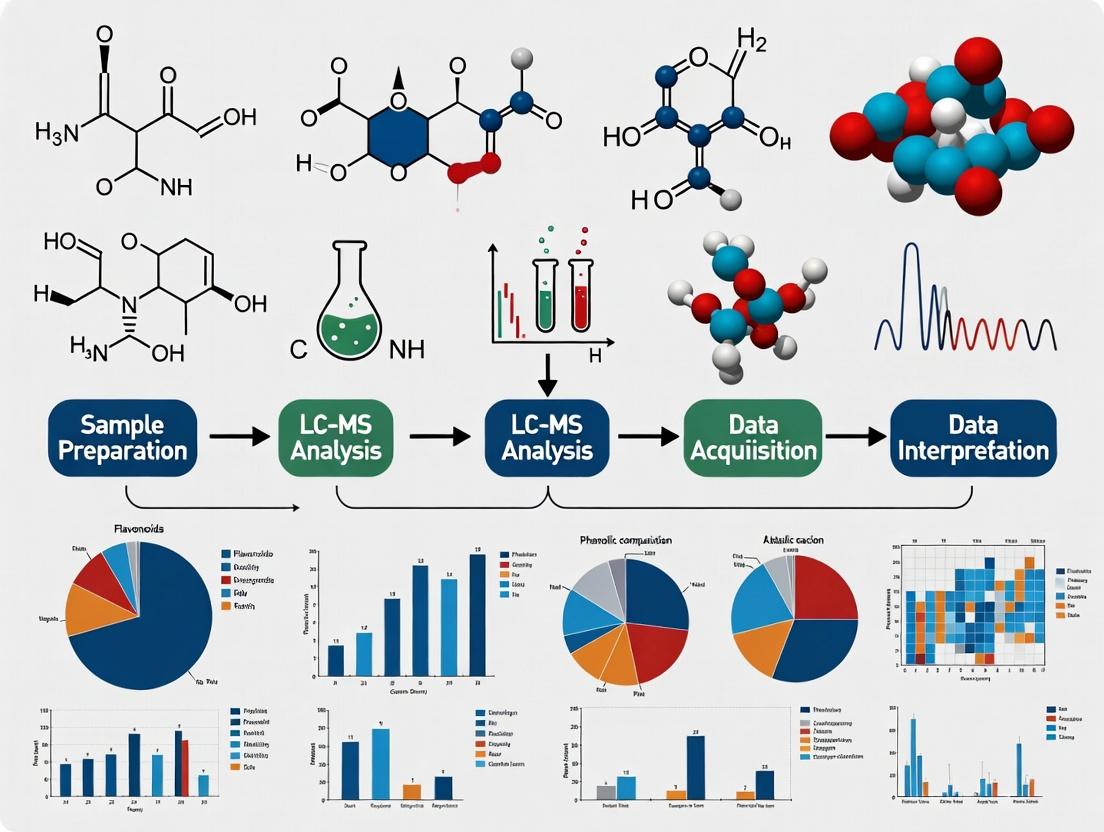
Visualization: Pathways and Workflows
Title: Phenolic Biosynthesis Pathways for Chemotaxonomy
Title: LC-MS Workflow for Phenolic Marker Discovery
The Scientist's Toolkit: Key Research Reagent Solutions
Table 3: Essential Materials for LC-MS Phenolic Chemotaxonomy
| Item/Reagent | Function & Rationale |
|---|---|
| Authenticated Plant Reference Material | Essential ground truth for building and validating chemotaxonomic models. Sourced from herbaria or certified suppliers. |
| LC-MS Grade Solvents (MeOH, ACN, H₂O) | Minimizes background noise, prevents ion suppression, and ensures reproducible chromatography. |
| Formic Acid (Optima LC-MS Grade) | Volatile ion-pairing agent (0.1% v/v) that enhances ionization efficiency and improves peak shape in acidic mobile phases. |
| Phenolic Standard Library | A curated set of >50 pure compounds (e.g., from Sigma, Extrasynthese) for absolute quantification and confirmation of marker identity. |
| Solid Phase Extraction (SPE) Cartridges (Strata-X, C18) | For sample clean-up to remove sugars and pigments that can foul the LC-MS system, crucial for complex extracts. |
| Lock Mass Solution | A solution of a known compound (e.g., leucine enkephalin) continuously infused for high-mass-accuracy correction during long runs. |
| Quality Control (QC) Pooled Sample | A pool of all study extracts injected repeatedly throughout the run to monitor system stability and for data normalization. |
| Retention Time Index Kit (e.g., FIA/Sheathflow) | A set of alkylphenones or other standards to calibrate retention times across instruments and laboratories. |
1.0 Introduction Accurate botanical origin confirmation is a critical, non-negotiable requirement in natural product research and phytopharmaceutical development. Variability in phenolic composition due to geographic, climatic, and genetic factors directly impacts safety, therapeutic efficacy, and the reproducibility of preclinical and clinical outcomes. This application note details a comprehensive LC-HRMS-based protocol for establishing a definitive phenolic chemical fingerprint to authenticate botanical material.
2.0 Key Data Summary: Impact of Geographic Origin on Marker Phenolics Table 1: Quantitative Variation of Key Phenolic Markers in *Echinacea purpurea Aerial Parts from Different Origins (µg/g dry weight). Data synthesized from recent literature and internal validation studies.*
| Phenolic Compound | Origin A (North America) | Origin B (Europe) | Origin C (Asia) | Reported Biological Activity |
|---|---|---|---|---|
| Cichoric Acid | 12,450 ± 1,230 | 8,570 ± 980 | 4,320 ± 650 | Immunomodulation, Antioxidant |
| Echinacoside | 1,230 ± 205 | 2,150 ± 310 | 980 ± 145 | Antioxidant, Neuroprotective |
| Chlorogenic Acid | 3,340 ± 420 | 2,890 ± 390 | 1,540 ± 230 | Anti-inflammatory, Metabolic |
| Cynarin | 85 ± 15 | 210 ± 35 | 45 ± 12 | Choleretic, Hepatoprotective |
| Total Phenolic Content (GAE) | 45.2 ± 3.1 mg/g | 38.7 ± 2.8 mg/g | 22.5 ± 2.1 mg/g | Aggregate Antioxidant Capacity |
3.0 Detailed Experimental Protocol: LC-HRMS Phenolic Fingerprinting
3.1 Sample Preparation (Solid-Liquid Extraction)
- Weighing: Precisely weigh 100.0 mg of lyophilized, homogenized botanical powder.
- Extraction: Transfer to a 15 mL centrifuge tube. Add 5.0 mL of extraction solvent (Methanol:Water:Formic Acid, 70:29:1, v/v/v).
- Sonication: Sonicate in an ice-water bath for 30 minutes (pulsed mode, 5s on/5s off).
- Centrifugation: Centrifuge at 12,000 x g for 15 minutes at 4°C.
- Filtration: Carefully collect the supernatant and filter through a 0.22 µm PTFE syringe filter into a 2 mL LC vial. Store at -80°C until analysis.
3.2 Instrumental Analysis (LC-HRMS Parameters)
- LC System: UHPLC with C18 reversed-phase column (100 x 2.1 mm, 1.7 µm particle size).
- Mobile Phase: A) 0.1% Formic acid in water; B) 0.1% Formic acid in acetonitrile.
- Gradient: 5% B to 30% B (0-15 min), 30% B to 95% B (15-20 min), hold 95% B (20-23 min), re-equilibrate (23-25 min).
- Flow Rate: 0.35 mL/min. Column Temp: 40°C. Injection Volume: 2 µL.
- MS System: High-Resolution Mass Spectrometer (Q-TOF or Orbitrap).
- Ionization: ESI negative mode. Mass Range: m/z 100-1500.
- Source Parameters: Capillary Voltage: -2.5 kV; Source Temp: 120°C; Desolvation Temp: 450°C; Cone Gas: 50 L/hr; Desolvation Gas: 800 L/hr.
3.3 Data Processing & Chemometric Analysis
- Raw Data Processing: Use vendor-neutral software (e.g., MZmine 3) for peak picking, alignment, and deconvolution.
- Normalization: Normalize peak areas to total ion current (TIC) or an internal standard (e.g., umbelliferone).
- Multivariate Analysis: Export aligned feature table for analysis in SIMCA or similar.
- PCA: Unsupervised pattern recognition to identify inherent clustering by origin.
- OPLS-DA: Supervised modeling to identify discriminant phenolic markers with high Variable Importance in Projection (VIP) scores.
4.0 Visualized Workflows and Pathways
Diagram Title: LC-MS Botanical Origin Verification Workflow
Diagram Title: Key Bioactivity Pathways of Phenolic Compounds
5.0 The Scientist's Toolkit: Essential Research Reagent Solutions Table 2: Key Materials and Reagents for Reproducible Phenolic Profiling
| Item | Function & Criticality | Example/Specification |
|---|---|---|
| Certified Reference Materials (CRMs) | Definitively identify and quantify target phenolics. Mandatory for method validation. | Cichoric acid, rutin, gallic acid from NIST or equivalent. |
| Stable Isotope-Labeled Internal Standards | Correct for ionization suppression/enhancement and extraction losses during LC-MS. | d3-Caffeic acid, 13C6-Quercetin for precise quantification. |
| LC-MS Grade Solvents & Additives | Minimize background noise, prevent ion source contamination, ensure run-to-run reproducibility. | LC-MS Grade Water, Acetonitrile, Methanol, Formic Acid (≥99.9%). |
| SPE Cartridges for Clean-up | Remove interfering sugars, chlorophyll, and lipids from complex botanical extracts. | Oasis HLB or C18 cartridges for selective phenolic retention. |
| UHPLC Column | Provide high-resolution separation of structurally similar phenolic isomers. | C18, 1.7-1.8µm, 100-150mm length, with phenyl or F5 phases for challenging separations. |
| Quality Control (QC) Pooled Sample | Monitor system stability, data quality, and reproducibility across analytical batches. | A homogeneous pool of all study extracts, injected at regular intervals. |
Application Notes
Liquid Chromatography-Mass Spectrometry (LC-MS) has become the cornerstone analytical technique for phenolic profiling in botanical origin confirmation research. Its unparalleled ability to separate complex matrices (LC) and provide sensitive, specific detection (MS) allows for the definitive identification and quantification of phenolic compounds, which serve as chemical fingerprints for plant species, geographic origin, and cultivation practices. This application note details its critical role within a thesis focused on validating the authenticity of medicinal botanicals.
Key Applications in Botanical Phenolic Profiling:
- Adulteration Detection: Differentiating between high-value botanicals (e.g., Vaccinium species, Ginkgo biloba) and cheaper substitutes by comparing unique phenolic signatures.
- Geographical Authentication: Correlating specific phenolic ratios or presence of marker compounds (e.g., specific flavonoids, phenolic acids) with geographic terroir.
- Standardization of Extracts: Quantifying active or marker phenolic compounds (e.g., curcuminoids, catechins, resveratrol) to ensure batch-to-batch consistency for pharmaceutical or nutraceutical development.
- Metabolite Profiling: Identifying and quantifying phenolic metabolites in biofluids for pharmacokinetic studies in drug development from botanical leads.
Quantitative Data Summary: LC-MS Analysis of Phenolics in Select Botanicals Table 1: Characteristic Phenolic Markers and Their LC-MS Parameters for Origin Confirmation
| Botanical Species | Target Phenolic Marker(s) | Primary Use / Significance | Typical Concentration Range (µg/g dry weight) | LC Retention Time (min) | MS Ionization Mode & Primary Ion [M-H]⁻ or [M+H]⁺ |
|---|---|---|---|---|---|
| Vaccinium myrtillus (Bilberry) | Delphinidin-3-O-galactoside | Authenticity vs. cheaper berries | 500 - 2500 | 8.2 | ESI⁻, 465.1 |
| Ginkgo biloba | Terpene Lactones (Ginkgolides) & Flavonol Glycosides | Standardization of extracts | 1000 - 5000 (for total flavonols) | 12.5 (for Quercetin-3-O-rutinoside) | ESI⁻, 609.1 |
| Curcuma longa (Turmeric) | Curcumin, Demethoxycurcumin, Bisdemethoxycurcumin | Adulteration detection & potency | 10,000 - 30,000 (for total curcuminoids) | 15.8 (Curcumin) | ESI⁺, 369.1 |
| Camellia sinensis (Green Tea) | Epigallocatechin Gallate (EGCG) | Bioactivity marker | 50,000 - 100,000 | 9.5 | ESI⁻, 457.1 |
| Hypericum perforatum (St. John’s Wort) | Hyperforin, Hypericin | Batch standardization | 2000 - 5000 (Hyperforin) | 22.1 (Hyperforin) | ESI⁺, 537.3 |
Experimental Protocols
Protocol 1: Comprehensive Phenolic Profiling for Botanical Fingerprinting
Objective: To generate a comprehensive phenolic profile from a botanical extract for origin confirmation.
Materials: Lyophilized plant material, methanol (LC-MS grade), formic acid (LC-MS grade), deionized water (18.2 MΩ·cm), acetonitrile (LC-MS grade), solid-phase extraction (SPE) cartridges (C18).
Instrumentation: UHPLC system coupled to a high-resolution Q-TOF or Orbitrap mass spectrometer.
Detailed Methodology:
Sample Preparation:
- Weigh 100 mg of finely powdered, lyophilized botanical material.
- Add 10 mL of 80% methanol in water (v/v) with 0.1% formic acid.
- Sonicate for 30 minutes at room temperature, then centrifuge at 10,000 x g for 10 minutes.
- Transfer supernatant. Repeat extraction on pellet and combine supernatants.
- Evaporate to dryness under a gentle nitrogen stream at 40°C.
- Reconstitute residue in 1 mL of 20% methanol in water with 0.1% formic acid. Filter through a 0.22 µm PTFE syringe filter prior to injection.
LC Conditions:
- Column: C18 reversed-phase column (100 x 2.1 mm, 1.7 µm particle size).
- Mobile Phase A: Water with 0.1% formic acid.
- Mobile Phase B: Acetonitrile with 0.1% formic acid.
- Gradient: 5% B to 95% B over 25 minutes, hold at 95% B for 3 minutes, re-equilibrate.
- Flow Rate: 0.3 mL/min. Column Temperature: 40°C. Injection Volume: 2 µL.
MS Conditions (ESI Negative/Ion Positive Switching):
- Ionization: Electrospray Ionization (ESI), dual polarity switching.
- Capillary Voltage: ±3.0 kV. Source Temperature: 150°C. Desolvation Temperature: 400°C.
- Cone Gas Flow: 50 L/hr. Desolvation Gas Flow: 800 L/hr.
- Full Scan Mode: m/z 100-1500 at 30,000 resolution (FWHM).
- Data-Dependent Acquisition (DDA): Top 3 most intense ions per scan fragmented with stepped collision energy (20, 40, 60 eV).
Data Analysis:
- Process raw data using metabolomics software (e.g., Progenesis QI, Compound Discoverer).
- Align peaks, deisotope, and perform compound identification by matching accurate mass (< 5 ppm) and MS/MS fragmentation patterns against online phenolic databases (e.g., Phenol-Explorer, MassBank).
Protocol 2: Targeted Quantification of Marker Phenolics Using LC-MS/MS (MRM)
Objective: To precisely quantify specific phenolic markers for batch standardization or adulteration testing.
Materials: As in Protocol 1. Also: Certified reference standards for target phenolics.
Instrumentation: UHPLC system coupled to a triple quadrupole (QQQ) mass spectrometer.
Detailed Methodology:
Sample & Standard Preparation:
- Prepare sample as in Protocol 1, Step 1.
- Prepare a series of calibration standards (e.g., 0.1, 1, 10, 100, 1000 ng/mL) for each target phenolic compound in a matrix-matched solvent.
LC Conditions: As described in Protocol 1, but optimized for the specific markers of interest.
MS/MS Conditions (Multiple Reaction Monitoring - MRM):
- Ionization: ESI in optimal polarity for each compound.
- Source Parameters: Optimized for maximum intensity of precursor ions.
- MRM Transitions: For each compound, define the precursor ion > product ion transition(s). Optimize collision energy for each transition.
- Example for EGCG: [M-H]⁻ 457.1 > 169.0 (Quantifier), 457.1 > 125.0 (Qualifier); Collision Energy: 25 eV.
- Dwell Time: 20-50 ms per transition.
Quantification:
- Integrate peak areas for the quantifier MRM transition for each analyte and standard.
- Generate a linear calibration curve (weighted 1/x).
- Calculate the concentration in the sample via extrapolation from the calibration curve, applying appropriate dilution factors.
Visualizations
Diagram Title: LC-MS Workflow for Botanical Phenolic Analysis
Diagram Title: LC-MS Role in Authentication Thesis Logic
The Scientist's Toolkit: Key Research Reagent Solutions
Table 2: Essential Materials for LC-MS Phenolic Profiling in Botanical Research
| Item / Reagent | Function & Importance in the Protocol | Key Considerations for Selection |
|---|---|---|
| LC-MS Grade Solvents (MeOH, ACN, Water) | Minimizes background chemical noise and ion suppression, ensuring high sensitivity and reproducible retention times. | Low UV absorbance, low volatile/ non-volatile residue, specific for LC-MS. |
| Acid Modifiers (Formic Acid, Acetic Acid) | Improves chromatographic peak shape (reduces tailing) and enhances ionization efficiency in ESI, especially for phenolics. | Typically used at 0.1%. Formic acid is common for positive mode; ammonium formate/acetate for negative. |
| Solid-Phase Extraction (SPE) Cartridges (C18) | Purifies and pre-concentrates samples, removing salts, sugars, and lipids that can foul the LC-MS system and complicate analysis. | Choice depends on analyte polarity. C18 is standard for medium-to-non-polar phenolics. |
| Certified Reference Standards | Enables definitive peak identification and accurate quantification. Critical for constructing calibration curves in targeted MRM assays. | Purity should be >95%. Preferably isotopically labeled internal standards for highest quantification accuracy. |
| UHPLC Columns (C18, 1.7-1.8µm) | Provides high-resolution separation of complex phenolic mixtures, resolving isomers (e.g., different glycosides) essential for fingerprinting. | Small particle size for high efficiency. Column chemistry (e.g., endcapped, polar-embedded) affects selectivity. |
| Synergy Filters (0.22 µm, PTFE or Nylon) | Removes particulate matter that could clog the UHPLC system and spectrometer capillary, protecting the instrument. | PTFE is chemically inert for organic solvents. Must be compatible with sample solvent. |
Within the framework of a doctoral thesis on Liquid Chromatography-Mass Spectrometry (LC-MS) phenolic profiling for botanical origin confirmation, the accurate identification and quantification of key phenolic classes are paramount. These compounds serve as chemical fingerprints, unique to plant species and influenced by geography, cultivar, and processing. This document provides detailed application notes and protocols for profiling four critical phenolic classes: hydroxybenzoic/cinnamic acids, flavonoids, lignans, and stilbenes, utilizing advanced LC-MS techniques.
The following table summarizes representative compounds, their mass ranges, and typical concentrations found in common botanical sources, based on recent literature and market analyses.
Table 1: Key Phenolic Classes for Botanical Profiling
| Phenolic Class | Core Structure | Representative Compounds (Examples) | Typical [M-H]⁻ m/z Range | Common Botanical Source | Reported Concentration Range (μg/g dry weight)* |
|---|---|---|---|---|---|
| Hydroxybenzoic Acids | C6-C1 | Gallic acid, Protocatechuic acid, Vanillic acid, Syringic acid | 137-185 | Green tea, berries, nuts | 50 - 5,000 |
| Hydroxycinnamic Acids | C6-C3 | Caffeic acid, Ferulic acid, p-Coumaric acid, Chlorogenic acid | 163-355 | Coffee, artichoke, cereals | 100 - 10,000 |
| Flavonoids | C6-C3-C6 | Quercetin (Flavonols), Cyanidin (Anthocyanidins), Naringenin (Flavanones), Epicatechin (Flavan-3-ols) | 271-611 | Citrus, cocoa, grapes, Ginkgo biloba | 200 - 20,000 |
| Lignans | (C6-C3)₂ | Secoisolariciresinol, Matairesinol, Pinoresinol | 357-419 | Flaxseed, sesame seeds, whole grains | 100 - 3,000 |
| Stilbenes | C6-C2-C6 | Resveratrol, Piceatannol, Pterostilbene | 227-271 | Grapes (skin), peanuts, blueberries | 0.1 - 100 |
*Concentration ranges are highly variable and source-dependent. Data synthesized from recent phytochemical surveys (2023-2024).
Detailed Experimental Protocols
Protocol 3.1: Standardized Extraction for Multi-Class Phenolic Profiling
Objective: To reproducibly extract the broad range of phenolic compounds from plant material. Materials: Freeze-dried plant powder (100 mg), 80% aqueous methanol (v/v) with 1% formic acid, ultrasonic bath, centrifuge, nitrogen evaporator. Procedure:
- Weigh 100.0 ± 0.5 mg of homogenized, freeze-dried botanical powder into a 15 mL polypropylene tube.
- Add 10 mL of cold extraction solvent (80% MeOH, 1% FA).
- Sonicate in an ice-water bath for 30 minutes (pulsed: 2 sec on, 1 sec off).
- Centrifuge at 10,000 x g for 15 minutes at 4°C.
- Decant and collect the supernatant.
- Re-extract the pellet with 5 mL of fresh solvent, repeat steps 3-5.
- Combine supernatants and evaporate to near-dryness under a gentle stream of nitrogen at 35°C.
- Reconstitute the residue in 1 mL of initial LC mobile phase (e.g., 2% acetonitrile in water, 0.1% formic acid). Vortex for 1 min, sonicate for 5 min.
- Filter through a 0.22 μm PTFE syringe filter into an LC vial. Store at -80°C until analysis.
Protocol 3.2: UHPLC-QTOF-MS Analysis for Untargeted Profiling
Objective: To separate and acquire high-resolution mass spectra for all phenolic classes in a single run. Chromatography:
- Column: C18 reversed-phase column (100 x 2.1 mm, 1.7 μm).
- Mobile Phase A: Water with 0.1% formic acid.
- Mobile Phase B: Acetonitrile with 0.1% formic acid.
- Gradient: 2% B to 40% B over 25 min, to 95% B at 28 min, hold for 2 min, re-equilibrate.
- Flow Rate: 0.35 mL/min. Column Temp: 40°C. Injection Volume: 2 μL.
Mass Spectrometry (Negative Ion Mode ESI):
- Instrument: QTOF mass spectrometer.
- Scan Range: m/z 100-1200.
- Capillary Voltage: 2500 V.
- Nebulizer Gas: 35 psi. Dry Gas: 10 L/min at 300°C.
- Collision Energy: 10 eV for MS1; 20-40 eV for MS2 (data-dependent acquisition).
- Reference Mass: Use a lock mass (e.g., hexakis(1H,1H,2H-difluoromethoxy)phosphazene) for real-time calibration.
Protocol 3.3: Targeted Quantification using LC-MS/MS (MRM)
Objective: To accurately quantify specific marker phenolics from each class. Chromatography: As in Protocol 3.2, but with a 15-min optimized gradient. Mass Spectrometry (MRM Mode):
- Instrument: Triple quadrupole MS.
- Ionization: Negative ESI for acids, positive/negative for flavonoids/lignans/stilbenes as optimized.
- Optimize compound-specific parameters (DP, CE, CXP) using pure standards. Quantification:
- Prepare a 6-point calibration curve (e.g., 0.01, 0.1, 1, 10, 100, 1000 ng/mL) for each phenolic standard.
- Use a stable isotope-labeled internal standard (e.g., d4-Resveratrol, 13C6-Caffeic acid) spiked into all samples and standards.
- Process data using peak area ratios (analyte/IS). Apply linear or quadratic regression with 1/x weighting.
Visualized Workflows and Pathways
Title: LC-MS Phenolic Profiling Workflow for Botanical Origin
Title: Biosynthetic Relationships of Key Phenolic Classes
The Scientist's Toolkit: Essential Research Reagents and Materials
Table 2: Key Reagent Solutions for LC-MS Phenolic Profiling
| Item | Function/Benefit in Profiling |
|---|---|
| Authenticated Botanical Reference Material | Essential for method validation and as a benchmark for authentic origin chemotypes. |
| Phenolic Compound Standard Library | Pure chemical standards for hydroxybenzoic/cinnamic acids, flavonoids, lignans, and stilbenes for peak annotation and quantification. |
| Stable Isotope-Labeled Internal Standards (e.g., 13C, 2H) | Correct for matrix effects and extraction losses during targeted LC-MS/MS quantification, ensuring accuracy. |
| LC-MS Grade Solvents (Acetonitrile, Methanol, Water) | Minimize background noise and ion suppression, ensuring reproducible chromatography and MS sensitivity. |
| Acid Modifiers (Formic, Acetic Acid) | Improve chromatographic peak shape (reduce tailing) and enhance ionization efficiency in ESI-MS, particularly in negative mode. |
| Solid Phase Extraction (SPE) Cartridges (C18, HLB) | For sample clean-up to remove sugars, pigments, and lipids that can interfere with analysis and foul the LC-MS system. |
| Quality Control (QC) Pooled Sample | A mixture of all study extracts, injected repeatedly throughout the batch to monitor instrument stability and data reproducibility in untargeted profiling. |
| Mass Spectrometric Databases (e.g., MassBank, GNPS, Phenol-Explorer) | Spectral libraries for matching acquired MS/MS fragmentation patterns to tentatively identify unknown phenolic compounds. |
Within the framework of LC-MS phenolic profiling for botanical origin confirmation, spectral libraries and curated databases serve as the cornerstone for accurate, reproducible, and high-throughput compound identification. These reference repositories transform raw LC-MS/MS data into actionable chemotaxonomic information. This document outlines the protocols for constructing orthogonal phenolic spectral libraries and provides methodologies for their application in verifying the geographic and species authenticity of botanical raw materials, a critical step in natural product drug development.
Key Application Notes:
- Authenticity & Adulteration Detection: By comparing the phenolic profile of an unknown sample against a validated reference library, researchers can identify non-compliant samples, detect adulterants, or confirm the declared botanical origin.
- Batch-to-Batch Consistency: Ensures the chemical fidelity of botanical extracts used in preclinical and clinical studies, linking bioactivity to specific phenolic compositions.
- Dereplication & Novelty Detection: Accelerates the identification of known phenolic compounds, allowing scientists to focus resources on characterizing novel or rare markers.
- Regulatory Compliance: Supports the documentation required by agencies (e.g., EMA, FDA) for the quality control of botanically-derived substances.
Protocols for Building a Reference Phenolic Spectral Library
Protocol: Generation of High-Quality Reference MS/MS Spectra
Objective: To acquire standardized, high-resolution MS/MS spectra for pure phenolic compounds to serve as library entries.
Materials & Reagents:
- Phenolic Reference Standards: A panel of authentic compounds representing major phenolic classes (e.g., flavonoids, phenolic acids, lignans, stilbenes).
- LC-MS/MS System: UHPLC coupled to a high-resolution tandem mass spectrometer (e.g., Q-TOF, Orbitrap).
- Mobile Phases: (A) 0.1% Formic acid in water; (B) 0.1% Formic acid in acetonitrile. LC-MS grade.
- Chromatography Column: Reversed-phase C18 column (e.g., 2.1 x 100 mm, 1.7-1.8 µm).
Procedure:
- Preparation: Individually dissolve each reference standard in a suitable solvent (e.g., methanol, DMSO) to a concentration of ~10 µg/mL.
- LC Conditions: Use a gradient elution (e.g., 5-95% B over 15 min). Maintain a constant flow rate (0.3 mL/min) and column temperature (40°C).
- MS Data Acquisition (DDA Mode):
- Operate in both positive and negative electrospray ionization (ESI) modes.
- Full MS scan range: m/z 100-1500.
- Use Data-Dependent Acquisition (DDA). Select the top 5 most intense ions per cycle for fragmentation.
- Set a dynamic exclusion window of 15 seconds.
- Apply stepped normalized collision energies (e.g., 20, 40, 60 eV) to capture fragmentation patterns across energy levels.
- Data Processing: Use vendor software to extract the consensus MS/MS spectrum for each compound, averaging spectra across the chromatographic peak and collision energies. Manually review spectrum quality.
Protocol: Creation of In-House Botanical Reference Material Profiles
Objective: To build a contextual library of phenolic profiles from authenticated botanical material of known origin.
Materials:
- Vouchered Plant Material: Samples with confirmed taxonomic identity (herbarium voucher) and documented geographic origin.
- Extraction Solvent: Methanol/Water (70:30, v/v) with 0.1% formic acid.
Procedure:
- Extraction: Weigh 100 mg of finely powdered plant material. Add 1 mL of extraction solvent. Sonicate for 30 minutes at room temperature. Centrifuge (13,000 x g, 10 min). Filter supernatant (0.22 µm PTFE) into an LC vial.
- LC-HRMS Analysis: Inject the extract using the LC-MS conditions from Protocol 2.1, but in Data-Independent Acquisition (DIA) or broad DDA mode to capture as many compounds as possible.
- Data Annotation: Process the data using software (e.g., Compound Discoverer, MS-DIAL). Annotate peaks by matching:
- Accurate mass (within 5 ppm).
- MS/MS spectra against the in-house library from Protocol 2.1.
- Retention time index (using a standard calibrant mixture).
- Isotopic pattern.
- Library Entry: For each authenticated sample, create a library entry containing the sample metadata (species, origin, collector) and the complete list of annotated phenolic compounds with their relative abundances (peak areas).
Table 1: Example Quantitative Summary of Phenolic Markers in Reference Botanical Materials
| Botanical Species (Origin) | Primary Phenolic Class | Key Marker Compound | Average Relative Abundance (Peak Area x10⁶) | Retention Time (min) | [M-H]⁻ (m/z) |
|---|---|---|---|---|---|
| Vaccinium myrtillus (Bulgaria) | Anthocyanins | Delphinidin-3-O-galactoside | 125.4 ± 10.2 | 4.52 | 465.1038 |
| Hypericum perforatum (Italy) | Acylphloroglucinols | Hyperforin | 893.7 ± 45.6 | 11.85 | 535.3176 |
| Camellia sinensis (Japan) | Flavan-3-ols | Epigallocatechin gallate | 654.2 ± 32.1 | 5.21 | 457.0776 |
| Curcuma longa (India) | Curcuminoids | Curcumin | 321.9 ± 25.8 | 7.88 | 367.1185 |
Protocols for Leveraging Libraries for Origin Confirmation
Protocol: Non-Targeted Phenotypic Profiling for Comparative Analysis
Objective: To compare an unknown botanical sample against a spectral database to determine its most likely origin.
Procedure:
- Sample Preparation & Analysis: Prepare and analyze the unknown sample identically to Protocol 2.2.
- Data Processing & Feature Alignment: Process the raw data to deconvolute and align all chromatographic peaks (features) across the unknown and the reference library profiles.
- Multivariate Statistical Analysis: Export the peak area table (features vs. samples) and import into statistical software (e.g., SIMCA, MetaboAnalyst).
- Perform Principal Component Analysis (PCA) for unsupervised pattern recognition.
- Use Orthogonal Projections to Latent Structures-Discriminant Analysis (OPLS-DA) to model differences between predefined groups (e.g., geographic origins).
- Marker Identification: Extract and identify the m/z and RT features with the highest contribution to the separation (VIP score > 1.5) by querying them against the spectral library.
Diagram Title: Workflow for Botanical Origin Confirmation
The Scientist's Toolkit: Research Reagent Solutions
Table 2: Essential Materials for LC-MS Phenolic Library Development
| Item | Function & Rationale |
|---|---|
| Authentic Phenolic Standards | Pure chemical references for unambiguous MS/MS spectrum acquisition and retention time calibration. |
| Certified Reference Plant Materials | Botanically and geographically vouchered samples to build ground-truthed, contextual spectral profiles. |
| Acquisition Software with DDA/DIA | Enables automated, reproducible MS/MS spectral acquisition for library building and unknown profiling. |
| Spectral Library Management Software | Platform (e.g., NIST MS Search, MS-DIAL, vendor-specific) to store, search, and manage custom libraries. |
| Chromatography QSR | A quality system suitability reference mix of phenolics to monitor LC-MS system performance daily. |
| Statistical Analysis Software | Essential for performing chemometric analyses (PCA, OPLS-DA) to interpret complex profiling data. |
From Sample to Spectrum: A Step-by-Step LC-MS Workflow for Phenolic Profiling
Within a broader thesis on Liquid Chromatography-Mass Spectrometry (LC-MS) phenolic profiling for botanical origin confirmation, the optimization of the initial extraction protocol is paramount. Phenolic compounds, including flavonoids, phenolic acids, and tannins, serve as critical chemotaxonomic markers. Their comprehensive recovery from complex botanical matrices is hindered by their diverse chemical structures, conjugation states (e.g., glycosylated, esterified), and susceptibility to degradation. This document provides detailed application notes and protocols for solvent selection, acid/alkaline hydrolysis, and quenching, aimed at maximizing the breadth and accuracy of phenolic profiles for subsequent LC-MS analysis in research and drug development.
Solvent Selection: A Tiered System for Comprehensive Recovery
Phenolic polarity spans a wide range. A single solvent is insufficient for comprehensive extraction. A tiered, sequential protocol is recommended.
Protocol 1.1: Sequential Solvent Extraction
Objective: To fractionate and extract phenolics based on polarity from a dried, homogenized botanical powder (e.g., Ginkgo biloba leaf, Vaccinium sp. berry). Materials: Lyophilized plant material (100 mg), ball mill, centrifuge, solvent evaporator (N₂ or vacuum). Reagents: See "Research Reagent Solutions" table.
Procedure:
- Defatting: To sample, add 1 mL of non-polar solvent (n-hexane). Vortex for 30 sec, sonicate in an ice bath for 10 min, and centrifuge at 10,000 × g for 5 min at 4°C. Discard supernatant (contains lipids, chlorophylls). Repeat once. Air-dry pellet.
- Medium-Polarity Extraction: To defatted pellet, add 1 mL of medium-polarity solvent (e.g., 80% aqueous methanol with 0.1% FA). Vortex, sonicate (ice bath, 15 min), centrifuge (10,000 × g, 10 min, 4°C). Transfer supernatant to a new tube. Repeat extraction twice on the pellet, pooling supernatants. This is Extract A (contains mid-polar aglycones, many glycosides).
- High-Polarity/Aqueous Extraction: To residual pellet, add 1 mL of high-polarity solvent (e.g., 50% aqueous acetone or water with 0.1% FA). Repeat sonication and centrifugation as in step 2, pooling supernatants. This is Extract B (contains highly polar phenolics, proanthocyanidins).
- Concentration: Evaporate Extracts A and B to dryness under a gentle stream of nitrogen or via vacuum centrifugation. Reconstitute each in 200 µL of initial LC-MS mobile phase (e.g., 2% acetonitrile in water with 0.1% FA), vortex thoroughly, filter through a 0.22 µm PTFE or nylon membrane, and transfer to an LC-MS vial.
Rationale: This sequential approach prevents solvent miscibility issues and selectively enriches different phenolic classes, reducing ion suppression in LC-MS.
Hydrolysis Protocols: Releasing Bound Phenolics
Many phenolics exist as soluble or insoluble conjugates. Hydrolysis is crucial for obtaining the "total phenolic" profile for chemotaxonomic comparison.
Protocol 2.1: Acid Hydrolysis for Anthocyanidins and Flavonoid Aglycones
Objective: To hydrolyze anthocyanins and flavonoid O-glycosides to their aglycone forms. Procedure:
- Take an aliquot of Extract A (or 5 mg of raw powder) in a screw-cap vial.
- Add 1 mL of methanol and 1 mL of 2 M hydrochloric acid (HCl).
- Flush vial headspace with nitrogen or argon, cap tightly.
- Heat at 90°C for 60 min in a dry bath or heating block.
- Immediate Quenching: Cool rapidly on ice. Neutralize carefully with 2 M sodium hydroxide (NaOH) to pH ~5-7. Critical: Perform this step within 1 minute of ending heating.
- Adjust final volume, filter, and analyze via LC-MS.
Protocol 2.2: Alkaline Hydrolysis for Phenolic Acids and Esters
Objective: To hydrolyze ester-bound phenolic acids (e.g., chlorogenic acids, hydroxycinnamates). Procedure:
- Take an aliquot of Extract B (or 5 mg of raw powder) in a vial.
- Add 2 mL of 2 M sodium hydroxide (NaOH). Flush with inert gas.
- Incubate at room temperature for 4 hours in the dark (to prevent oxidation).
- Immediate Quenching: Acidity immediately with concentrated hydrochloric acid (HCl) or formic acid (FA) to pH ~2-3 to re-protonate acids and stop hydrolysis.
- Extract liberated phenolic acids with ethyl acetate (3 x 1 mL). Pool ethyl acetate layers, evaporate, and reconstitute in mobile phase for LC-MS.
Quenching: Critical for Stabilization
Quenching is not merely stopping a reaction; it is a stabilization step to prevent post-hydrolysis degradation and oxidation.
Key Principles:
- Speed: Transfer reaction vial to ice bath immediately after the timed incubation.
- pH Control: Rapidly adjust pH to a stable range for the target analytes (typically acidic for LC-MS).
- Antioxidants: Consider adding 0.1% w/v of reducing agents like ascorbic acid or ethylenediaminetetraacetic acid (EDTA) to the quenching solution to chelate metals and prevent oxidation.
- Cold Solvent Dilution: For some protocols, diluting the reaction mixture 10-fold with ice-cold acidified solvent (e.g., methanol with 0.1% FA) is effective.
Protocol 3.1: Standardized Quenching Solution
Preparation: 0.1 M Hydrochloric Acid (HCl) in ice-cold 50% aqueous methanol, containing 0.1% ascorbic acid. Prepare fresh and keep on ice. Application: Add a 2x volume of quenching solution directly to the hot hydrolysis mixture at t = 0 min post-incubation. Mix vigorously, then proceed to neutralization/pH adjustment as required.
Data Presentation: Solvent Efficiency Comparison
Table 1: Recovery Efficiency of Phenolic Classes Using Different Extraction Solvents from Vaccinium myrtillus (Standardized to 100 mg DW)
| Phenolic Class (Example Compound) | 100% Methanol | 70% Aqueous Acetone | 80% Aqueous Methanol + 0.1% FA | Sequential Protocol (Hexane/80% MeOH/50% Acetone) |
|---|---|---|---|---|
| Anthocyanins (Cyanidin-3-glucoside) | 4.2 ± 0.3 mg/g | 5.1 ± 0.4 mg/g | 4.8 ± 0.2 mg/g | 4.9 ± 0.3 mg/g |
| Flavonol Glycosides (Quercetin-3-rutinoside) | 2.1 ± 0.2 mg/g | 2.5 ± 0.2 mg/g | 2.8 ± 0.3 mg/g | 2.8 ± 0.2 mg/g |
| Phenolic Acids (Chlorogenic acid) | 1.5 ± 0.1 mg/g | 1.7 ± 0.2 mg/g | 2.2 ± 0.2 mg/g | 2.0 ± 0.2 mg/g |
| Proanthocyanidins (DP > 4) | 8.5 ± 0.9 mg/g | 12.3 ± 1.1 mg/g | 9.2 ± 0.8 mg/g | 11.8 ± 1.0 mg/g |
| Total Identified Phenolics | 16.3 mg/g | 21.6 mg/g | 19.0 mg/g | 21.5 mg/g |
| LC-MS Signal Suppression (Matrix Effect, %) | -35% | -25% | -20% | -15% |
Table 2: Impact of Hydrolysis & Quenching on Phenolic Yield (% Increase vs. Non-Hydrolyzed Extract)
| Phenolic Type | Acid Hydrolysis (90°C, 60 min) | Alkaline Hydrolysis (RT, 4h) | Quenching Delay (2 min vs. Immediate) Effect on Yield |
|---|---|---|---|
| Anthocyanidins (Cyanidin) | +950% (from glycosides) | N/A | -40% (Degradation) |
| Flavonoid Aglycones (Quercetin) | +700% (from glycosides) | +5% | -25% |
| Esterified Phenolic Acids (Caffeic acid) | +10% | +300% | -60% (Oxidation) |
| Total Liberated Aglycones | +820% | +310% | N/A |
The Scientist's Toolkit: Research Reagent Solutions
Table 3: Essential Materials for Phenolic Extraction and Hydrolysis
| Item | Function & Rationale |
|---|---|
| Formic Acid (FA), LC-MS Grade | Acidifier in extraction solvents; suppresses analyte ionization, improves chromatography peak shape, and stabilizes acidic phenolics. |
| Hydrochloric Acid (HCl), 2 M Solution | Reagent for acid hydrolysis of glycosidic bonds. Must be high-purity to avoid metal contamination. |
| Sodium Hydroxide (NaOH), 2 M Solution | Reagent for alkaline hydrolysis of ester bonds. Prepare fresh with degassed water to minimize carbonate formation. |
| Ascorbic Acid | Reducing agent added to quenching solutions and sometimes extraction solvents to prevent oxidative degradation of labile phenolics. |
| Ethylenediaminetetraacetic Acid (EDTA) | Metal chelator added to extraction buffers (1-10 mM) to inhibit polyphenol oxidase (PPO) activity. |
| PTFE or Nylon Syringe Filters (0.22 µm) | For final extract filtration prior to LC-MS to remove particulate matter that could damage instrumentation. |
| Inert Atmosphere (N₂/Ar) Canister | For purging sample headspace during hydrolysis and solvent evaporation to prevent oxidation. |
| pH Meter with Micro-Electrode | Critical for accurate pH adjustment during quenching and sample reconstitution for reproducible LC-MS analysis. |
Workflow and Pathway Visualizations
Diagram Title: Comprehensive Phenolic Extraction and Hydrolysis Workflow
Diagram Title: Hydrolysis Pathway and Quenching Role
This application note details the development of a robust, high-resolution liquid chromatography method coupled to mass spectrometry (LC-MS) for the separation of complex phenolic compounds. The method is designed to support botanical origin confirmation research, where precise phenolic profiling serves as a chemical fingerprint to authenticate plant-derived materials. Optimization of column chemistry, mobile phase, and gradient elution is critical to resolving structurally similar phenolic acids, flavonoids, and their isomers.
Column Chemistry Selection
The stationary phase is the primary determinant of selectivity for phenolic compounds. Based on current literature and comparative studies, the following columns were evaluated.
Table 1: Evaluation of HPLC Column Chemistry for Phenolic Separations
| Column Type | Stationary Phase Chemistry | Key Advantages for Phenolics | Common Trade Names/Examples |
|---|---|---|---|
| C18 | Octadecylsilane (ODS) bonded silica | Excellent retentivity for most phenolics; wide pH range (2-8). | Agilent ZORBAX Eclipse Plus C18, Waters Acquity UPLC BEH C18 |
| Phenyl-Hexyl | Phenyl-propyl bonded silica | π-π interactions with aromatic rings; enhanced shape selectivity for isomers. | Phenomenex Luna Omega Polar C18, Supelco Ascentis Express Phenyl-Hexyl |
| Pentafluorophenyl (PFP) | Pentafluorophenylpropyl bonded silica | Multiple interaction modes (dipole-dipole, π-π, hydrophobic); superior isomer separation. | Restek Raptor Biphenyl, Thermo Scientific Accucore PFP |
| HILIC | Bare silica or polar functionalized silica | Retains very polar phenolics (e.g., glycosides); orthogonal mechanism to RPLC. | Waters Acquity UPLC BEH HILIC, Merck SeQuant ZIC-HILIC |
Protocol 1: Column Screening Experiment
- Equipment: LC-MS system with a quaternary pump, column oven, and PDA/MS detector.
- Test Columns: Install and condition each column from Table 1 (dimensions: 100 x 2.1 mm, 1.7-2.6 µm particle size).
- Test Analytes: Prepare a 10 µg/mL standard mix containing: caffeic acid, ferulic acid, quercetin-3-O-glucoside, kaempferol, rutin, and epicatechin.
- Initial Conditions: Mobile Phase A: 0.1% Formic acid in water. B: 0.1% Formic acid in acetonitrile. Isocratic hold at 5% B for 1 min, then gradient to 95% B over 10 min. Flow rate: 0.3 mL/min. Temperature: 35°C.
- Analysis: Compare chromatograms for peak capacity, resolution of critical pairs (e.g., caffeic vs. ferulic acid), peak symmetry, and MS response.
Mobile Phase Optimization
Mobile phase composition affects ionization efficiency (for MS) and chromatographic selectivity.
Table 2: Effect of Mobile Phase Modifiers on Phenolic Separation and MS Signal
| Modifier (in Water & ACN) | Typical Conc. | Impact on Separation | Impact on ESI-MS Signal (Negative Mode) |
|---|---|---|---|
| Formic Acid (FA) | 0.1% | Improves peak shape for acidic phenolics; common for general use. | Moderate signal suppression for some phenolics. |
| Acetic Acid (AA) | 0.1-1% | Slightly less acidic than FA; can alter selectivity. | Less suppression than FA for many acids; good choice. |
| Ammonium Formate | 5-10 mM | Provides buffering capacity; essential for reproducibility. | Can enhance [M-H]- signals; compatible with MS. |
| Ammonium Acetate | 5-10 mM | Buffers at near-neutral pH; useful for anthocyanins (positive mode). | Good compatibility; may reduce sensitivity for strong acids. |
Protocol 2: Modifier and pH Optimization
- Preparation: Prepare Mobile Phase A with four different modifiers: (i) 0.1% FA, (ii) 0.5% AA, (iii) 10 mM Ammonium Formate (pH ~3.5), (iv) 10 mM Ammonium Acetate (pH ~6.8). Use ACN with 0.1% modifier as Mobile Phase B.
- System: Use the best column from Protocol 1.
- Gradient: 5-95% B over 15 min.
- Evaluation: Monitor the resolution between a critical pair (e.g., two isomeric flavonoid glycosides) and the total ion chromatogram (TIC) peak area for a mid-polarity phenolic like quercetin.
Gradient Elution Profile Development
A tailored gradient is required to elute a wide range of phenolic polarities from simple acids to polymeric flavonoids.
Table 3: Optimized Gradient Profile for Comprehensive Phenolic Profiling
| Time (min) | % Mobile Phase B (0.1% FA in ACN) | Flow Rate (mL/min) | Purpose |
|---|---|---|---|
| 0.0 | 5 | 0.30 | Equilibration, retention of very polar compounds. |
| 2.0 | 5 | 0.30 | Isocratic hold for organic acids & simple phenolics. |
| 15.0 | 30 | 0.30 | Shallow ramp for separation of flavonoid glycosides. |
| 25.0 | 50 | 0.30 | Steeper ramp for separation of aglycones. |
| 30.0 | 95 | 0.30 | Elution of most non-polar compounds (e.g., prenylated flavonoids). |
| 32.0 | 95 | 0.30 | Column cleaning. |
| 32.1 | 5 | 0.30 | Quick return to initial conditions. |
| 35.0 | 5 | 0.30 | Column re-equilibration. |
Protocol 3: Gradient Steepness Optimization
- Design: Using the chosen column and mobile phase, test three gradient times (10, 20, and 30 min) from 5% to 95% B.
- Calculation: Calculate the peak capacity (P) for each run: P = 1 + (tG / w), where tG is gradient time and w is average peak width at base.
- Selection: Choose the gradient that provides P > 150 while maintaining baseline resolution for the earliest eluting critical pair.
The Scientist's Toolkit: Research Reagent Solutions
Table 4: Essential Materials for LC-MS Phenolic Method Development
| Item | Function/Description |
|---|---|
| Phenolic Compound Standards | Certified reference materials for method calibration, identification, and peak assignment. |
| MS-Grade Water & Acetonitrile | Ultra-pure, low-UV absorbance, and minimal ion contamination to ensure baseline stability and high MS sensitivity. |
| Ammonium Formate (MS-Grade) | Provides volatile buffering for consistent retention times and enhanced MS analyte ionization. |
| Formic Acid (Optima LC-MS Grade) | A common acidic modifier to improve chromatographic peak shape and act as a proton donor in ESI. |
| C18 Solid Phase Extraction (SPE) Cartridges | For sample clean-up of crude botanical extracts to remove pigments, lipids, and salts that can foul the column/MS. |
| Column Regeneration Kit | Includes seals and tools for maintaining column performance; necessary for analyzing complex plant extracts. |
Diagrams
Workflow for LC-MS Method Development
Analyte-Stationary Phase Interactions
This protocol is framed within a broader thesis investigating Liquid Chromatography-Mass Spectrometry (LC-MS) phenolic profiling for botanical origin confirmation. Accurate identification of phenolic compounds—crucial markers for plant taxonomy, authenticity, and bioactivity—is highly dependent on the optimization of MS ionization, polarity, and fragmentation parameters. This application note provides a detailed methodological guide for tuning these key parameters to enhance sensitivity, coverage, and confidence in phenolic compound identification for research and drug development.
Key Parameter Comparison and Quantitative Data
Table 1: Comparative Performance of ESI and APCI for Major Phenolic Classes
| Phenolic Class | Example Compounds | Recommended Ionization | Optimal Polarity | Relative Sensitivity (ESI vs. APCI)* | Key Fragmentation Ions (m/z) |
|---|---|---|---|---|---|
| Simple Phenolic Acids | Gallic, Caffeic, Ferulic acid | ESI | Negative (-) | ESI > APCI (10-20x) | [M-H]⁻; 169 (gallic), 179 (caffeic) |
| Flavonoids (Aglycones) | Quercetin, Kaempferol, Luteolin | ESI | Negative (-) | ESI > APCI (5-15x) | [M-H]⁻; 301 (quercetin), 285 (kaempferol) |
| Flavonoid Glycosides | Rutin, Hesperidin | ESI | Negative (-) | ESI >> APCI (50-100x) | [M-H]⁻; loss of 162 (hexose), 146 (rhamnose) |
| Condensed Tannins | Procyanidin B2 | ESI | Positive (+) | ESI > APCI (3-5x) | [M+H]⁺; 289 (retro-Diels-Alder fragment) |
| Volatile Phenolics | Eugenol, Thymol | APCI | Positive (+) | APCI > ESI (2-5x) | [M+H]⁺; 164 (eugenol), 151 (thymol) |
| Lignans | Secoisolariciresinol | APCI/ESI | Positive (+) | Comparable | [M+H]⁺; 361, 165 |
*Sensitivity comparison is instrument and compound-dependent; values represent typical order-of-magnitude differences observed in optimized workflows.
Table 2: Optimized Polarity Switching Timetable for Broad Phenolic Screening
| Time Segment (min) | Ionization Mode | Polarity | Collision Energy (eV) | Targeted Compound Class |
|---|---|---|---|---|
| 0.0 - 2.0 | ESI | Positive | 10 (Low) | Proanthocyanidins, basic phenolics |
| 2.0 - 25.0 | ESI | Negative | 10-20 (Ramped) | Primary window for acids, flavonoids, glycosides |
| 25.0 - 30.0 | ESI | Positive | 35 (High) | Post-elution column clean-up & high-mass tannins |
| 30.0 - 35.0 | APCI | Positive | 20 | Late-eluting, less polar phenolics (e.g., alkylphenols) |
Detailed Experimental Protocols
Protocol 1: Tuning Ionization Source Parameters for ESI and APCI
Objective: To optimize source conditions for maximum ion yield of phenolic compounds.
Materials: Standard mixture of phenolic acids (gallic, caffeic), flavonoid aglycones (quercetin), and glycosides (rutin) at 1 µg/mL in 50:50 methanol/water with 0.1% formic acid.
ESI Optimization Steps:
- Infusion: Directly infuse standard mix at 10 µL/min via syringe pump.
- Key Parameters: Sequentially tune:
- Capillary Voltage: Test 2.5 - 4.0 kV (positive) and 2.0 - 3.5 kV (negative). Optimal typically ~3.0 kV for negative mode phenolics.
- Nebulizer Gas Pressure: 30-50 psi. Optimize for stable total ion current (TIC).
- Drying Gas Flow/Temperature: 8-12 L/min; 300-350°C. Higher temperatures aid desolvation for glycosides.
- Sheath Gas/Heater: If available, set 10-12 L/min and 350-400°C for improved ion transmission.
- Monitor: The [M-H]⁻ ion signal for quercetin (m/z 301) or rutin (m/z 609). Aim for maximum intensity and stability.
APCI Optimization Steps:
- Infusion: As above. Use a higher flow rate (50-100 µL/min) if needed.
- Key Parameters:
- Corona Needle Current: 3-8 µA. Start at 4 µA for phenolic standards.
- Vaporizer Temperature: Critical. Test 350-500°C. Optimal for phenolics often 400-450°C to prevent thermal degradation.
- Nebulizer/Drying Gas: Similar to ESI.
- Monitor: The [M+H]⁺ ion signal for a less polar phenolic like eugenol (m/z 165).
Protocol 2: Implementing Data-Dependent Acquisition (DDA) with Polarity Switching
Objective: To acquire both MS1 and MS2 spectra for unknowns in a single chromatographic run.
LC Conditions: C18 column (2.1 x 100 mm, 1.7 µm). Gradient: 5-95% B over 25 min (A=0.1% Formic Acid in H₂O, B=0.1% Formic Acid in Acetonitrile). Flow: 0.3 mL/min.
MS Setup (Q-TOF or Orbitrap):
- MS1 Survey Scan: 100-1500 m/z. Resolution >30,000.
- Polarity Switching: Enable fast negative/positive switching. Use the timetable from Table 2.
- DDA Criteria:
- Top 3-5 most intense ions per cycle.
- Intensity threshold: 5,000 counts.
- Dynamic exclusion: 15 sec.
- Fragmentation:
- Collision Energy Ramping: Apply a compound-class-dependent CE.
- Phenolic acids: 10-20 eV
- Flavonoid glycosides: 25-40 eV (to observe aglycone and cross-ring cleavages)
- Aglycones: 30-50 eV (for rich RDA fragment patterns)
- Isolation Width: 1.3-2.0 m/z.
- Collision Energy Ramping: Apply a compound-class-dependent CE.
Protocol 3: Fragmentation Pattern Library Generation for Identification
Objective: To create an in-house spectral library for targeted botanical confirmation.
Procedure:
- Analyze Authentic Standards: Inject at least 20 phenolic standards relevant to your botanical system (e.g., rosmarinic acid for Lamiaceae) at 0.1, 1, and 10 µg/mL.
- Acquire MS/MS at Multiple CEs: For each standard, collect fragmentation spectra at three collision energies (e.g., 15, 30, 45 eV).
- Data Processing: Use software (e.g., Agilent MassHunter, Thermo Compound Discoverer) to:
- Extract precursor m/z, retention time, and all fragment ions.
- Generate a consensus MS/MS spectrum for each compound.
- Export spectra in .msp or .json format for library building.
- Library Application: Use this library to search against unknown peaks in botanical extracts. A match score >80% (with RT tolerance ±0.2 min) provides high-confidence identification.
Visualizations
Title: LC-MS Phenolic Profiling Decision Workflow
Title: Key Flavonoid Fragmentation Pathways
The Scientist's Toolkit: Research Reagent Solutions
| Item/Category | Function in Phenolic LC-MS Analysis | Example Product/Brand |
|---|---|---|
| Phenolic Acid & Flavonoid Standards | Essential for tuning, calibration, fragmentation library generation, and quantification. | Sigma-Aldrich Phytochemical Library; Extrasynthese Native Compound Standards. |
| LC-MS Grade Solvents & Additives | Minimize background noise, enhance ionization efficiency, and ensure column longevity. | Fisher Optima LC/MS; Honeywell CHROMASOLV Plus. |
| Acid Additives (Volatile) | Modifies mobile phase pH to control ionization state. Formic/Acetic Acid for positive mode; Ammonium Formate for negative mode. | Fluka MS Grade Formic Acid. |
| Solid Phase Extraction (SPE) Cartridges | Pre-concentrate and clean up botanical extracts, removing salts and non-phenolics. | Waters Oasis HLB; Phenomenex Strata-X. |
| UHPLC Columns (C18) | Core separation media. High efficiency (1.7-1.8 µm particles) for resolving complex phenolic mixtures. | Waters ACQUITY UPLC BEH C18; Agilent ZORBAX RRHD Eclipse Plus C18. |
| ESI & APCI Probe Inserts | Consumable parts requiring regular cleaning/replacement to maintain optimal ion source sensitivity. | Manufacturer-specific (e.g., Thermo, Agilent, Sciex) ESI/APCI capillaries, nebulizers. |
| Mass Calibration Solution | Ensures mass accuracy (< 5 ppm) critical for elemental composition assignment. | Agilent ESI-L Low Concentration Tuning Mix; Thermo Pierce LTQ Velos ESI Positive Ion Calibration Solution. |
| Internal Standards (Isotope Labeled) | Correct for matrix effects and ionization suppression/enhancement in quantitative assays. | e.g., ¹³C₆-Quercetin, D₆-Caffeic Acid (available from Cambridge Isotope Laboratories). |
Within the framework of LC-MS phenolic profiling for botanical origin confirmation, selecting the optimal data acquisition strategy is paramount. Phenolic compounds serve as reliable chemical fingerprints for authenticity and geographical tracing. This application note details three core mass spectrometry acquisition modes—Full Scan, Targeted (SIM/MRM), and Data-Dependent Analysis (DDA)—contrasting their capabilities, applications, and protocols within botanical research.
The choice of strategy balances comprehensiveness, sensitivity, and specificity. The following table summarizes key performance metrics.
Table 1: Comparison of LC-MS Data Acquisition Strategies for Phenolic Profiling
| Parameter | Full Scan | Targeted SIM/MRM | Data-Dependent Analysis (DDA) |
|---|---|---|---|
| Primary Objective | Untargeted profiling, discovery of markers | High-precision quantification of known compounds | Untargeted identification of components in a sample |
| Ionization Mode | Typically ESI+ and ESI- | ESI+ or ESI- optimized per analyte | Typically ESI+ and ESI- |
| Scan Speed | Moderate (1-2 Hz) | Very High (all dwell time on few ions) | Cyclical: MS1 scan (moderate) + successive MS2 scans (fast) |
| Sensitivity | Lower (signal spread over wide m/z range) | Highest (signal focused on specific m/z) | Moderate for MS1, compound-dependent for MS2 |
| Selectivity | Low | Very High | High (via MS2 fragmentation) |
| Dynamic Range | ~3 orders of magnitude | ~4-5 orders of magnitude | ~3 orders of magnitude |
| Ideal for Botanical Research | Initial screening, finding unknown phenolic patterns | Validating known marker phenolics (e.g., rosmarinic acid, quercetin) | Obtaining structural data for unknown phenolic compounds |
| Key Limitation | Poor sensitivity for trace analytes, no structural confirmation | Requires prior knowledge; cannot discover unknowns | Stochastic; may miss low-abundance ions in complex matrices |
Detailed Methodologies & Protocols
Protocol: Full Scan Profiling for Initial Botanical Screening
Objective: To acquire a comprehensive metabolic profile of a botanical extract for pattern recognition and origin discrimination.
Materials:
- LC-MS system with a quadrupole or time-of-flight (TOF) mass analyzer.
- C18 reversed-phase column (e.g., 2.1 x 100 mm, 1.7 µm).
- Solvents: Water with 0.1% formic acid (A), Acetonitrile with 0.1% formic acid (B).
- Standardized botanical extract (e.g., Origanum vulgare), 1 mg/mL in methanol:water (1:1).
Procedure:
- LC Conditions: Gradient: 5% B to 95% B over 20 min, hold 2 min, re-equilibrate. Flow: 0.3 mL/min. Column Temp: 40°C.
- MS Conditions (Q-TOF example):
- Ionization: ESI positive and negative modes, separate runs.
- Mass Range: m/z 50–1200.
- Scan Rate: 1.5 spectra/sec.
- Capillary Voltage: 3.0 kV (ESI+), 2.5 kV (ESI-).
- Nebulizer Gas: 30 psi. Drying Gas: 10 L/min, 325°C.
- Data Analysis: Process total ion chromatograms (TICs) and extracted ion chromatograms (EICs) of known phenolic masses. Use principal component analysis (PCA) on aligned peak lists to differentiate origins.
Protocol: Targeted MRM Quantification of Phenolic Markers
Objective: To precisely quantify specific phenolic acids and flavonoids that discriminate between geographic origins of a botanical (e.g., lavender).
Materials:
- Triple quadrupole LC-MS/MS system.
- C18 reversed-phase column (2.1 x 50 mm, 1.8 µm).
- Solvents: As in 3.1.
- Analytic Standards: Caffeic acid, rosmarinic acid, luteolin, apigenin (10 µg/mL stock solutions).
- Internal Standard: Deuterated quercetin (d3-Quercetin).
Procedure:
- LC Conditions: Fast gradient: 10% B to 80% B over 8 min. Flow: 0.4 mL/min.
- MS/MS Method Development:
- Directly infuse individual standards (100 ng/mL) to optimize precursor ion, fragmentor voltage, and select 2–3 optimal product ions per compound.
- Optimize collision energies for each transition.
- MRM Method: Create a timed MRM table. Example transitions (positive mode):
- Rosmarinic acid: m/z 361 → 163 (CE: 22 V), 361 → 197 (CE: 18 V)
- Luteolin: m/z 287 → 153 (CE: 25 V), 287 → 135 (CE: 30 V)
- Dwell Time: 20-50 ms per transition.
- Quantification: Prepare a 5-point calibration curve (1–500 ng/mL) with constant internal standard. Inject sample extracts in triplicate. Use peak area ratios (analyte/IS) for quantification.
Protocol: DDA for Structural Elucidation of Unknown Phenolics
Objective: To automatically acquire MS/MS spectra for the most abundant ions in a complex botanical extract (e.g., green tea polyphenols).
Materials:
- LC-MS system with tandem capability (Q-TOF, Orbitrap, or ion trap).
- Column and solvents as in 3.1.
- Green tea (Camellia sinensis) extract.
Procedure:
- LC Conditions: As per 3.1 to separate compounds.
- MS1 Survey Scan: Full scan from m/z 100–1000 at resolution >30,000 (for Orbitrap/TOF).
- DDA Criteria:
- Intensity Threshold: Top 10 most intense ions per cycle.
- Charge State Exclusion: +1 only (for small molecules).
- Dynamic Exclusion: Exclude previously fragmented ions for 30 sec.
- Isolation Width: m/z 1.5 (unitless for Q-TOF, 2 m/z for ion trap).
- MS2 Acquisition: Fragment selected precursors using stepped collision energies (e.g., 20, 35, 50 eV). Scan rate: 5–10 Hz.
- Data Analysis: Use software to deconvolute MS2 spectra, propose molecular formulas, and search against spectral libraries (e.g., MassBank, NIST) for phenolic compounds.
Visualized Workflows & Relationships
Title: DDA Acquisition Cycle for Phenolic ID
Title: Strategy Selection Based on Research Goal
The Scientist's Toolkit: Essential Research Reagents & Materials
Table 2: Key Reagents and Materials for LC-MS Phenolic Profiling
| Item | Function in Research | Example/Brand |
|---|---|---|
| Phenolic Acid & Flavonoid Standards | Calibration and positive identification for targeted MRM; retention time locking. | Rosmarinic acid, chlorogenic acid, quercetin (Sigma-Aldrich, Extrasynthese) |
| Stable Isotope-Labeled Internal Standards | Correct for matrix effects and ionization variability during quantification. | d3-Quercetin, 13C6-Caffeic acid (Cambridge Isotope Laboratories) |
| LC-MS Grade Solvents & Additives | Minimize background noise, ensure reproducible ionization and chromatography. | Acetonitrile, Methanol, Water, Formic Acid (Fisher Optima, Honeywell) |
| Solid Phase Extraction (SPE) Cartridges | Clean-up and pre-concentration of phenolic compounds from complex botanical matrices. | Strata-X (Phenomenex), Oasis HLB (Waters) |
| UHPLC Columns (C18, Phenyl) | High-resolution separation of structurally similar phenolic isomers. | Waters ACQUITY BEH C18, Thermo Scientific Accucore Phenyl-Hexyl |
| Mass Spectral Libraries | Database matching for putative identification of unknowns from DDA data. | NIST MS/MS, MassBank, Metlin |
| Q-TOF or Orbitrap Mass Calibrant | Ensure high mass accuracy (<5 ppm) for reliable formula assignment in Full Scan/DDA. | ESI-L Low Concentration Tuning Mix (Agilent), Pierce LTQ Velos ESI Positive Ion Calibration Solution (Thermo) |
Application Notes: LC-MS Phenolic Profiling for Botanical Authentication
Within the thesis framework of LC-MS phenolic profiling for botanical origin confirmation, phenolic and other signature secondary metabolites serve as chemical fingerprints to combat adulteration in the botanical supply chain. The following case studies illustrate targeted and untargeted approaches.
Table 1: Quantitative Marker Compounds for Authentication of Key Botanicals
| Botanical | Target Analytic(s) | Typical Concentration Range in Authentic Material | Common Adulterant / Issue | Distinguishing LC-MS Feature |
|---|---|---|---|---|
| Panax ginseng (Asian) | Ginsenosides Rg1, Re, Rb1, Rf | Rg1: 1.5-4.2 mg/g; Rf: 0.5-1.8 mg/g | Panax quinquefolius (American) | Presence of ginsenoside Rf (Asian); Presence of pseudoginsenoside F11 (American) |
| Curcuma longa (Turmeric) | Curcuminoids (Curcumin, DMC, BDMC) | Total: 20-50 mg/g | Adulteration with synthetic curcumin or cheaper Curcuma species | Ratios of Curcumin:DMC:BDMC; Detection of synthetic impurities (e.g., cis-isomers) |
| Ginkgo biloba | Flavonol glycosides, Terpene lactones | Flavonols: 24-32 mg/g; Lactones: 2-6 mg/g | Addition of pure rutin/quercetin or extraction residues | Specific glycosylation pattern (e.g., kaempferol-3-O-rutinoside); Presence of ginkgolic acids (toxic, removed in extracts) |
| Polyherbal Formulation | Multiple botanical-specific markers | Variable | Omission of high-cost ingredients, substitution | Detection/Negation of all expected marker ions; Chemometric pattern matching of full profile |
Experimental Protocols
Protocol 1: Untargeted Phenolic Profiling for Herbal Formulation Verification
- Sample Preparation: Weigh 100 mg of powdered formulation. Extract with 5 mL of methanol:water (70:30, v/v) containing 0.1% formic acid in an ultrasonic bath for 30 min at 25°C. Centrifuge at 10,000 x g for 10 min. Filter supernatant through a 0.22 μm PTFE syringe filter.
- LC-MS Analysis:
- Column: C18 reversed-phase (100 x 2.1 mm, 1.7 μm).
- Mobile Phase: (A) 0.1% Formic acid in water; (B) 0.1% Formic acid in acetonitrile.
- Gradient: 5% B to 95% B over 25 min, hold 5 min.
- MS: High-resolution Q-TOF in negative electrospray mode. Data acquired in full-scan (m/z 100-1500) with auto-MS/MS.
- Data Processing: Use software (e.g., MZmine, XCMS) for peak picking, alignment, and deconvolution. Build a reference library from authenticated single-botanical extracts. Perform PCA or OPLS-DA to identify formulation outliers.
Protocol 2: Targeted Quantification of Ginsenosides for Panax Species Differentiation
- Sample Preparation: Extract 200 mg of powdered root with 10 mL of 70% methanol under reflux for 1 hour. Evaporate an aliquot to dryness and reconstitute in 1 mL of LC-MS grade methanol/water (1:1).
- LC-MS/MS Analysis:
- Column: HSS T3 C18 (150 x 2.1 mm, 1.8 μm).
- Mobile Phase: (A) Water; (B) Acetonitrile, both with 10 mM ammonium acetate.
- Gradient: 10% B to 35% B (15 min), to 100% B (18 min).
- MS: Triple Quadrupole in negative MRM mode. Key transitions: Rf (845.5→799.5), F11 (845.5→475.4).
- Quantification: Use external calibration curves (0.1-100 μg/mL) for ginsenosides Rg1, Re, Rb1, and Rf. The presence and ratio of Rf to F11 confirm Asian vs. American origin.
Visualizations
Title: Untargeted Phenolic Profiling Workflow
Title: Decision Logic for Ginseng Speciation
The Scientist's Toolkit: Research Reagent Solutions
| Item / Reagent | Function in LC-MS Phenolic Profiling |
|---|---|
| Hybrid Quadrupole-TOF (Q-TOF) Mass Spectrometer | Provides high-resolution, accurate-mass data for untargeted fingerprinting and unknown compound identification. |
| Triple Quadrupole (QQQ) Mass Spectrometer | Enables highly sensitive and selective targeted quantification using Multiple Reaction Monitoring (MRM). |
| UPLC/HPLC C18 Reverse-Phase Column (e.g., 1.7-1.8 μm particle size) | Separates complex mixtures of phenolic compounds based on hydrophobicity. |
| MS-Grade Methanol, Acetonitrile, & Formic Acid | Ensure low background noise, prevent ion suppression, and enhance ionization efficiency (as mobile phase/additive). |
| Reference Standard Compounds (e.g., Curcumin, Ginsenosides, Ginkgolides) | Critical for constructing calibration curves, verifying retention times, and confirming MS/MS fragmentation patterns. |
| Chemometric Software (e.g., SIMCA, MetaboAnalyst) | Performs multivariate statistical analysis (PCA, OPLS-DA) on large MS datasets to identify discriminatory markers. |
Solving LC-MS Pitfalls: Troubleshooting Signal, Separation, and Identification Challenges in Phenolic Analysis
Within the broader thesis on LC-MS Phenolic Profiling for Botanical Origin Confirmation, a critical analytical challenge is the poor ionization efficiency of many phenolic compounds, particularly glycosylated flavonoids, phenolic acids, and certain aglycones. This poor ionization leads to low signal intensity, reduced sensitivity, and unreliable quantification, compromising the chemometric models used for origin authentication. This application note details synergistic strategies combining post-column additive infusion and electrospray ionization (ESI) source parameter optimization to robustly enhance phenolic signals in negative ion mode LC-MS.
Table 1: Impact of Post-Column Additives on Signal Intensity of Key Phenolic Classes
| Additive (0.1% v/v in IPA, 20 µL/min) | Flavonoid Glycosides (% Increase) | Phenolic Acids (% Increase) | Aglycones (% Change) | Notes |
|---|---|---|---|---|
| No Additive (Control) | 0% | 0% | 0% | Baseline in 0.1% Formic Acid. |
| Ammonium Hydroxide (0.1%) | 45% | 220% | -15% | Great for acids, suppresses some aglycones. |
| Diethylamine (DEA, 0.1%) | 180% | 150% | 5% | Most effective for glycosides; requires careful source cleaning. |
| Triethylamine (TEA, 0.1%) | 120% | 90% | -10% | Less effective than DEA for glycosides. |
| Aniline (0.05%) | 250% | 40% | 20% | Highest boost for glycosides; toxic, use with caution. |
Table 2: Optimized ESI Source Parameters for Negative Mode Phenolic Analysis
| Parameter | Typical Value | Recommended Optimized Range | Effect on Signal |
|---|---|---|---|
| Capillary Voltage (kV) | -2.5 | -2.8 to -3.2 | ↑ Higher voltage strengthens field, but can increase in-source fragmentation. |
| Source Temperature (°C) | 150 | 100 - 120 | ↓ Lower temp reduces thermal degradation of labile glycosides. |
| Desolvation Gas Flow (L/hr) | 800 | 600 - 700 | ↓ Lower flow may improve ionization efficiency for mid-polarity phenolics. |
| Cone Voltage / Fragmentor (V) | 40 | 20 - 30 | ↓ Lower voltage minimizes unwanted in-source collision-induced dissociation. |
| Nebulizer Gas (psi) | 45 | 35 - 40 | Optimize for stable spray; too high can cool droplets excessively. |
Experimental Protocols
Protocol 3.1: Post-Column Additive Infusion Setup Objective: To introduce a basic additive post-column to enhance deprotonation in the ESI source. Materials: HPLC system, T-connector, syringe pump (or secondary LC pump), low-dead-volume PEEK tubing, additive stock solution. Procedure:
- Connect the outlet of the LC column to one arm of a low-dead-volume T-connector.
- Connect a syringe pump (or a secondary LC pump) loaded with the additive solution to the second arm of the T-connector. Use 50-100 µL syringe for pump.
- Connect the third arm of the T-connector to the MS inlet using the shortest possible length of PEEK tubing (e.g., 15 cm, 0.005" ID).
- Set the LC flow rate (e.g., 0.3 mL/min) and the additive infusion rate (e.g., 20 µL/min). Ensure total flow is within the optimal range for the ESI source.
- Prepare additive stock (e.g., 1% v/v Diethylamine in Isopropanol). Dilute in-line to final concentration (e.g., 0.1%) by the combined flow.
- Equilibrate the system with mobile phase and additive flow for at least 15 minutes before analysis.
Protocol 3.2: Systematic ESI Source Parameter Optimization Objective: To empirically determine the optimal source parameters for maximum [M-H]⁻ signal of target phenolics. Materials: Standard mixture of representative phenolics (e.g., chlorogenic acid, rutin, quercetin), LC-MS system with tunable ESI source. Procedure:
- Inject the standard mixture using a generic gradient and a fixed, moderate additive infusion.
- Select one parameter (e.g., Capillary Voltage). While continuously infusing the standard, step through a predefined range (e.g., -2.5, -2.8, -3.0, -3.2 kV).
- Monitor the extracted ion chromatogram (EIC) peak area or height for 2-3 key analyte ions in real-time. Allow signal to stabilize for 30-60 seconds at each step.
- Record the signal response at each step. Reset the parameter to the value that yielded the maximum stable signal.
- Repeat steps 2-4 for the next parameter (e.g., Source Temperature), keeping others fixed at their newly optimized values.
- The recommended order is: Nebulizer Gas → Desolvation Gas Temp/Flow → Source Temperature → Capillary Voltage → Cone Voltage.
- Validate final parameters with a full chromatographic run of a complex botanical extract (e.g., Ginkgo biloba or green tea extract).
Visualization of Workflows and Concepts
Title: LC-MS Workflow with Additive Infusion for Phenolics
Title: Ionization Problems and Corresponding Solutions
The Scientist's Toolkit: Key Research Reagent Solutions
Table 3: Essential Materials for Phenolic Ionization Enhancement
| Item | Function & Rationale |
|---|---|
| Diethylamine (DEA), HPLC Grade | Primary Additive: A volatile base that drastically increases deprotonation efficiency of phenolic glycosides by acting as a gas-phase proton acceptor, boosting [M-H]⁻ signal. |
| Syringe Pump (or 2nd LC Pump) | Infusion Device: Provides precise, pulseless addition of additive solution post-column at µL/min flow rates. |
| Low-Dead-Volume PEEK T-Connector | Fluidic Mixing: Minimizes band broadening while allowing thorough mixing of column eluent with additive prior to ESI. |
| Phenolic Standard Mix | Optimization Calibrant: Contains representative acids, glycosides, and aglycones for systematic parameter tuning. |
| ESI Tuning & Calibration Solution | Source Baseline: Standard solution (e.g., Agilent Tuning Mix) used to calibrate and baseline the MS before phenolic-specific optimization. |
| Botanical Reference Material (e.g., NIST Green Tea) | Validation Matrix: Complex, real-world sample for final method validation and assessing signal enhancement gains. |
Within the research thesis on LC-MS Phenolic Profiling for Botanical Origin Confirmation, the primary analytical challenge is the separation of structurally similar phenolic compounds (e.g., glycosylated flavonoids, phenolic acids, isomers) present in complex botanical extracts. Co-elution leads to inaccurate quantification and misidentification, while peak tailing reduces sensitivity and resolution, compromising the definitive chemical fingerprint required for origin authentication. This document details advanced Liquid Chromatography (LC) solutions to address these specific issues, enabling robust, high-resolution separations as a prerequisite for accurate MS detection and multivariate data analysis.
Advanced LC Solutions: Mechanisms & Applications
A. Stationary Phase Engineering Novel stationary phase chemistries are critical for resolving co-elution.
- Core-Shell (Fused-Core) Particles: Provide high efficiency with lower backpressure than sub-2µm fully porous particles. Ideal for scaling methods from conventional HPLC to UHPLC platforms.
- Phenyl-Hexyl and Biphenyl Phases: Offer orthogonal selectivity to C18 phases through π-π interactions with aromatic rings of phenolic compounds, effectively separating isomers.
- Polar-Embedded and HILIC Phases: Mitigate peak tailing for acidic phenols (e.g., hydroxycinnamic acids) by reducing secondary interactions with residual silanols. HILIC is particularly useful for early-eluting, polar phenolic glycosides.
B. Mobile Phase Optimization & Additives Tailoring the mobile phase addresses both co-elution and tailing.
- Acidic Modifiers: Formic acid (0.1%) or acetic acid is standard. For difficult separations, trifluoroacetic acid (TFA at 0.01-0.05%) can dramatically improve peak shape for acidic analytes but may suppress MS ionization.
- Ammonium Salts: Ammonium formate or acetate (e.g., 2-10 mM) buffers the pH and provides a volatile MS-compatible solution. The ammonium ion can passivate silanol sites, reducing tailing.
- Temperature Optimization: Increasing column temperature (40-60°C) improves kinetics, reducing tailing and often enhancing selectivity for isomers.
Table 1: Comparison of Stationary Phases for Phenolic Separation
| Stationary Phase Type | Key Mechanism | Best For Resolving | Typical Reduction in Peak Tailing Factor* |
|---|---|---|---|
| Standard C18 | Hydrophobicity | General flavonoids | Baseline (Reference) |
| Phenyl-Hexyl | π-π interactions | Isomeric flavones (e.g., luteolin vs. apigenin) | ~15% |
| Polar-Embedded (e.g., C18-amide) | Hydrophobicity + H-bonding | Phenolic acids (e.g., chlorogenic acid) | ~40% |
| HILIC | Hydrophilicity | Polar glycosides (e.g., rutin) | Up to 60% for early eluters |
| Fused-Core C18 | Kinetic efficiency | All classes, for faster analysis | ~25% (vs. fully porous 5µm) |
*Representative average improvement based on published method comparisons.
Detailed Experimental Protocols
Protocol 1: Screening for Optimal Selectivity in Botanical Extracts Objective: Identify the best stationary phase to resolve co-eluting peaks in a Ginkgo biloba leaf extract (focus on flavonol glycosides and terpene lactones).
- Sample Prep: Accurately weigh 100 mg of dried, powdered leaf. Extract with 10 mL of methanol:water (70:30, v/v) in an ultrasonic bath for 30 min. Centrifuge at 10,000 x g for 10 min. Filter supernatant through a 0.22 µm PVDF syringe filter.
- LC Conditions (Scouting Gradient):
- Columns: Install four 100 x 2.1 mm, 2.7 µm columns in series or parallel: C18, Phenyl-Hexyl, Polar-Embedded, HILIC.
- Mobile Phase A: Water with 0.1% Formic Acid.
- Mobile Phase B: Acetonitrile with 0.1% Formic Acid.
- Gradient: 5% B to 95% B over 15 min (HILIC: start at 95% B to 5% B).
- Flow Rate: 0.4 mL/min.
- Temperature: 45°C.
- Injection: 2 µL.
- Analysis: Monitor at 260 nm and 350 nm. Compare chromatograms, noting the number of baseline-resolved peaks (Resolution, Rs > 1.5) and peak asymmetry factors (As) for major targets.
Protocol 2: Systematic Optimization to Minimize Peak Tailing Objective: Optimize mobile phase for sharp, symmetrical peaks of rosmarinic acid and salvianolic acids in a Salvia miltiorrhiza (Danshen) extract.
- Standard Solution: Prepare 10 µg/mL solutions of rosmarinic acid and lithospermic acid in methanol.
- LC Conditions (C18 Column, 100 x 2.1 mm, 1.7 µm):
- Test three different mobile phase systems:
- System 1: A: 0.1% FA in H₂O, B: 0.1% FA in ACN.
- System 2: A: 10 mM Ammonium Formate (pH ~3), B: ACN.
- System 3: A: 0.01% TFA in H₂O, B: 0.01% TFA in ACN.
- Use an isocratic method of 30% B for 5 min for direct comparison.
- Flow: 0.3 mL/min, Temp: 40°C, Injection: 1 µL.
- Test three different mobile phase systems:
- Evaluation: Calculate peak asymmetry factor (As) at 10% peak height for each analyte in each system. System 3 (TFA) will typically yield As closest to 1.0 but requires post-column infusion for MS compatibility if used.
Table 2: Key Research Reagent Solutions
| Item | Function in Phenolic LC-MS Profiling |
|---|---|
| Acetonitrile (LC-MS Grade) | Primary organic modifier; low UV cutoff and excellent MS compatibility. |
| Formic Acid (Optima LC-MS Grade) | Most common acidic modifier; promotes protonation, good for ESI+ and ESI-. |
| Ammonium Formate (LC-MS Grade) | Volatile buffer; stabilizes pH, suppresses silanol activity, improves peak shape. |
| Trifluoroacetic Acid (HPLC Grade) | Strong ion-pairing agent; excellent for peak symmetry of acids but can cause ion suppression. |
| Methanol (LC-MS Grade) | Extraction solvent and alternative modifier; different selectivity vs. ACN. |
| Deionized Water (≥18.2 MΩ·cm) | Base for aqueous mobile phase; must be ultrapure to avoid background ions. |
| Solid Phase Extraction (SPE) Cartridges (C18, HLB) | Clean-up and pre-concentration of crude botanical extracts to reduce matrix interference. |
| Reference Standard Mix (e.g., phenolic acids, flavonoids) | Essential for method development, peak identification, and calculating response factors. |
Integrated Workflow for Botanical Profiling
Diagram Title: LC-MS Botanical Profiling Optimization Workflow
Case Study: Green Tea Catechins
Green tea contains multiple catechin isomers (e.g., epicatechin, catechin, epigallocatechin gallate) that are prone to co-elution and tailing.
- Problem: Baseline separation of (-)-epicatechin (EC) and (+)-catechin (C) on standard C18.
- Solution: Employ a phenyl-hexyl column with a water-acetonitrile gradient acidified with 0.01% TFA. The π-π interactions with the phenyl ring differentiate the stereochemistry, while TFA ensures symmetric peaks.
- Result: Resolution (Rs) improved from 0.8 to 2.1. Peak asymmetry (As) for EGCG improved from 1.8 to 1.1, leading to more accurate integration and quantification for the origin profiling model.
Protocol 3: Final Method for Catechin Separation
- Column: Phenyl-Hexyl, 150 x 3.0 mm, 2.7 µm.
- Mobile Phase A: Water with 0.01% TFA. B: Acetonitrile with 0.01% TFA.
- Gradient: 5% B to 22% B over 25 min.
- Flow: 0.4 mL/min. Temp: 35°C.
- Detection: ESI-MS in negative mode, MRM transitions for EC (289>245), C (289>245), EGCG (457>169).
The accurate profiling of phenolic compounds using Liquid Chromatography-Electrospray Ionization Mass Spectrometry (LC-ESI-MS) is a cornerstone of modern botanical origin confirmation research. A core challenge is the presence of matrix effects, where co-eluting compounds from complex botanical extracts alter the ionization efficiency of target analytes, leading to signal suppression or enhancement. This compromises quantitative accuracy, method robustness, and the reliability of the phenolic "fingerprint" used for authentication. These Application Notes detail practical strategies for diagnosing, managing, and correcting matrix effects to ensure data integrity within a broader phytochemical validation thesis.
Diagnosis and Assessment of Matrix Effects
The first step is a quantitative assessment of matrix effects. The most accepted protocol is the post-column infusion method and the post-extraction spike method.
Table 1: Methods for Assessing Matrix Effect Magnitude
| Method | Protocol Summary | Calculation | Interpretation |
|---|---|---|---|
| Post-Column Infusion | Continuous infusion of analyte into LC eluent post-column while injecting blank matrix extract. | Visual inspection of signal stability in chromatogram. | A stable signal indicates no matrix effect. Suppression/Enhancement appears as a dip/peak correlated with matrix elution. |
| Post-Extraction Spike | Compare analyte response in neat solvent vs. spiked post-extraction into blank matrix. | ME% = (Peak Areaspiked matrix / Peak Areaneat standard − 1) × 100 | ME% = 0: No effect. ME% < 0: Suppression. ME% > 0: Enhancement. |
| Internal Standard (IS) Calibration | Use stable isotope-labeled internal standards (SIL-IS) for each analyte. | Compare calibration slope in matrix vs. solvent. | Slope ratio (matrix/solvent) ≈ 1 indicates compensated matrix effects. Deviation indicates residual effect. |
Protocol 2.1: Post-Extraction Spike for Quantitative ME%
- Prepare Samples: a. Neat Standard: Prepare analyte at low, mid, and high concentrations in mobile phase. b. Blank Matrix: Extract representative botanical sample (e.g., Ginkgo biloba leaf) using standard protocol without the target phenolics. c. Spiked Matrix: Spike the blank matrix extract with analytes at identical concentrations as the neat standard.
- LC-MS Analysis: Analyze all samples in random order with at least n=5 replicates.
- Data Analysis: Calculate ME% for each concentration. Report mean ME% and relative standard deviation (RSD). An |ME%| > 20% is typically considered significant.
Strategies for Suppression/Enhancement Management
Table 2: Strategic Approaches to Mitigate Matrix Effects
| Strategy Category | Specific Technique | Mechanism of Action | Advantages | Limitations |
|---|---|---|---|---|
| Sample Preparation | Selective Solid-Phase Extraction (SPE) | Removes non-target matrix components pre-injection. | High selectivity; can concentrate analytes. | Method development intensive; may lose some analytes. |
| QuEChERS (Quick, Easy, Cheap, Effective, Rugged, Safe) | Dispersive SPE cleanup removes sugars, acids, lipids. | Fast, high-throughput for many botanicals. | May be less selective than cartridge SPE. | |
| Chromatography | Improved Separation (Longer runs, gradient optimization) | Increases temporal resolution between analytes and matrix interferences. | Universal, reduces ESI competition. | Increases analysis time; may not fully resolve all interferences. |
| Hydrophilic Interaction LC (HILIC) | Different retention mechanism separates polar phenolics from different matrix classes. | Excellent for polar phenolic glycosides. | Long column equilibration; method transfer challenges. | |
| Internal Standardization | Stable Isotope-Labeled Internal Standards (SIL-IS) | Co-elutes with analyte, experiences identical ME, perfect compensation. | Gold standard for quantitation. | Expensive; synthetically challenging for many phenolics. |
| Structural or Analog IS | Similar chemistry to analyte, partial compensation. | More available and affordable. | May not perfectly co-elute; compensation can be incomplete. | |
| Calibration | Matrix-Matched Calibration | Calibrators prepared in blank matrix mimic sample ME. | Directly compensates for consistent ME. | Requires abundant, consistent blank matrix. |
| Standard Addition | Analyte is spiked at varying levels into the sample itself. | Compensates for ME without needing blank matrix. | Labor-intensive; sample consumption high. | |
| ESI Source & MS | Nano-ESI or Microflow LC | Reduced flow rates increase ionization efficiency, can alter ME landscape. | Increased sensitivity; sometimes reduced ME. | More technically demanding; clogging risk. |
| Alternative Ionization (e.g., APCI) | Gas-phase ionization less susceptible to some liquid-phase ME. | Can bypass certain suppression mechanisms. | Not suitable for non-volatile or thermally labile phenolics. |
Protocol 3.1: dSPE Cleanup for Phenolic Extracts (Modified QuEChERS)
- Extraction: Homogenize 1g dried botanical powder with 10 mL acidified methanol/water (80:20, v/v, 0.1% formic acid). Vortex and centrifuge.
- dSPE Cleanup: Transfer 1 mL supernatant to a 2 mL tube containing 150 mg MgSO₄ (dries) and 50 mg PSA (Primary Secondary Amine, removes sugars, fatty acids). Vortex vigorously for 1 min.
- Centrifugation: Centrifuge at 12,000 rpm for 5 min.
- Filtration & Analysis: Filter the supernatant through a 0.22 µm PTFE syringe filter into an LC vial. Analyze alongside uncleaned extract to assess ME reduction via Protocol 2.1.
The Scientist's Toolkit: Key Research Reagent Solutions
Table 3: Essential Materials for Managing Matrix Effects in Phenolic Profiling
| Item | Function & Rationale |
|---|---|
| Stable Isotope-Labeled Standards (SIL-IS) (e.g., [¹³C₆]-Quercetin, [D₆]-Caffeic Acid) | Ideal internal standards that co-elute with native analytes, providing perfect compensation for ionization suppression/enhancement during quantitation. |
| PSA (Primary Secondary Amine) Sorbent | A key dSPE sorbent for removing polar matrix interferences like sugars, organic acids, and some pigments from phenolic extracts. |
| C18 SPE Cartridges | For selective offline cleanup and concentration of medium-to-low polarity phenolics (aglycones, some glycosides) from complex botanical matrices. |
| Formic Acid (LC-MS Grade) | Mobile phase additive (typically 0.1%) to promote protonation [M+H]⁺ in positive ESI mode and improve chromatographic peak shape for acidic phenolics. |
| Ammonium Acetate / Ammonium Formate Buffers | Volatile buffers for mobile phase pH control, crucial for reproducible retention of phenolic acids and flavonoids in reversed-phase LC. |
| PFP (Pentafluorophenyl) LC Column | Stationary phase offering alternative selectivity to C18, often better separating isomeric phenolic compounds (e.g., flavonoid glycosides) from each other and matrix. |
| In-Line Filter (0.5 µm) & Guard Column | Protects the analytical column from particulate matter and retained matrix components, preserving separation performance and reducing long-term signal drift. |
Visualized Workflows and Relationships
Decision Workflow for Managing Matrix Effects
Mechanisms of Ion Suppression and Enhancement in ESI
Within the context of LC-MS phenolic profiling for botanical origin confirmation, the accurate identification and quantification of marker compounds are paramount. A significant analytical challenge arises from the presence of isobaric (same nominal mass) and isomeric (same exact mass, different structure) compounds prevalent in plant metabolomes. This application note details protocols for leveraging orthogonal data dimensions—MS/MS spectral libraries and chromatographic retention time (RT)—to resolve these interferences, ensuring high-confidence annotations essential for robust chemotaxonomic analysis.
Key Experimental Protocols
Protocol 2.1: Optimized LC-MS/MS Method for Phenolic Separation
Objective: Achieve baseline separation of critical isomeric pairs (e.g., quercetin-3-O-glucoside vs. quercetin-4'-O-glucoside) while generating high-quality MS/MS spectra.
Materials & Method:
- Column: Kinetex F5 (Pentafluorophenyl) 2.6 µm, 100 x 2.1 mm. Provides orthogonal selectivity to C18 for phenolic isomers.
- Mobile Phase:
- A: 0.1% Formic acid in water (LC-MS grade).
- B: 0.1% Formic acid in acetonitrile (LC-MS grade).
- Gradient: 5% B to 40% B over 25 min, then to 95% B by 30 min, hold for 5 min. Re-equilibration: 10 min.
- Flow Rate: 0.3 mL/min. Column Temp: 40°C.
- Injection Volume: 2 µL (partial loop mode).
- MS Parameters (Q-TOF or Tandem Quadrupole):
- Ionization: ESI Negative mode (preferred for phenolics).
- Capillary Voltage: 2500 V.
- Source Temp: 150°C; Desolvation Temp: 400°C.
- Data Acquisition: Full scan (m/z 100-1500) + data-dependent acquisition (DDA). Top 3 most intense ions per cycle fragmented with collision energies ramped from 20 to 40 eV.
Protocol 2.2: Establishment of a In-House Spectral & RT Library
Objective: Create a reference database for targeted botanical markers.
- Analyze authentic standards (minimum 10-20 µg/mL) in triplicate using Protocol 2.1.
- Record the average retention time (RT) and its standard deviation (RSD < 2%).
- Acquire MS/MS spectra at three collision energies (e.g., 15, 30, 45 eV). Merge spectra to create a representative fragmentation pattern.
- Populate a database with: Compound Name, Formula, Exact Mass, Adduct(s) Observed (e.g., [M-H]-), Average RT, RT Tolerance Window (typically ± 0.2 min or 3*SD), and the reference MS/MS spectrum.
- Use software (e.g., Skyline, MassHunter) to import and manage the library.
Protocol 2.3: Data Analysis for Interference Resolution
Objective: Apply orthogonal filters to unknown feature annotations.
- Feature Detection: Use software (e.g., MS-DIAL, XCMS) to deconvolute LC-MS data. Align features by m/z (5-10 ppm tolerance) and RT.
- Level 1 Identification (Confident): Match to in-house library requiring: i) m/z match < 5 ppm, ii) RT within pre-defined tolerance window, iii) MS/MS spectral match (Dot Product score > 0.80).
- Level 2 Identification (Probable): For features without an RT standard, use public MS/MS libraries (e.g., GNPS, MassBank) requiring high spectral similarity (Dot Product > 0.85) and plausible adduct formation.
- Isomeric Discrimination: For co-eluting isobars (e.g., m/z 463.088 [M-H]-), inspect diagnostic fragment ions. Kaempferol-3-O-glucoside yields a primary fragment at m/z 285 ([aglycone-H]-), while its isomer may show a different fragmentation pattern or relative abundance.
Data Presentation
Table 1: Quantitative Recovery of Phenolic Isomers in a Spiked Matrix
Analysis of key isomers in a blank tea extract matrix (n=6). Chromatographic separation achieved prior to MS/MS quantification.
| Compound Pair | Nominal Mass | Spiked Concentration (ng/mL) | Mean Recovered Concentration (ng/mL) | Accuracy (%) | RSD (%) | Resolution (Rs) |
|---|---|---|---|---|---|---|
| Quercetin-3-O-Glc / Quercetin-4'-O-Glc | 464.1 | 50.0 | 48.7 / 49.1 | 97.4 / 98.2 | 2.1 / 1.8 | 1.5 |
| Kaempferol-3-O-Rut / Kaempferol-7-O-Rut | 594.1 | 50.0 | 52.3 / 47.9 | 104.6 / 95.8 | 3.2 / 2.7 | 2.1 |
| (E)- / (Z)- Resveratrol | 228.1 | 100.0 | 96.5 / 102.3 | 96.5 / 102.3 | 4.1 / 3.5 | 1.8 |
| Catechin / Epicatechin | 290.1 | 200.0 | 194.2 / 205.8 | 97.1 / 102.9 | 1.9 / 2.3 | 1.2 |
Table 2: Confidence Levels for Phenolic Annotation
Hierarchical annotation scheme based on available orthogonal data.
| Confidence Level | Identification Criteria | Required Evidence | Example in Botanical Profiling |
|---|---|---|---|
| Level 1 | Confident | RT match to authentic standard (± 0.2 min), MS/MS match (Dot Prod > 0.80), exact mass (ppm < 5). | Quantification of rutin in Sophora japonica. |
| Level 2 | Probable | MS/MS match to public library (Dot Prod > 0.85), exact mass, plausible RT based on logP prediction. | Annotation of a quercetin hexoside isomer in Ginkgo biloba. |
| Level 3 | Tentative | Exact mass match to molecular formula, characteristic MS/MS fragments for compound class. | Putative annotation of a procyanidin dimer. |
| Level 4 | Unknown | Distinct m/z and RT, but no spectral or formula data. | Uncharacterized feature requiring further study. |
The Scientist's Toolkit
| Research Reagent / Material | Function in Overcoming Interferences |
|---|---|
| Pentafluorophenyl (F5) LC Column | Provides orthogonal retention mechanism to C18, enhancing separation of isomeric phenolic compounds based on dipole-dipole and pi-pi interactions. |
| Authentic Phenolic Standards | Essential for building in-house RT/MS/MS libraries, enabling Level 1 identification and calibration for quantification. |
| LC-MS Grade Acids (Formic, Acetic) | Improves chromatographic peak shape and ionization efficiency in ESI, critical for reproducible RT and sensitivity. |
| Stable Isotope-Labeled Internal Standards | Corrects for matrix effects and ion suppression, ensuring accurate quantification despite co-eluting interferences. |
| Hybrid Quadrupole-TOF or Orbitrap MS | Delivers high-resolution and accurate mass (HRAM) measurements for formula assignment, paired with MS/MS capability for structural elucidation. |
| C18 Trap Cartridges for SPE | Pre-concentrates and cleans up complex botanical extracts, removing salts and non-polar interferences that complicate the analysis. |
Visualized Workflows and Pathways
Orthogonal Data Filtering Strategy
Botanical Origin Confirmation Workflow
Confirming the botanical origin of plant-derived materials, such as those used in herbal supplements, pharmaceuticals, and food products, is critical for ensuring authenticity, safety, and efficacy. Liquid Chromatography-Mass Spectrometry (LC-MS) phenolic profiling has emerged as a powerful technique for this purpose, generating complex chromatographic data rich in chemical fingerprints. However, the accurate extraction of meaningful information from this data is contingent upon overcoming three principal data processing hurdles: consistent peak integration, effective baseline correction, and the resolution of co-eluting compounds through deconvolution. This application note details protocols and solutions for these challenges within a research workflow aimed at establishing definitive phenolic markers for botanical authentication.
Core Data Processing Challenges & Quantitative Comparisons
Table 1: Impact of Data Processing Methods on Phenolic Compound Quantitation
| Processing Step | Common Method | Key Challenge in Phenolic Profiling | Typical Impact on Concentration (RSD%) | Recommended Software/Tool |
|---|---|---|---|---|
| Baseline Correction | Polynomial Fitting | Variable baseline drift in complex botanical extracts. | 5-15% | MZmine 3, XCMS Online, Thermo Freestyle |
| Peak Integration | Traditional (Apex to Valley) | Inconsistent integration of asymmetrical phenolic peaks. | 10-25% | Skyline, MarkerView, Agilent MassHunter |
| Peak Deconvolution | Multivariate Curve Resolution (MCR) | Resolving isobaric and co-eluting flavonoids & phenolic acids. | 15-30% (if unresolved) | ADAP-GC/LC, MS-DIAL, PARAFAC2 algorithms |
Table 2: Performance Metrics of Deconvolution Algorithms for Overlapping Flavonoid Peaks
| Algorithm | Principle | Resolution Success Rate* (%) | Computational Demand | Best For |
|---|---|---|---|---|
| Multivariate Curve Resolution-Alternating Least Squares (MCR-ALS) | Iterative optimization of concentration & spectral profiles. | 85-92 | High | Complex clusters with spectral differences. |
| Fast Fourier Transform (FFT) Filtering | Frequency domain noise reduction & component separation. | 70-80 | Low | Pre-processing for slight co-elution. |
| Independent Component Analysis (ICA) | Statistical separation of independent source signals. | 75-85 | Medium | Peaks with orthogonal fragmentation patterns. |
| Gaussian Mixture Modeling (GMM) | Fits multiple Gaussian distributions to the peak shape. | 80-88 | Medium-High | Well-defined, symmetrical overlapping peaks. |
*Success Rate: Defined as the accurate extraction of ≥95% pure chromatographic and spectral profiles for each component in a test set of known binary/ternary phenolic mixtures.
Experimental Protocols
Protocol 3.1: Optimized Baseline Correction for LC-MS Phenolic Profiles
Objective: To remove systematic baseline drift and noise without distorting low-abundance phenolic signals. Materials:
- Raw LC-MS data file (.raw, .d, .mzML format)
- MZmine 3 (Open Source) or proprietary vendor software (e.g., Compound Discoverer 4.0) Procedure:
- Data Import: Import the LC-MS run into the processing software.
- Chromatogram Building: Generate a Base Peak Chromatogram (BPC) and Total Ion Chromatogram (TIC).
- Baseline Estimation: Apply the "Baseline Correction" module.
- Method Selection: Choose "Asymmetric Least Squares (AsLS)" or "Top-Hat Filter".
- Parameter Optimization:
- For AsLS: Set λ (smoothness) to 10^5 - 10^7 and p (asymmetry) to 0.001 - 0.01.
- For Top-Hat: Set structural element width to ~1.5x the average peak width at base.
- Validation: Visually inspect corrected chromatogram to ensure baseline is flat near zero and no peak material has been subtracted. Compare integrated area of a stable internal standard (e.g., chlorogenic acid-d3) before and after correction. RSD should be <5%.
Protocol 3.2: Robust Peak Integration for Asymmetrical Phenolic Peaks
Objective: To achieve consistent and accurate integration of target phenolic compound peaks across multiple samples. Materials:
- Baseline-corrected LC-MS data.
- Skyline software (for targeted analysis) or MarkerView (for untargeted). Procedure:
- Peak Detection: Use the "Chromatogram Peak Picker" with settings adjusted for phenolic compounds.
- Peak Width: Set to 70-80% of the average observed peak width.
- Noise Level: Determine from a silent region of the chromatogram (typically 3-5x the RMS noise).
- Integration Method Selection:
- For symmetrical peaks: Use the "Apex to Valley" or "Gaussian Fit" method.
- For asymmetrical/tailing peaks (common for phenolics): Use the "Integration by Second Derivative" or "Summation of Data Points" method, which is less sensitive to tailing.
- Manual Review & Alignment: Manually review all integrated peaks. Adjust integration boundaries to ensure consistency across all samples. Use retention time alignment if drift >0.1 min.
- Export Data: Export peak area and height lists for statistical analysis.
Protocol 3.3: Deconvolution of Co-Eluting Phenolic Compounds via MCR-ALS
Objective: To resolve the pure chromatographic and spectral profiles of two or more co-eluting phenolic compounds. Materials:
- LC-MS/MS data of the overlapping peak region (.mzML format).
- MATLAB or Python with MCR-ALS toolbox (or software like MS-DIAL). Procedure:
- Data Segmentation: Isolate the retention time window containing the overlapping peaks.
- Building the Data Matrix (D): Create a 2D matrix
D(m/z × time points) of ion intensities within the window. - Initial Estimation: Estimate the number of components (n) using Singular Value Decomposition (SVD). Obtain initial spectral or concentration profiles via Evolving Factor Analysis (EFA).
- ALS Optimization: Iteratively solve
D = C * S^T + Eusing the MCR-ALS algorithm.- Apply constraints: Non-negativity (concentration & spectra), unimodality (concentration profiles).
- Use pure MS/MS spectra (if available) as initial spectral estimates
S.
- Resolution & Validation: Evaluate resolved profiles
CandS. Use correlation with pure standards or fragmentation pattern matching to confirm identity. Calculate lack-of-fit and percent of variance explained (should be >95%).
Visualized Workflows and Relationships
Title: LC-MS Phenolic Data Processing Workflow
Title: Deconvolution Process via MCR-ALS
The Scientist's Toolkit: Key Research Reagent Solutions
Table 3: Essential Materials for LC-MS Phenolic Profiling & Data Processing
| Item | Function in Research | Example Product/Supplier |
|---|---|---|
| Phenolic Compound Standards | Authentication and calibration for target quantification; provides pure spectral libraries for deconvolution. | Sigma-Aldrich Phytochemical Library, ChromaDex Reference Standards. |
| Stable Isotope-Labeled Internal Standards | Corrects for matrix effects and ionization variability during peak integration; essential for precise quantification. | Chlorogenic acid-d3, Quercetin-d3 (Cambridge Isotope Laboratories). |
| LC-MS Grade Solvents & Buffers | Ensures minimal background noise, reducing baseline artifacts and spurious peaks. | Fisher Chemical Optima LC/MS, Honeywell CHROMASOLV. |
| Specialized Data Processing Software | Executes advanced baseline correction, peak integration, and deconvolution algorithms. | MZmine 3 (open source), Thermo Fisher Compound Discoverer, Agilent MassHunter. |
| High-Resolution LC-MS System | Generates the high-fidelity data required for resolving isobaric and co-eluting phenolics. | Thermo Q-Exactive series, Agilent 6546 Q-TOF, Waters Xevo G3. |
| Validated Botanical Reference Extracts | Serves as positive controls and training sets for developing processing parameters and marker discovery. | NIST Standard Reference Materials (e.g., SRM 3254 Green Tea), American Herbal Pharmacopoeia standards. |
Benchmarking the Method: Validation, Comparative Analysis, and Establishing Definitive Proof of Origin
Within the context of developing and accrediting an LC-MS/MS method for phenolic profiling to confirm the botanical origin of medicinal plants, validation is a mandatory step. This ensures the method is reliable, reproducible, and fit for its intended purpose. This document details application notes and protocols for evaluating the five core validation parameters, framed within a research thesis aiming to differentiate Vaccinium species (e.g., bilberry vs. blueberry) based on their phenolic fingerprint.
Validation Parameters: Application Notes & Protocols
Specificity
Application Note: Specificity is the ability to assess the analyte unequivocally in the presence of other components, such as matrix interferences or co-eluting phenolic compounds. In botanical profiling, it confirms that the target biomarkers (e.g., delphinidin-3-O-galactoside, chlorogenic acid) are resolved from other phenolic compounds and matrix peaks.
Experimental Protocol:
- Samples: Analyze (a) a blank solvent (methanol/water 80:20 v/v), (b) a placebo matrix (a phenolic-free model plant matrix), (c) a standard solution of target analytes, and (d) the authentic botanical sample (Vaccinium myrtillus extract).
- LC-MS/MS Conditions:
- Column: C18 (100 x 2.1 mm, 1.8 µm).
- Mobile Phase: (A) 0.1% Formic acid in water; (B) 0.1% Formic acid in acetonitrile.
- Gradient: 5-95% B over 20 min.
- MS: Targeted Multiple Reaction Monitoring (MRM) mode. Monitor at least two MRM transitions per analyte (quantifier and qualifier).
- Acceptance Criterion: The analyte peak in the spiked sample and authentic sample must be chromatographically resolved (resolution >1.5) and free from co-eluting interference at the same retention time (±0.1 min) in the blank and placebo. The qualifier-to-quantifier ion ratio must be within ±30% of that from the standard.
Diagram Title: Specificity Assessment Workflow in LC-MS
Sensitivity (LOD & LOQ)
Application Note: Sensitivity defines the lowest amount of an analyte that can be reliably detected (LOD) and quantified (LOQ). This is critical for detecting trace-level biomarker phenolics that may be characteristic of a specific botanical origin.
Experimental Protocol:
- Procedure: Prepare a serial dilution of a mixed phenolic standard (e.g., anthocyanins, flavonols, phenolic acids) in the calibration range. Inject each concentration in triplicate.
- LOD Calculation: Signal-to-Noise (S/N) method. LOD is the concentration yielding S/N ≥ 3.
- LOQ Calculation: Defined as the concentration yielding S/N ≥ 10 and a precision (RSD) of ≤20% and accuracy of 80-120%.
- Acceptance Criterion: The LOQ must be at or below the minimum required level for differentiating between botanical species based on key marker compounds.
Table 1: Example LOD and LOQ for Key Phenolic Markers
| Analyte | Class | LOD (ng/mL) | LOQ (ng/mL) | MRM Transition (Quantifier) |
|---|---|---|---|---|
| Chlorogenic Acid | Phenolic Acid | 0.5 | 1.5 | 353>191 |
| Quercetin-3-O-glucoside | Flavonol | 0.2 | 0.6 | 463>300 |
| Delphinidin-3-O-galactoside | Anthocyanin | 1.0 | 3.0 | 465>303 |
| Procyanidin B2 | Flavan-3-ol | 2.0 | 6.0 | 577>425 |
Linearity
Application Note: Linearity tests the method's ability to produce results directly proportional to analyte concentration. A linear response across a defined range is essential for accurate quantification of varied phenolic concentrations across different botanical samples.
Experimental Protocol:
- Calibration Standards: Prepare at least six non-zero calibration standards in triplicate, covering the expected range (e.g., LOQ to 500 µg/mL).
- Analysis: Inject in random order. Plot peak area vs. concentration.
- Statistical Evaluation: Perform least-squares linear regression. Calculate correlation coefficient (r), slope, intercept, and residual plot.
- Acceptance Criterion: r ≥ 0.995. The residual plot should show random scatter around zero.
Precision
Application Note: Precision expresses the closeness of agreement between independent test results under prescribed conditions. It includes repeatability (intra-day) and intermediate precision (inter-day, inter-analyst). This ensures the phenolic profile is consistently measurable.
Experimental Protocol:
- Sample Preparation: Prepare three different concentrations (low, mid, high) of a QC sample from a homogeneous V. myrtillus extract.
- Repeatability: One analyst analyzes each QC level six times within one day.
- Intermediate Precision: A second analyst repeats the procedure on a different day with freshly prepared solvents and standards.
- Calculation: Express precision as %RSD for the concentration of each major analyte at each level.
- Acceptance Criterion: Repeatability RSD ≤ 5%; Intermediate Precision RSD ≤ 10%.
Table 2: Precision Data for Key Analytics (Mid-Level QC)
| Analyte | Nominal Conc. (µg/mL) | Repeatability (%RSD, n=6) | Intermediate Precision (%RSD, n=12) |
|---|---|---|---|
| Chlorogenic Acid | 50.0 | 1.8 | 3.2 |
| Quercetin-3-O-glucoside | 25.0 | 2.1 | 4.5 |
| Delphinidin-3-O-galactoside | 30.0 | 3.0 | 6.1 |
| Procyanidin B2 | 15.0 | 2.5 | 5.7 |
Robustness
Application Note: Robustness measures the method's capacity to remain unaffected by small, deliberate variations in procedural parameters. It identifies critical steps in the LC-MS phenolic profiling workflow.
Experimental Protocol (Youden's Ruggedness Test):
- Select 7 Parameters: Slight variations are made to: (A) Column Temperature (±2°C), (B) Flow Rate (±0.05 mL/min), (C) Mobile Phase pH (±0.1), (D) Gradient Start %B (±1%), (E) Injection Volume (±1 µL), (F) Autosampler Temp (±2°C), (G) MS Drying Gas Temp (±10°C).
- Experimental Design: Perform 8 experiments, each combining high (+) or low (-) values for each parameter according to a predefined matrix.
- Response Measurement: Analyze a mid-level QC in each condition. Monitor responses: retention time (Rt), peak area, and resolution of a critical pair.
- Evaluation: Calculate the effect of each parameter variation. A large effect indicates a critical parameter requiring strict control.
Diagram Title: Robustness Test Design & Evaluation
The Scientist's Toolkit: Research Reagent Solutions
Table 3: Essential Materials for LC-MS Phenolic Profiling Method Validation
| Item | Function/Description |
|---|---|
| Hybrid Quadrupole-TOF or Tandem Quadrupole MS | High-resolution accurate mass (HRAM) for untargeted profiling or sensitive MRM for targeted quantification of phenolic compounds. |
| UHPLC-grade Solvents (Acetonitrile, Methanol, Water) | Minimal background interference, essential for high-sensitivity LC-MS analysis. |
| Acid Modifiers (Formic Acid, Trifluoroacetic Acid) | Improves chromatographic peak shape and ionization efficiency for phenolic compounds in ESI. |
| Stable Isotope-Labeled Internal Standards (e.g., 13C-labeled Quercetin) | Corrects for matrix effects and losses during sample preparation, improving accuracy. |
| Certified Reference Material (CRM) of Botanical Matrix (e.g., NIST Green Tea) | Provides a validated matrix for assessing method accuracy and recovery during validation. |
| Phenolic Compound Certified Reference Standards | Pure, characterized compounds for positive identification, calibration, and calculation of recovery. |
| Solid-Phase Extraction (SPE) Cartridges (C18, HLB) | For sample clean-up and pre-concentration of phenolic compounds from complex plant extracts. |
| In-line Solvent Degasser & Column Oven | Ensures mobile phase consistency and stable chromatographic retention times, critical for precision. |
This application note, framed within a broader thesis on LC-MS phenolic profiling for botanical origin confirmation, provides a comparative analysis of key analytical techniques. The authentication and quality control of botanicals demand precise, high-throughput phenolic fingerprinting. While HPLC-UV, HPTLC, and NMR are established methods, Liquid Chromatography-Mass Spectrometry (LC-MS) offers superior performance for comprehensive profiling. This document details the comparative advantages and provides protocols to implement LC-MS for definitive botanical origin research.
Comparative Performance Data
The following tables summarize key performance metrics for phenolic profiling, based on current literature and standard laboratory validations.
Table 1: General Method Comparison for Phenolic Profiling
| Parameter | LC-MS | HPLC-UV | HPTLC | NMR |
|---|---|---|---|---|
| Detection Limit | Low pg-fg (excellent) | Low ng (good) | Mid-high ng (moderate) | µg-mg (poor) |
| Selectivity | Extremely High (via m/z) | Moderate (λ-based) | Moderate (Rf-based) | High (chemical shift) |
| Throughput | High (fast acquisition) | Moderate | High (parallel samples) | Very Low (long acquisition) |
| Structural Elucidation | MS/MS provides fragmentation pathways | Limited to λ & Rt | Limited (co-chromatography) | Direct, definitive structure |
| Quantitation | Excellent (with stable isotope IS) | Good (external calibration) | Semi-quantitative | Quantitative (absolute qNMR) |
| Sample Prep Complexity | Moderate | Moderate | Low | High (extensive purification) |
Table 2: Representative Data for Key Phenolics in Vitis vinifera (Grape) Extract
| Compound | LC-MS (LOD, pg) | HPLC-UV (LOD, ng) | HPTLC (LOD, ng) | NMR (LOD, µg) | LC-MS ID Confidence (MS/MS Fragments) |
|---|---|---|---|---|---|
| Gallic Acid | 5 | 2 | 50 | 10 | 125→97, 79, 81 |
| Catechin | 10 | 5 | 100 | 25 | 289→245, 205, 179 |
| Quercetin-3-glucoside | 2 | 10 | 30 | 15 | 463→301 (aglycone loss) |
| Resveratrol | 1 | 3 | 20 | 5 | 227→185, 159, 143 |
Detailed Experimental Protocols
Protocol 1: High-Throughput LC-MS Phenolic Profiling for Botanical Authentication
Objective: To generate a comprehensive phenolic fingerprint for origin confirmation. Materials: See "The Scientist's Toolkit" below. Procedure:
- Extraction: Weigh 100 mg of powdered botanical sample. Add 10 mL of methanol/water/formic acid (70:29:1, v/v/v). Sonicate for 30 min at 25°C. Centrifuge at 12,000 x g for 10 min. Filter supernatant through a 0.22 µm PTFE syringe filter.
- LC Conditions:
- Column: C18 (100 x 2.1 mm, 1.8 µm).
- Mobile Phase: A) 0.1% Formic acid in water; B) 0.1% Formic acid in acetonitrile.
- Gradient: 5% B to 95% B over 18 min, hold 2 min, re-equilibrate.
- Flow Rate: 0.3 mL/min. Column Temp: 40°C. Injection: 2 µL.
- MS Conditions:
- Ionization: ESI, negative ion mode.
- Scan Range: m/z 100-1500.
- Data-Dependent Acquisition (DDA): Top 5 most intense ions per scan fragmented using CID.
- Source Parameters: Capillary Voltage, -3.0 kV; Drying Gas Temp, 325°C; Nebulizer, 35 psi.
- Data Analysis: Align chromatograms using cheminformatics software. Create a peak list with m/z, Rt, and intensity. Perform multivariate analysis (PCA, OPLS-DA) on the dataset to discriminate botanical origins.
Protocol 2: Comparative Analysis Using HPLC-UV (for Benchmarking)
Objective: To generate UV-based phenolic profiles for comparison with LC-MS data. Procedure:
- Use the same extract as Protocol 1.
- HPLC-UV Conditions:
- Column: C18 (250 x 4.6 mm, 5 µm).
- Mobile Phase: A) 2% Acetic acid in water; B) Acetonitrile.
- Gradient: 5% B to 60% B over 40 min.
- Flow Rate: 1.0 mL/min. Detection: 280 nm and 320 nm.
- Injection: 20 µL.
- Quantify against external standards of gallic acid, catechin, and quercetin.
Visualization of Workflows & Relationships
Diagram Title: LC-MS Phenolic Profiling Workflow for Botanical Authentication
Diagram Title: Ranking of Analytical Techniques by Performance Criteria
The Scientist's Toolkit: Essential Research Reagents & Materials
Table 3: Key LC-MS Reagent Solutions for Phenolic Profiling
| Item | Function & Rationale |
|---|---|
| LC-MS Grade Solvents (ACN, MeOH, Water) | Minimizes chemical noise and ion suppression, ensuring high-sensitivity MS detection. |
| Formic Acid (Optima LC/MS Grade) | Volatile ion-pairing agent (0.1%) to improve chromatographic peak shape and enhance ionization in ESI. |
| Phenolic Acid & Flavonoid Standards | For constructing calibration curves, validating methods, and tentative identification via Rt and m/z matching. |
| Stable Isotope-Labeled Internal Standards (e.g., 13C-Caffeic Acid) | Essential for precise and accurate quantitation, correcting for matrix effects and recovery losses. |
| Solid-Phase Extraction (SPE) Cartridges (C18, HLB) | For sample clean-up and pre-concentration of phenolics from complex botanical matrices. |
| U/HPLC Column (C18, 1.7-1.8 µm, 100-150 mm) | Provides high-resolution separation of complex phenolic mixtures prior to MS detection. |
| ESI Tuning & Calibration Solution | For optimal mass accuracy and sensitivity of the MS instrument prior to analysis. |
This document provides detailed application notes and protocols for the use of Principal Component Analysis (PCA), Partial Least Squares-Discriminant Analysis (PLS-DA), and Orthogonal Partial Least Squares-Discriminant Analysis (OPLS-DA) within a broader thesis research framework focusing on Liquid Chromatography-Mass Spectrometry (LC-MS) phenolic profiling for botanical origin confirmation. The ability to verify the botanical source of raw materials is critical in pharmaceuticals, nutraceuticals, and food safety. Multivariate statistical analysis of complex LC-MS datasets is the cornerstone for identifying discriminatory phenolic compounds and constructing robust classification models.
Core Multivariate Methods: Principles and Application
Principal Component Analysis (PCA)
- Purpose: An unsupervised method used for exploratory data analysis, dimensionality reduction, and outlier detection. It identifies the main sources of variance in the dataset without using class information.
- Role in Botanical Discrimination: The initial step to observe natural clustering of samples based on phenolic profiles, assess data quality, and identify any outliers before supervised modeling.
Partial Least Squares-Discriminant Analysis (PLS-DA)
- Purpose: A supervised method that finds components (latent variables) that maximize the covariance between the spectral data (X) and the class membership matrix (Y). It is used for classification and feature selection.
- Role in Botanical Discrimination: Builds a predictive model to classify unknown botanical samples and identifies phenolic ions (Variables of Importance in Projection, VIP) that most contribute to the separation between pre-defined botanical groups.
Orthogonal Partial Least Squares-Discriminant Analysis (OPLS-DA)
- Purpose: A supervised extension of PLS-DA that separates the systematic variation in X into two parts: 1) variation correlated to Y (predictive), and 2) variation orthogonal (uncorrelated) to Y. This simplifies model interpretation.
- Role in Botanical Discrimination: Provides clearer visualization and enhanced interpretability by isolating the class-discriminatory variation from other structured noise (e.g., batch effects), leading to more reliable biomarker (phenolic compound) discovery.
Detailed Experimental Protocols
Protocol 3.1: LC-MS Phenolic Profiling for Multivariate Analysis
Objective: Generate high-quality, reproducible LC-MS data suitable for PCA, PLS-DA, and OPLS-DA. Materials: Botanical specimens (e.g., Ginkgo biloba, Hypericum perforatum, Vaccinium spp.), LC-MS system (Q-TOF or Orbitrap preferred), C18 chromatographic column, extraction solvents (methanol, acidified water). Procedure:
- Sample Preparation: Homogenize 100 mg of dried botanical material. Extract with 1.0 mL of 70% methanol/30% water (v/v) with 0.1% formic acid in an ultrasonic bath for 30 min at 25°C. Centrifuge at 14,000 x g for 10 min. Filter supernatant through a 0.22 µm PVDF membrane.
- LC-MS Analysis: Inject 5 µL of filtrate.
- Chromatography: Gradient elution (A: 0.1% Formic acid in water; B: 0.1% Formic acid in acetonitrile). Flow rate: 0.3 mL/min. Run time: 20-25 min.
- Mass Spectrometry: Operate in negative electrospray ionization (ESI-) mode for most phenolics. Data-Dependent Acquisition (DDA) or Data-Independent Acquisition (DIA) for MS/MS. Mass range: 100-1500 m/z.
- Quality Control: Inject a pooled QC sample (mix of all extracts) every 5-10 analytical runs to monitor system stability.
Protocol 3.2: Data Preprocessing for Multivariate Analysis
Objective: Convert raw LC-MS files into a standardized data matrix. Software: XCMS Online, MS-DIAL, or similar. Procedure:
- Feature Detection: Perform peak picking, alignment, and gap filling across all samples.
- Matrix Creation: Export a peak table with rows as samples, columns as metabolite features (defined by m/z and retention time), and cells as peak intensity/area.
- Data Cleaning: Remove features with >30% missing values in QC or biological groups. Impute remaining missing values with 1/5 of the minimum positive value for its feature.
- Normalization: Apply probabilistic quotient normalization (PQN) to correct for overall signal drift.
- Scaling: Apply Pareto scaling (mean-centered and divided by the square root of the standard deviation) to reduce the influence of high-intensity ions while preserving data structure.
Protocol 3.3: Model Building, Validation, and Interpretation
Objective: Construct and validate robust discriminant models. Software: SIMCA, MetaboAnalyst, R (ropls, mixOmics packages). Procedure:
- PCA: Apply to the preprocessed, scaled data. Examine scores plot (PC1 vs. PC2) for natural clustering and outliers.
- PLS-DA/OPLS-DA Model Training: Use class labels (botanical source) as the Y-variable. Split data into training (70%) and test (30%) sets. Build the model on the training set.
- Model Validation:
- Cross-Validation: Use 7-fold cross-validation to determine the optimal number of components and avoid overfitting. Evaluate R²Y (goodness of fit) and Q² (goodness of prediction).
- Permutation Test: Perform 200 random permutations of the Y-vector. The original model's R² and Q² intercepts should be significantly higher than those from permuted models (p < 0.05).
- External Validation: Predict the hold-out test set. Calculate accuracy, sensitivity, and specificity.
- Biomarker Identification: From the validated OPLS-DA model, extract VIP scores. Select features with VIP > 1.5. Tentatively identify these features by matching their accurate mass and MS/MS fragments to databases (e.g., Metlin, GNPS, Phenol-Explorer). Confirm with analytical standards.
Data Presentation & Results
Table 1: Comparative Performance of Multivariate Models in Discriminating Three Vaccinium Species
| Model | Components (Predictive/Orthogonal) | R²X(cum) | R²Y(cum) | Q²(cum) | CV-Accuracy | Test Set Accuracy |
|---|---|---|---|---|---|---|
| PCA | 3 (N/A) | 0.72 | N/A | N/A | N/A | N/A |
| PLS-DA | 3 (N/A) | 0.68 | 0.95 | 0.88 | 92% | 89% |
| OPLS-DA | 1+2 | 0.65 | 0.94 | 0.90 | 94% | 91% |
Table 2: Key Discriminatory Phenolic Compounds Identified by OPLS-DA (VIP > 1.8)
| Tentative Identification | m/z [M-H]⁻ | RT (min) | VIP Score | Putative Role in Discrimination |
|---|---|---|---|---|
| Cyanidin-3-O-galactoside | 449.1082 | 8.2 | 2.4 | Marker for V. myrtillus |
| Chlorogenic Acid | 353.0878 | 6.5 | 2.1 | High in V. macrocarpon |
| Hyperoside | 463.0876 | 10.1 | 1.9 | Marker for V. corymbosum |
| Procyanidin B2 | 577.1352 | 9.8 | 1.9 | Contributes to separation of all species |
Visualized Workflows
Diagram 1: Botanical Discrimination via LC-MS & Multivariate Analysis
Diagram 2: OPLS-DA Validation & Interpretation Steps
The Scientist's Toolkit
Table 3: Research Reagent & Software Solutions for Botanical Discrimination Studies
| Item / Solution | Function / Purpose |
|---|---|
| LC-MS Grade Solvents (Methanol, Acetonitrile, Water with 0.1% Formic Acid) | Ensure high-purity mobile phases for optimal chromatographic separation and ionization efficiency, minimizing background noise. |
| Phenolic Compound Analytical Standards (e.g., Quercetin, Chlorogenic Acid, Catechin) | Essential for method development, calibration, and confirmation of metabolite identities suggested by statistical models. |
| Solid Phase Extraction (SPE) Cartridges (C18, Phenyl) | Optional clean-up step to concentrate phenolic compounds and remove interfering matrix components (e.g., sugars, chlorophyll). |
| Pooled Quality Control (QC) Sample | A mixture of all study extracts, injected repeatedly. Critical for monitoring LC-MS system stability during long batches and for data preprocessing. |
| Metabolite Databases (Phenol-Explorer, Metlin, GNPS) | Spectral libraries for tentative identification of discriminatory phenolic features based on accurate mass and MS/MS fragmentation patterns. |
Multivariate Analysis Software (SIMCA, MetaboAnalyst 5.0, R packages ropls, mixOmics) |
Platforms for performing PCA, PLS-DA, OPLS-DA, and associated validation tests (cross-validation, permutation). |
| Data Preprocessing Platforms (XCMS Online, MS-DIAL, MZmine 3) | Convert raw vendor LC-MS files into a feature intensity matrix suitable for statistical analysis. |
Within botanical origin confirmation research, Liquid Chromatography-Mass Spectrometry (LC-MS) phenolic profiling serves as a cornerstone for establishing identity, authenticity, and traceability. The transition from a raw analytical "fingerprint" to a legally and regulatorily defensible dossier requires a meticulous, documented chain of evidence. This application note outlines the integrated protocols and documentation strategies necessary to construct an unassailable case for botanical origin, critical for FDA Botanical Drug Development (Botanical Review Team, BRT) guidance, EMA herbal medicine submissions, and DSHEA-related GRAS/NDI notifications.
Core Analytical Protocol: LC-MS Phenolic Profiling for Defensibility
This standardized protocol is designed to generate forensically valid data.
2.1 Materials & Sample Preparation
- Plant Material: Voucher specimens must be deposited in a recognized herbarium, with certificate cited. All working samples are traceable to this voucher (Chain of Custody Form AN-001).
- Extraction: Precisely weigh 100.0 ± 0.1 mg of dried, homogenized botanical powder. Add 10.0 mL of methanol:water (70:30, v/v) with 0.1% formic acid. Sonicate (30°C, 30 min), centrifuge (10,000 × g, 10 min), filter (0.22 μm PTFE). Prepare in triplicate.
- Reference Standards: A minimum of 12 phenolic compounds spanning subclasses (e.g., hydroxycinnamic acids, flavonols, flavan-3-ols) are analyzed concurrently for quantification and spectral library building.
2.2 Instrumental Parameters (UHPLC-Q-TOF-MS)
- Column: C18 (100 x 2.1 mm, 1.7 μm)
- Gradient: Water (A) and Acetonitrile (B), both with 0.1% Formic Acid. 5% B to 95% B over 18 min.
- Flow Rate: 0.3 mL/min
- MS: Electrospray Ionization (ESI), negative mode. Mass range: m/z 100-1500. Collision energies: 20, 40 eV for MS/MS.
2.3 Data Processing & Metabolite Annotation
- Use aligned peak tables (Retention Time ± 0.1 min, m/z ± 5 ppm). Annotate using:
- Level 1: Match to in-house standard RT and MS/MS.
- Level 2: Match to public spectral library (e.g., GNPS, MassBank).
- Level 3: Putative class based on m/z and fragmentation patterns.
- Documentation Requirement: All processing parameters (S/N threshold, alignment tolerance) and annotation confidence levels must be recorded in the Laboratory Information Management System (LIMS).
Quantitative Data Presentation: The Comparative Profile
Table 1: Phenolic Marker Quantification in Echinacea purpurea Aerial Parts vs. E. angustifolia Root (μg/g dry weight, n=6)
| Phenolic Compound (Class) | E. purpurea (Mean ± SD) | E. angustifolia (Mean ± SD) | p-value (t-test) | Key Differentiator |
|---|---|---|---|---|
| Cichoric Acid (CAF) | 24560 ± 1250 | 420 ± 85 | <0.001 | Primary Marker for E. purpurea |
| Echinacoside (Phen. Gly.) | 15 ± 5 | 18500 ± 950 | <0.001 | Primary Marker for E. angustifolia |
| Cynarin (DCAF) | 120 ± 25 | 8500 ± 420 | <0.001 | Confirmatory for E. angustifolia |
| Chlorogenic Acid (CAF) | 1850 ± 110 | 650 ± 90 | <0.001 | Supportive |
| Rutin (Flav. Gly.) | 3200 ± 205 | 120 ± 30 | <0.001 | Supportive for E. purpurea |
Table 2: Statistical Model Performance for Origin Classification
| Model | Features Used | Accuracy (%) | Specificity (%) | Sensitivity (%) | Cross-Validated R² |
|---|---|---|---|---|---|
| PLS-DA | Top 20 Phenolic Peaks | 98.7 | 99.1 | 98.2 | 0.94 |
| Random Forest | Full Profile (≥150 peaks) | 99.5 | 99.8 | 99.3 | 0.96 |
| Linear SVM | 5 Key Markers (Table 1) | 97.2 | 96.5 | 97.8 | 0.91 |
The Scientist's Toolkit: Essential Research Reagents & Materials
Table 3: Key Research Reagent Solutions for Defensible Phenolic Profiling
| Item | Function & Rationale for Defensibility |
|---|---|
| Certified Reference Standards | Enables Level 1 identification and absolute quantification. Certificates of Analysis (CoA) are mandatory for regulatory submission. |
| Isotopically Labeled Internal Standards (e.g., Caffeic Acid-d3) | Corrects for extraction efficiency and matrix-induced ionization suppression, improving data accuracy. |
| SRM 3251 (Cinnamon) or In-House QC Extract | Provides longitudinal system suitability data. Documents instrumental performance over the study period. |
| Voucher Specimen & Herbarium Certificate | Legally anchors the botanical identity. The foundational document for all claims of origin. |
| Chain of Custody (CoC) Forms | Documents every transfer of the physical sample, critical for audit trails and forensic defensibility. |
| Validated SOPs (e.g., SOP-LC-005) | Standardizes every action from weighing to reporting, ensuring reproducibility and compliance with GLP principles. |
From Data to Documentation: The Defensible Workflow
(Title: Workflow for Building a Defensible Botanical Origin Case)
Regulatory Submission Protocol: Assembling the Dossier
Protocol 6.1: Compilation of the Technical Document
- Cover Sheet & Executive Summary: Clearly state botanical Latin name, part used, and primary chemical differentiating claim.
- Volume 1: Identity & Characterization:
- Herbarium certificate and macroscopic/microscopic description.
- LC-MS Phenolic Profile Report: Include Tables 1 & 2. Provide annotated chromatograms and representative MS/MS spectra for key markers.
- Method validation data (specificity, LOD/LOQ, precision, accuracy).
- Volume 2: Supporting Stability & Batch Data:
- Demonstrate phenolic profile stability under proposed storage conditions.
- Provide QC data from multiple batches/lots to establish natural variability ranges.
- Volume 3: Cross-Referenced Compliance:
- Map all data to relevant regulatory guidelines (e.g., FDA Guidance for Industry: Botanical Drugs, EMA's HMPC guidelines).
- Include a complete audit trail and references to all SOPs used.
The Validation Pathway: Connecting Chemistry to Claim
(Title: Scientific & Regulatory Validation Pathway)
The verification of botanical origin via phenolic profiling using Liquid Chromatography-Mass Spectrometry (LC-MS) is a critical research area with implications for pharmaceuticals, nutraceuticals, and food safety. A core challenge lies in the transferability of complex multi-analyte methods between laboratories and instrument platforms. This application note details standardized protocols for inter-laboratory comparisons (ILCs) and proficiency testing (PT) to ensure the reliability and reproducibility of phenolic profiling data, a fundamental requirement for robust scientific conclusions in botanical origin research.
Quantitative Data from Recent Collaborative Studies
Table 1: Summary of Key Performance Indicators from Recent LC-MS Phenolic Profiling PT Schemes
| PT Scheme & Year | Analyte Classes | No. of Labs | Target CV% (Horwitz) | Achieved Mean CV% | Key Challenge Identified |
|---|---|---|---|---|---|
| CAMAG Ginkgo biloba PT (2023) | Flavonol glycosides, Terpene lactones | 24 | <15% | 12.8% | Standard hydrolysis efficiency |
| AOAC Vaccinium spp. PT (2024) | Anthocyanins, Procyanidins | 32 | <20% | 18.2% | Anthocyanin isomer separation |
| PhytoQuest Herbal Extract PT (2023) | Phenolic acids, Flavones, Lignans | 18 | <16% | 14.5% | Ion suppression in complex matrices |
| EMPA Food Profiling PT (2024) | Hydroxycinnamic acids, Stilbenes | 28 | <18% | 22.1%* | Internal standard selection & recovery |
*Value exceeding target CV indicates need for method harmonization.
Table 2: Common Phenolic Marker Stability Under Different Storage Conditions for PT Materials
| Phenolic Class (Example) | -20°C, Dark (6 months) | 4°C, Dark (1 month) | 20°C, Ambient Light (1 week) | Recommended PT Shipment Condition |
|---|---|---|---|---|
| Anthocyanins (Cyanidin-3-glucoside) | 98.5% ± 2.1% | 95.2% ± 3.5% | 75.4% ± 8.7% | Dry ice, opaque vial |
| Flavonol Glycosides (Rutin) | 99.8% ± 1.0% | 99.1% ± 1.5% | 97.3% ± 2.2% | -20°C with desiccant |
| Phenolic Acids (Chlorogenic acid) | 99.2% ± 1.8% | 97.8% ± 2.4% | 89.5% ± 5.1% | -20°C, inert atmosphere |
| Stilbenes (Resveratrol) | 96.7% ± 3.0% | 92.1% ± 4.2% | 80.3% ± 7.9% | -80°C, amber vial |
Experimental Protocols
Protocol 3.1: Preparation of Homogeneous and Stable PT Material for Phenolic Profiling
- Objective: To produce a stable, homogeneous botanical reference material for distribution in a proficiency testing scheme.
- Materials: Source botanicals (verified identity), liquid nitrogen, freeze-dryer, cryogenic mill, 100-mesh sieve, amber glass vials with PTFE-lined caps, argon gas, humidity-controlled glove box (<10% RH).
- Procedure:
- Homogenization: Snap-freeze 500g of authenticated plant material in liquid nitrogen. Mill to a fine powder using a pre-chilled cryogenic mill. Sieve through a 100-mesh sieve. Repeat grinding and sieving of the coarse fraction.
- Homogeneity Testing: Using a validated LC-MS method, analyze 10 randomly selected sub-samples (taken using a validated sample thief) for 5 target phenolic markers. Calculate the between-bottle variance (CVb) using ANOVA. Accept if CVb < 1/3 of the target inter-laboratory CV.
- Stability Assessment: Store PT units at -20°C (long-term), +4°C (short-term), and +20°C (accelerated). Test at 0, 1, 3, and 6 months. Stability is confirmed if no statistically significant trend (p < 0.05) is observed over time at the recommended storage temperature (-20°C).
- Packaging: Weigh aliquots (e.g., 250 mg ± 5 mg) into amber vials in a low-humidity environment. Flush vials with argon before sealing. Label with unique, anonymized codes.
Protocol 3.2: Core LC-MS/MS Method for Quantitative Phenolic Profiling in ILCs
- Objective: A harmonized, transferable LC-MS/MS method for the quantification of key phenolic acids, flavonoids, and lignans.
- Materials: UHPLC system (e.g., Waters, Agilent, Thermo), Triple Quadrupole MS (QQQ), C18 column (100 x 2.1 mm, 1.7-1.8 μm), 0.22 μm PVDF syringe filters, stable isotope-labeled internal standards (e.g., Quercetin-d3, Caffeic acid-d3).
- Chromatography: Mobile Phase A: 0.1% Formic acid in water. Mobile Phase B: 0.1% Formic acid in acetonitrile. Gradient: 5% B to 30% B over 15 min, to 95% B at 16 min, hold 2 min. Column Temp: 40°C. Flow Rate: 0.35 mL/min. Injection Volume: 2 μL.
- Mass Spectrometry: ESI negative/positive ion switching mode. Capillary Voltage: 3.0 kV. Source Temp: 150°C. Desolvation Temp: 500°C. Cone/Desolvation Gas: Nitrogen. Data acquired in Multiple Reaction Monitoring (MRM) mode. Two transitions per analyte (quantifier/qualifier).
- Calibration & Quantification: Prepare a 7-point calibration curve using authentic standards bracketing expected concentrations. Include a blank and zero sample (with IS). Use isotope dilution where possible; otherwise, use the closest eluting/similar structure internal standard for correction.
Protocol 3.3: Data Submission, Analysis, and z-Score Calculation for PT
- Objective: To standardize the evaluation of laboratory performance in a PT scheme.
- Procedure for Participating Labs:
- Analyze the PT sample in triplicate alongside a freshly prepared calibration curve over two separate days.
- Report the mean concentration for each assigned analyte, the associated measurement uncertainty (if available), and the specific LC-MS instrument and column used.
- Procedure for PT Provider (Statistical Analysis):
- Robust Statistics: Calculate the assigned value (Xpt) as the robust mean (Algorithm A, ISO 13528) of all participant results after outlier removal (Cochran and Grubbs tests at p=0.05).
- Standard Deviation for Proficiency (σpt): Set using fitness-for-purpose criteria, typically a target relative standard deviation (e.g., 15-20% based on historical data or the Horwitz equation).
- z-Score Calculation: For each laboratory i and analyte, compute: z = (Xlab - Xpt) / σ_pt
- Interpretation: |z| ≤ 2.0 = Satisfactory; 2.0 < |z| < 3.0 = Questionable (Warning); |z| ≥ 3.0 = Unsatisfactory (Action required).
Visualization: Workflows and Relationships
Title: Proficiency Testing Scheme Workflow for LC-MS Methods
Title: Relationships Between Tools for Method Transferability
The Scientist's Toolkit: Key Research Reagent Solutions
Table 3: Essential Materials for Reliable LC-MS Phenolic Profiling in Collaborative Studies
| Item | Function & Rationale |
|---|---|
| Stable Isotope-Labeled Internal Standards (SIL-IS) | Correct for analyte losses during extraction and matrix-induced ionization suppression/enhancement in MS. Essential for high-precision quantitation in ILCs. (e.g., Quercetin-d3, Caffeic acid-d3). |
| Certified Reference Materials (CRMs) for Botanicals | Provide a matrix-matched material with assigned values for key phenolics. Used for method validation, bias assessment, and as a primary control in PT schemes (e.g., NIST SRM 3254 Ginkgo biloba). |
| Ultra-Pure Phenolic Analytical Standards | High-purity (>95% by HPLC) standards are required for unambiguous identification (retention time, MS/MS spectrum) and accurate calibration curve generation. |
| Quality Control (QC) Pooled Sample | A homogeneous, well-characterized bulk extract of the target botanical. Run repeatedly across sequences to monitor instrument stability, repeatability, and long-term reproducibility. |
| Standardized Extraction Kits | Pre-measured solvents and buffers provided in PT schemes to eliminate extraction variability as a source of inter-laboratory discrepancy. |
| MRM Transition Library | A curated, vendor-agnostic digital library of optimized compound-specific MS parameters (precursor ion, product ions, collision energies) to ensure consistent identification across instrument platforms. |
Conclusion
LC-MS phenolic profiling has emerged as an indispensable, powerful tool for the unambiguous confirmation of botanical origin, addressing a fundamental need for quality assurance in research and pharmaceutical development. By understanding the foundational chemotaxonomic principles (Intent 1), implementing a robust and optimized methodological workflow (Intent 2), proactively troubleshooting analytical challenges (Intent 3), and rigorously validating results through comparative statistical analysis (Intent 4), scientists can generate definitive, reproducible proof of material identity. Future directions involve the integration of high-resolution mass spectrometry (HRMS) with informatics and machine learning for automated fingerprint matching, the expansion of shared spectral libraries, and the direct correlation of specific phenolic profiles with clinical bioactivity. This progression will further solidify phenolic fingerprinting as a cornerstone of quality control, enabling safer, more efficacious, and reliably sourced natural product-based therapies.