Optimized VIGS Protocol for Plant Gene Function: A Comprehensive Guide from Principles to Biomedical Applications
This article provides a comprehensive resource for researchers utilizing Virus-Induced Gene Silencing (VIGS) for functional genomics in plants.
Optimized VIGS Protocol for Plant Gene Function: A Comprehensive Guide from Principles to Biomedical Applications
Abstract
This article provides a comprehensive resource for researchers utilizing Virus-Induced Gene Silencing (VIGS) for functional genomics in plants. It covers the foundational principles of VIGS, including its mechanism as a post-transcriptional gene silencing (PTGS) process exploiting plant antiviral defense [citation:2][citation:3]. Detailed methodological protocols are presented for diverse plant species, from model organisms to crops, highlighting optimized delivery methods such as Agrobacterium-mediated inoculation and novel techniques like root wounding-immersion [citation:4][citation:6]. The guide addresses critical troubleshooting parameters—including plant genotype, developmental stage, environmental conditions, and vector selection—that significantly impact silencing efficiency [citation:3][citation:7][citation:8]. Furthermore, it outlines robust validation strategies and explores emerging applications, such as the use of ultra-short synthetic oligonucleotides (vsRNAi) for high-throughput screening [citation:5]. This synthesis of established and cutting-edge VIGS methodologies aims to accelerate gene function characterization and facilitate the discovery of valuable genetic traits for crop improvement and biomedical research.
Understanding VIGS: Core Principles and Mechanisms of Plant Gene Silencing
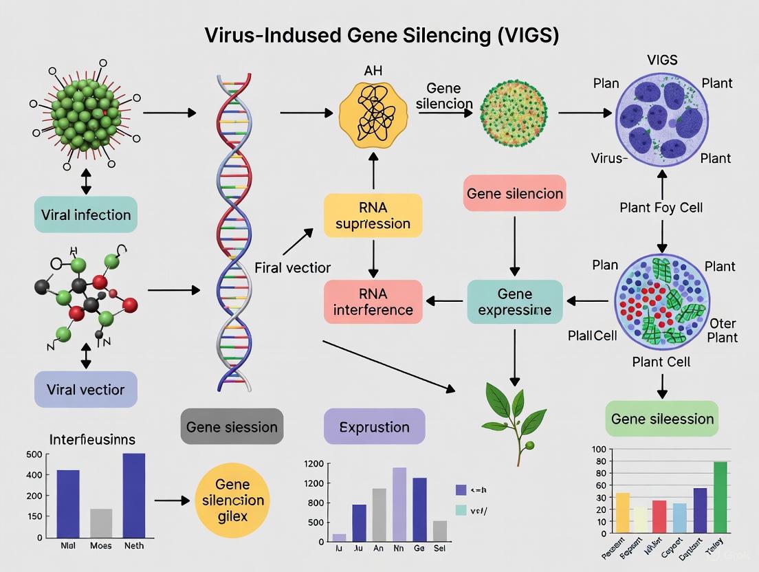
Virus-induced gene silencing (VIGS) is an RNA-mediated reverse genetics technique that leverages the plant's innate antiviral defense machinery to achieve targeted downregulation of endogenous genes [1]. This powerful functional genomics tool allows researchers to analyze gene function by introducing recombinant viral vectors containing fragments of plant gene transcripts, which triggers sequence-specific degradation of homologous cellular mRNAs [2] [3]. First demonstrated by Kumagai et al. in 1995 using a Tobacco mosaic virus vector to silence the phytoene desaturase (PDS) gene in Nicotiana benthamiana, VIGS has since evolved into an indispensable approach for plant functional genomics [2].
The fundamental biological basis of VIGS lies in the mechanism of post-transcriptional gene silencing (PTGS), an epigenetic phenomenon that represents one of the plant's key antiviral defense systems [1] [2]. When a recombinant virus carrying a plant gene fragment infects the host, the plant's cellular machinery processes the viral double-stranded RNA replication intermediates into 21-24 nucleotide small interfering RNAs (siRNAs) via Dicer-like enzymes [1]. These siRNAs are then incorporated into the RNA-induced silencing complex (RISC), which guides the sequence-specific degradation of complementary viral and endogenous mRNA transcripts [1] [2]. This sophisticated cellular defense mechanism is harnessed in VIGS to transiently knock down expression of targeted plant genes, enabling functional characterization through observable phenotypic changes [2].
Figure 1: Core Mechanism of Virus-Induced Gene Silencing (VIGS)
The VIGS Toolbox: Vectors, Methods, and Reagents
Viral Vector Systems for Diverse Applications
The effectiveness of VIGS depends critically on selecting appropriate viral vectors tailored to specific plant hosts and research objectives. Numerous viral vectors have been developed, falling into three main categories: RNA viruses, DNA viruses, and satellite virus-based systems [2]. Each vector type exhibits distinct advantages and limitations in terms of host range, silencing efficiency, insert size capacity, and symptom development [2].
RNA virus-based vectors, particularly Tobacco Rattle Virus (TRV), represent the most widely utilized VIGS system, especially for Solanaceae family plants [2]. TRV's bipartite genome organization requires two plasmid constructs: TRV1, encoding replicase and movement proteins, and TRV2, containing the coat protein gene and a multiple cloning site for inserting target sequences [2]. Other significant RNA vectors include Barley Stripe Mosaic Virus (BSMV) for monocots like wheat and barley, Cucumber Mosaic Virus (CMV), and Alfalfa Mosaic Virus (AMV) [3] [4]. DNA virus-based vectors, primarily geminiviruses such as Cotton Leaf Crumple Virus (CLCrV) and African Cassava Mosaic Virus (ACMV), offer alternative platforms with distinct replication mechanisms and potentially different host compatibility [2].
Table 1: Major Viral Vector Systems Used in VIGS
| Vector Name | Virus Type | Host Range | Key Features | Optimal Insert Size |
|---|---|---|---|---|
| Tobacco Rattle Virus (TRV) | RNA virus | Broad (especially Solanaceae) | Efficient systemic movement; mild symptoms; targets meristematic tissues | 200-1500 bp [2] [5] |
| Barley Stripe Mosaic Virus (BSMV) | RNA virus | Monocots (wheat, barley) | Effective in cereal crops; tripartite genome | ~185-500 bp [3] [4] |
| Tobacco Mosaic Virus (TMV) | RNA virus | Dicots | First VIGS vector developed; robust replication | 200-500 bp |
| Cucumber Mosaic Virus (CMV) | RNA virus | Broad | Wide host range; useful for diverse species | Varies by construct |
| Geminiviruses (CLCrV, ACMV) | DNA virus | Species-specific | Different replication mechanism; potential for larger inserts | Varies by construct [2] |
Essential Research Reagents and Solutions
Successful implementation of VIGS requires carefully selected research reagents and molecular tools. The core components include viral vectors, Agrobacterium strains for delivery, specialized growth media, and selection antibiotics [6] [4].
Table 2: Essential Research Reagent Solutions for VIGS
| Reagent/Resource | Function/Purpose | Examples/Specifications |
|---|---|---|
| Viral Vectors | Delivery of target gene fragments to trigger silencing | TRV (pYL156, pYL192), BSMV (pα, pβ, pγ), TMV-based [6] [4] |
| Agrobacterium tumefaciens Strains | Plant transformation and vector delivery | GV3101, LBA4404 [6] [5] |
| Antibiotics | Selection of transformed bacteria | Kanamycin (50-100 µg/mL), Rifampicin (25-50 µg/mL), Gentamicin (25 µg/mL) [6] [4] |
| Induction Buffer | Activation of Agrobacterium for plant infiltration | 10 mM MES, 10 mM MgCl₂, 200 µM acetosyringone [6] |
| Growth Media | Bacterial and plant cultivation | LB broth/agar (bacteria), John Innes compost (plants) [4] |
| In Vitro Transcription Kits | RNA transcript synthesis for some viral vectors | mMessage mMachine T7 kit [4] |
| Restriction Enzymes | Vector linearization and insert cloning | PacI, MluI, SpeI, SmaI [4] |
Established VIGS Protocols and Methodologies
TRV-Mediated VIGS in Dicotyledonous Plants
The TRV-based VIGS protocol has been optimized for numerous dicot species, particularly within the Solanaceae family, including pepper (Capsicum annuum L.), tomato, and tobacco [2]. The standard procedure begins with the selection of a 200-300 bp gene-specific fragment with careful attention to avoid off-target silencing through bioinformatic analysis using tools like the SGN VIGS Tool [5]. This fragment is then cloned into the TRV2 vector using restriction enzymes or recombination-based cloning [6].
For Agrobacterium-mediated delivery, the recombinant TRV2 and helper TRV1 plasmids are transformed into Agrobacterium tumefaciens strain GV3101 [6]. Single colonies are inoculated into liquid LB medium containing appropriate antibiotics (kanamycin 50 µg/mL, gentamicin 25 µg/mL) and grown overnight at 28°C [6]. The bacterial cultures are then diluted in induction buffer (10 mM MES, 10 mM MgCl₂, 200 µM acetosyringone) to an OD₆₀₀ of 0.8-1.2 and incubated for 3-4 hours at room temperature [6] [5]. For plant inoculation, the TRV1 and TRV2 cultures are mixed in a 1:1 ratio, and the mixture is infiltrated into plant tissues using a needleless syringe [2]. For pepper plants, 7-10-day-old seedlings with fully expanded cotyledons are ideal for infiltration, with researchers puncturing superficial wounds on the abaxial side of each cotyledon before flooding with the Agrobacterium mixture [6]. After infiltration, plants should be maintained under high humidity for 24 hours, then transferred to standard growth conditions (22-25°C, 14-16 hour photoperiod) for phenotypic observation, which typically appears within 2-4 weeks post-infiltration [2] [6].
BSMV-Mediated VIGS in Monocotyledonous Plants
For monocot species like barley and wheat, Barley Stripe Mosaic Virus (BSMV) has been established as an effective VIGS vector [3] [4]. The BSMV protocol differs significantly from TRV-based methods due to its tripartite RNA genome (α, β, and γ RNAs) and utilization of in vitro transcription rather than Agrobacterium infiltration for initial infection [4]. Target gene fragments (typically 150-500 bp) are cloned into the γb gene of the BSMV γ RNA immediately downstream of the termination codon [3] [4]. The recombinant plasmids are linearized with appropriate restriction enzymes (MluI for pα, pγ, and pγ-derivatives; SpeI for pβ), followed by in vitro transcription using the mMessage mMachine T7 kit to generate capped RNA transcripts [4].
For plant inoculation, the three RNA components (α, β, and γ with insert) are mixed in equal proportions and diluted in GP buffer (50 mM glycine, 30 mM K₂HPO₄, pH 9.2, 1% bentonite, 1% celite) [4]. Seven to ten-day-old barley or wheat seedlings are rub-inoculated with this mixture onto the second leaf [3] [4]. Inoculated plants should be maintained under controlled environmental conditions (16-h light at 24°C during day, 20°C at night) for optimal viral spread and silencing efficiency [3]. Silencing phenotypes in wheat typically become visible approximately 10 days post-inoculation, often manifesting as striped patterns parallel to leaf veins rather than complete leaf bleaching [3].
Figure 2: Standard VIGS Experimental Workflow
Optimization Strategies for Enhanced Silencing Efficiency
Critical Factors Influencing VIGS Success
Multiple factors significantly impact the efficiency and reproducibility of VIGS experiments. Proper optimization of these parameters is essential for achieving consistent and interpretable results. Key considerations include plant developmental stage, environmental conditions, Agrobacterium concentration (for TRV-based systems), and insert design [2] [5].
Plant developmental stage at inoculation critically affects silencing efficiency. Research in Camellia drupifera capsules demonstrated that optimal VIGS effects occurred at specific developmental stages: early stages (~69.80% efficiency for CdCRY1) and mid stages (~90.91% efficiency for CdLAC15) [5]. For seedling infiltration, 7-14-day-old plants typically yield the best results across species [6] [4]. Environmental conditions, particularly temperature, profoundly influence viral spread and silencing efficacy. Maintaining plants at 20-25°C post-inoculation generally optimizes results, while higher temperatures can reduce silencing efficiency and increase viral symptom severity [3]. Agrobacterium concentration also requires careful optimization; OD₆₀₀ values of 0.8-1.5 for TRV-based systems typically provide optimal results without causing excessive phytotoxicity [6] [5].
Table 3: Optimization Parameters for Enhanced VIGS Efficiency
| Parameter | Optimal Conditions/Range | Impact on Silencing Efficiency |
|---|---|---|
| Plant Developmental Stage | 7-14 days (seedlings); species-specific for tissues | Younger tissues generally more amenable; affects systemic spread [6] [5] |
| Temperature | 20-25°C post-inoculation | Higher temperatures reduce efficiency and increase symptoms [3] |
| Agrobacterium OD₆₀₀ (TRV) | 0.8-1.5 | Lower concentrations reduce efficiency; higher cause phytotoxicity [6] [5] |
| Insert Size | 200-500 bp | Smaller fragments may reduce efficiency; larger may affect viral stability [5] |
| Insert Position in Vector | Downstream of coat protein or γb stop codon | Critical for proper processing and siRNA generation [3] [4] |
| Photoperiod | Species-dependent (e.g., 14:10 L:D for cotton) | Affects plant physiology and viral replication [6] |
| Inoculation Method | Agroinfiltration, rub-inoculation, pericarp cutting immersion | Tissue-dependent efficiency [5] |
Advanced Enhancement Techniques
Several advanced strategies can further improve VIGS efficiency, particularly in recalcitrant species or for challenging targets. Co-expression of viral suppressors of RNA silencing (VSRs) represents a powerful approach to enhance transient silencing. Proteins such as P19 from Tomato Bushy Stunt Virus and HC-Pro from Potato Virus Y can inhibit aspects of the plant's silencing machinery, allowing for more robust viral accumulation and spread before defense mechanisms activate [2]. However, this approach requires careful optimization, as excessive suppression can eliminate the silencing effect altogether.
For difficult-to-transform tissues, such as lignified capsules in woody plants, alternative infiltration methods can dramatically improve results. Research in Camellia drupifera demonstrated that pericarp cutting immersion achieved approximately 93.94% infiltration efficiency, significantly outperforming peduncle injection or direct pericarp injection methods [5]. Additionally, employing tissue-specific or inducible promoters to drive viral vector expression can provide spatial and temporal control over silencing, enabling functional analysis of genes whose constitutive silencing might be lethal [2].
Validation and Applications in Functional Genomics
Molecular Validation of Silencing Efficiency
Rigorous validation of target gene knockdown is essential for interpreting VIGS phenotypes accurately. Reverse-transcription quantitative PCR (RT-qPCR) represents the gold standard for quantifying silencing efficiency at the transcript level [6]. Proper experimental design for validation includes appropriate sampling timing (typically 2-4 weeks post-inoculation), tissue selection (often young systemic leaves where silencing is most pronounced), and selection of stable reference genes for normalization [6].
Critical to accurate RT-qPCR validation is the choice of appropriate reference genes, which must demonstrate stable expression across experimental conditions. A comprehensive 2025 study in cotton evaluated six candidate reference genes under VIGS and herbivory stress conditions, finding that commonly used references GhUBQ7 and GhUBQ14 were the least stable, while GhACT7 and GhPP2A1 provided the most consistent expression [6]. Normalization with inappropriate reference genes can obscure real biological effects; in the cotton study, normalization with unstable GhUBQ7 reduced sensitivity to detect significant upregulation of GhHYDRA1 in aphid-infested plants, while normalization with stable GhACT7/GhPP2A1 clearly revealed this expression change [6].
Diverse Research Applications
VIGS has enabled functional characterization of genes involved in diverse biological processes across numerous plant species. In pepper (Capsicum annuum L.), VIGS has successfully identified genes governing fruit quality traits including color, biochemical composition, and pungency, as well as resistance to bacterial, oomycete, and insect pathogens [2]. The technology has also elucidated genes regulating plant architecture, development, and responses to abiotic stresses such as temperature extremes, salt, and osmotic stress [2].
In wheat, BSMV-VIGS has proven invaluable for dissecting disease resistance pathways, successfully silencing genes such as Lr21 (a nucleotide binding site-leucine-rich repeat class resistance gene), RAR1, SGT1, and HSP90 to demonstrate their essential roles in leaf rust resistance [3]. More recently, VIGS has evolved beyond transient knockdowns to induce heritable epigenetic modifications, with studies demonstrating that VIGS can trigger RNA-directed DNA methylation (RdDM) leading to stable epigenetic alleles that persist over multiple generations [1]. This expanding application space solidifies VIGS as a versatile platform not only for rapid gene functional analysis but also for epigenetic studies and crop improvement.
Post-transcriptional gene silencing (PTGS) is a homology-dependent RNA degradation mechanism that serves as a potent innate immune response against viruses in plants [7]. This sequence-specific process inactivates aberrant or highly expressed RNAs in the cytoplasm, functioning as an adaptive antiviral response that can target viral RNA exclusively in the cytoplasmic compartment [7] [8]. As a fundamental component of the RNA interference (RNAi) pathway, PTGS represents a conserved evolutionary defense strategy that plants employ to recognize and degrade viral pathogens, with parallel mechanisms identified across diverse eukaryotic species [7] [9].
The significance of PTGS extends beyond its natural antiviral function to become the foundational mechanism underlying Virus-Induced Gene Silencing (VIGS), a powerful reverse genetics tool that enables researchers to study gene function by transiently knocking down target gene expression [2] [1]. VIGS leverages the plant's PTGS machinery, utilizing recombinant viral vectors to trigger systemic suppression of endogenous plant genes, leading to visible phenotypic changes that facilitate gene characterization without the need for stable transformation [2]. This application has become particularly valuable for functional genomics in non-model plants and crops where stable genetic transformation remains challenging [2].
Molecular Mechanisms of Antiviral PTGS
The antiviral PTGS pathway initiates when the plant immune system recognizes viral double-stranded RNA (dsRNA) replicative intermediates as pathogen-associated molecular patterns (PAMPs) [9]. These vRI-dsRNAs are cleaved by Dicer-like (DCL) enzymes, which process them into 21-24 nucleotide virus-derived small interfering RNAs (vsiRNAs) characterized by 2-nucleotide 3' overhangs [9] [1]. These vsiRNAs are then loaded into the RNA-induced silencing complex (RISC), where the Argonaute (AGO) protein, particularly AGO2, uses the guide strand to identify complementary viral RNA sequences through perfect base-pairing [9]. The PIWI domain of AGO2, which contains an RNase H-like fold, subsequently cleaves and degrades the target viral RNAs, thereby achieving antiviral immunity [9].
Amplification of the silencing signal occurs through host RNA-dependent RNA polymerases (RdRPs), which exponentially produce additional viral dsRNAs that serve as substrates for DCL processing, generating secondary vsiRNAs that enhance the robustness and systemic spread of the immune response [9] [1]. This amplification mechanism creates a powerful RNA-based immune memory that can effectively limit viral replication and spread throughout the plant [9].
Table 1: Core Components of the Plant PTGS Antiviral Pathway
| Component | Structure/Features | Function in Antiviral PTGS |
|---|---|---|
| Dicer-like (DCL) | HEL, DUF283, PAZ, RNase III, dsRBD domains [9] | Recognizes and cleaves viral dsRNA into 21-24nt vsiRNAs [9] |
| Argonaute (AGO) | N, PAZ, MID, PIWI domains; AGO2 has RNase H activity [9] | Slices target viral RNA guided by vsiRNAs within RISC [9] |
| vsiRNAs | 21-24 nucleotides with 2-nt 3' overhangs [9] [1] | Sequence-specific guides for viral RNA recognition and degradation |
| RISC | Multi-protein complex with AGO at core [9] [1] | Effector complex that executes viral RNA cleavage |
| RdRP | RNA-dependent RNA polymerase [9] | Amplifies silencing signal by synthesizing secondary dsRNA |
Figure 1: Molecular Pathway of PTGS-Mediated Antiviral Defense. The mechanism initiates with viral dsRNA recognition and progresses through vsiRNA biogenesis to targeted viral RNA degradation.
Viral Suppressors of RNA Silencing (VSRs) and Immune Evasion
Through evolutionary arms races, viruses have developed sophisticated counter-defense strategies in the form of viral suppressors of RNA silencing (VSRs) that target distinct stages of the PTGS pathway [7] [9]. These VSRs employ diverse molecular tactics to evade host immunity, including dsRNA binding, vsiRNA sequestration, interference with DCL or AGO functions, and manipulation of intracellular signaling networks [9] [10]. The cucumber mosaic cucumovirus (CMV) 2b protein exemplifies this adaptive strategy, functioning as a virulence determinant that localizes to the nucleus via an arginine-rich nuclear localization signal (²²KRRRRR²⁷) to suppress PTGS initiation [7]. Nuclear targeting is essential for the 2b protein's suppressor activity, suggesting that PTGS may be blocked at the nuclear level despite primarily targeting RNA in the cytoplasm [7].
Other well-characterized VSRs include the P1/HC-Pro polyprotein encoded by tobacco etch virus, which suppresses PTGS at the post-transcriptional level [8], and the P19 protein of Tomato bushy stunt virus, which is counteracted by host PRMT6 through arginine methylation that blocks P19-siRNA binding [9]. The multifunctionality of VSR proteins enables fine-tuning of plant-virus interactions, with many VSRs performing additional roles in viral replication, movement, and packaging while simultaneously suppressing host defense mechanisms [10].
Table 2: Characterized Viral Suppressors of RNA Silencing (VSRs)
| VSR Protein | Virus Origin | Mechanism of Action | Cellular Localization |
|---|---|---|---|
| 2b | Cucumber mosaic virus (CMV) [7] | Blocks PTGS initiation; requires nuclear import [7] | Nucleus [7] |
| P1/HC-Pro | Tobacco etch virus [8] | Suppresses established PTGS at post-transcriptional level [8] | Cytoplasm [8] |
| P19 | Tomato bushy stunt virus [9] | Sequesters vsiRNAs; inhibited by host PRMT6 methylation [9] | Cytoplasm [9] |
| Multiple VSRs | Diverse plant viruses [10] | Target DCL, AGO, vsiRNAs, RdRPs; often multifunctional [10] | Various compartments [10] |
Virus-Induced Gene Silencing (VIGS): Protocol and Applications
VIGS Experimental Workflow
VIGS harnesses the plant's PTGS machinery to silence endogenous genes through recombinant viral vectors, enabling high-throughput functional genomics without stable transformation [2]. The core protocol begins with the selection of an appropriate viral vector system based on the host plant species and experimental requirements, with Tobacco Rattle Virus (TRV) being particularly versatile for Solanaceae family plants due to its broad host range and efficient systemic movement [2]. The target gene fragment (typically 200-500 bp) is then cloned into the viral vector, ensuring minimal off-target effects through careful sequence verification [2] [1].
For plants amenable to agroinfiltration, such as Nicotiana benthamiana, recombinant Agrobacterium tumefaciens strains carrying the VIGS constructs are cultured to an OD₆₀₀ of 0.5-2.0, harvested, and resuspended in infiltration medium [2]. The bacterial suspension is infiltrated into expanded leaves using a needleless syringe, with optimal results obtained when plants are at the 3-4 leaf stage [2]. Following inoculation, plants are maintained under controlled environmental conditions (22-25°C, 16h light/8h dark photoperiod) for 2-4 weeks to allow systemic silencing establishment before phenotypic analysis [2].
Figure 2: VIGS Experimental Workflow. The process involves vector preparation, plant inoculation, and phenotypic analysis phases for gene function characterization.
Optimization Strategies and Technical Considerations
Successful VIGS implementation requires careful optimization of multiple parameters. The choice of viral vector significantly impacts silencing efficiency, with different vectors exhibiting varying host ranges, symptom severity, and persistence [2]. TRV vectors are preferred for many applications due to minimal symptom development and effective meristem targeting, while Bean Pod Mottle Virus (BPMV) works well for legumes, and Barley Stripe Mosaic Virus (BSMV) is effective in monocots [2] [1].
Insert design critically influences silencing specificity and efficiency, with optimal fragments of 200-500 bp exhibiting low self-complementarity and positioned away from highly conserved domains to minimize off-target effects [2]. Environmental parameters, particularly temperature, profoundly impact VIGS efficiency, with most systems performing optimally at 22-25°C, while higher temperatures can attenuate silencing [2]. The incorporation of viral suppressors of RNA silencing (VSRs) like P19 or C2b can enhance VIGS efficacy by transiently overwhelming the plant's silencing machinery, though this must be carefully balanced against potential cytotoxic effects [2].
Table 3: VIGS Vector Systems and Their Applications
| Vector System | Virus Type | Host Range | Key Features | Optimal Applications |
|---|---|---|---|---|
| TRV (Tobacco Rattle Virus) [2] | RNA virus | Broad (Solanaceae) [2] | Bipartite genome; minimal symptoms; targets meristems [2] | High-throughput screening; developmental studies [2] |
| BSMV (Barley Stripe Mosaic Virus) [1] | RNA virus | Monocots [1] | Efficient in cereals; moderate symptoms | Cereal functional genomics [1] |
| CMV (Cucumber Mosaic Virus) [2] | RNA virus | Very broad [2] | Strong silencing signal; can cause severe symptoms [2] | Difficult-to-silence targets [2] |
| CLCrV (Cotton Leaf Crumple Virus) [2] | DNA virus (geminivirus) | Limited host range [2] | DNA-based; prolonged silencing [2] | Extended silencing duration studies [2] |
The Scientist's Toolkit: Essential Research Reagents and Materials
Table 4: Essential Research Reagents for PTGS and VIGS Studies
| Reagent/Material | Specifications | Experimental Function |
|---|---|---|
| Viral Vectors | TRV (pTRV1, pTRV2), BSMV, CMV, CLCrV [2] | Delivery of target gene fragments to trigger PTGS |
| Agrobacterium Strains | GV3101, LBA4404, AGL1 [2] | Delivery of viral vectors to plant tissues |
| Targetron Vectors | Programmable group II intron systems [11] | Specific gene knockout in host receptors |
| DCL Antibodies | Specific to DCL1, DCL2, DCL3, DCL4 [9] | Monitoring DCL protein expression and localization |
| AGO Antibodies | Specific to AGO1, AGO2, AGO4 [9] | RISC complex immunodetection and quantification |
| sRNA Library Prep Kits | High-throughput sequencing compatible | vsiRNA and miRNA profiling |
| Infiltration Buffers | 10mM MES, 10mM MgCl₂, 150μM acetosyringone [2] | Enhanced Agrobacterium-mediated delivery |
| Phagemid Systems | M13 origin + ColE1 plasmid backbone [11] | Mobilizable gene therapy vector delivery |
Emerging Applications and Future Perspectives
Recent advances have expanded PTGS applications beyond traditional functional genomics to include heritable epigenetic modifications through virus-induced transcriptional gene silencing (ViTGS) [1]. This approach utilizes viral vectors carrying sequences homologous to gene promoters rather than coding regions, inducing RNA-directed DNA methylation (RdDM) that can be stably inherited over multiple generations [1]. The Bond et al. (2015) study demonstrated that TRV:FWAᵗʳ infection leads to transgenerational epigenetic silencing of the FWA promoter sequence in Arabidopsis, establishing VIGS as a tool for creating stable epigenetic variants [1].
Therapeutic applications of PTGS mechanisms are emerging in both plant and animal systems, with synthetic vsiRNAs designed against conserved viral genomic regions showing promise as RNAi-based immunotherapies [9]. Wu et al. (2025) identified a position-dependent 3 bp motif (RWM) that enhances Dicer processing efficiency, enabling design of vsiRNAs with potent antiviral activity against coronaviruses and influenza viruses in vitro and in vivo [9]. Engineering of transmissible antiviral defenses using benign viral vectors represents another frontier, with prototype systems successfully implemented in E. coli using bacteriophage M13 and phagemid vectors to confer resistance to lethal phage T5 [11].
The integration of VIGS with multi-omics technologies and genome-editing platforms is accelerating crop improvement programs, particularly for species like pepper (Capsicum annuum L.) where stable transformation remains challenging [2]. High-throughput VIGS screening has enabled systematic functional characterization of genes governing fruit quality, pathogen resistance, abiotic stress tolerance, and plant architecture, providing valuable molecular insights for marker-assisted breeding [2]. As these technologies mature, PTGS-based approaches will continue to illuminate fundamental biological processes while enabling innovative strategies for crop protection and therapeutic intervention.
Virus-Induced Gene Silencing (VIGS) is a powerful reverse genetics tool that leverages the plant's innate antiviral RNA interference (RNAi) machinery to silence endogenous genes [1]. This process is orchestrated by a core set of molecular players that act in sequence to process viral RNA into silencing signals and execute sequence-specific gene repression [12]. When a recombinant virus carrying a fragment of a plant gene infiltrates the host cell, it triggers a cascade of molecular events centered on Dicer-like enzymes (DCLs), small interfering RNAs (siRNAs), and the RNA-induced silencing complex (RISC) [1]. This sophisticated cellular defense system, repurposed for functional genomics, allows researchers to rapidly knock down gene expression and investigate gene function in planta [2]. Understanding these key components—their identities, functions, and interactions—is fundamental to designing effective VIGS experiments and interpreting their results.
Core Molecular Components and Mechanisms
The process of VIGS is a specialized application of the post-transcriptional gene silencing (PTGS) pathway. The following diagram illustrates the coordinated sequence of events from viral infection to target gene silencing.
Dicer-like (DCL) Enzymes
Dicer-like (DCL) enzymes are multi-domain RNases that act as the initiation point for the RNAi cascade. They function as molecular sensors that recognize and cleave long double-stranded RNA (dsRNA) precursors, which are produced as viral replication intermediates or through the activity of host RNA-dependent RNA polymerases (RDRPs) [1] [12]. In the model plant Arabidopsis thaliana, four DCL enzymes (DCL1-4) coordinate the biogenesis of different small RNA classes, with DCL2, DCL3, and DCL4 playing primary roles in antiviral defense [12].
Table: Dicer-like Enzymes in Plant RNAi
| Enzyme | Primary Function | siRNA Product Length | Key Role in VIGS |
|---|---|---|---|
| DCL4 | Antiviral defense; Processes RDRP-derived dsRNA [12] | 21 nt [12] | Primary processor of viral RNA; generates 21-nt siRNAs for systemic silencing [12]. |
| DCL2 | Backup antiviral defense; Processes viral dsRNA [12] | 22 nt [12] | Compensates when DCL4 is absent; generates 22-nt siRNAs that can trigger secondary siRNA production [12] [13]. |
| DCL3 | Heterochromatin formation; Nuclear RNAi [12] | 24 nt [12] | Generates 24-nt heterochromatic siRNAs; can reinforce silencing through RNA-directed DNA methylation (RdDM) [1]. |
The specific DCL enzyme involved in a VIGS response can depend on the host plant species and the type of viral vector used. For instance, research using a novel Turnip crinkle virus (TCV)-based VIGS vector in Arabidopsis demonstrated the primary involvement of DCL4 in the antiviral silencing pathway [13].
Small Interfering RNAs (siRNAs)
The cleavage of long dsRNA by DCL enzymes produces small interfering RNAs (siRNAs), the sequence-specific guides of the RNAi system. These are short, double-stranded RNA molecules typically 21 to 24 nucleotides in length [1] [12]. One key feature of the siRNA pathway is amplification. Plant RNA-dependent RNA polymerases (RDRPs) can use the cleavage products of targeted mRNAs as templates to synthesize new dsRNA, which is subsequently processed by DCLs into secondary siRNAs. This process, known as transitivity, amplifies the silencing signal and ensures its systemic spread throughout the plant [12].
The RISC Complex
The RNA-induced silencing complex (RISC) is the effector complex that executes gene silencing. The core catalytic component of RISC is an Argonaute (AGO) protein [1] [12]. During RISC assembly, the siRNA duplex is loaded and the passenger strand is discarded. The remaining guide strand directs the complex to complementary mRNA sequences. The AGO protein, often possessing Slicer activity, then cleaves the target mRNA, leading to its degradation [1] [14]. Different AGO proteins have specialized functions. For example, studies in Arabidopsis have shown that AGO2 is involved in the VIGS response and antiviral defense, particularly in certain mutant backgrounds [13]. In the nematode C. elegans, the RDE-1 protein (an AGO homolog) is essential for initiating RNAi, while secondary AGO proteins like CSR-1 guide the destruction of target transcripts [14].
Advanced VIGS Protocols and Applications
The following experimental protocol provides a detailed methodology for implementing a highly efficient VIGS procedure using a root inoculation technique.
Protocol: Root Wounding-Immersion Method for High-Efficiency VIGS
Background: This protocol describes a root wounding–immersion method for efficient VIGS in a variety of plants, including Nicotiana benthamiana, tomato, pepper, eggplant, and Arabidopsis thaliana [15]. This method is suitable for high-throughput functional genomics screening as it allows for the inoculation of large batches of plants in a short time with high efficiency.
Table: Key Reagents for Root Wounding-Immersion VIGS
| Reagent/Solution | Function/Description | Critical Parameters |
|---|---|---|
| pTRV1 & pTRV2 Vectors | Binary T-DNA vectors containing the bipartite Tobacco Rattle Virus (TRV) genome [15]. | TRV1 encodes replication/movement proteins. TRV2 carries the target gene insert for silencing. |
| Agrobacterium tumefaciens GV1301 | Bacterial strain used to deliver TRV vectors into plant cells [15]. | Contains helper plasmids for T-DNA transfer. |
| Acetosyringone | A phenolic compound that induces the Agrobacterium Vir genes essential for T-DNA transfer [15]. | Final concentration of 150-200 µM in the infiltration solution is critical. |
| Infiltration Solution | Buffer for suspending Agrobacterium before inoculation [15]. | 10 mM MgCl₂, 10 mM MES (pH 5.6), 150 µM acetosyringone. |
| Antibiotics (Kanamycin, Rifampicin) | Selective agents to maintain binary vectors and the Agrobacterium strain [15]. | Concentrations: 50 µg/mL Kanamycin, 25 µg/mL Rifampicin. |
Step-by-Step Procedure:
Plant Material Preparation:
- Germinate seeds of your target species (e.g., tomato, N. benthamiana) and grow seedlings until they develop 3-4 true leaves (approximately 3 weeks old) [15].
Agrobacterium Culture Preparation:
- Inoculate Agrobacterium strain GV1301 harboring pTRV1 and a second culture harboring pTRV2 (containing your gene of interest) in LB broth with appropriate antibiotics.
- Incubate overnight at 28°C with shaking at 200 rpm [15].
- The next day, pellet the bacteria and resuspend in infiltration solution (10 mM MgCl₂, 10 mM MES pH 5.6, 150 µM acetosyringone) to a final OD₆₀₀ = 0.8 [15].
- Incubate the suspensions in the dark at 28°C for 3-4 hours to induce Vir gene expression [15].
Inoculum Mixing:
- Mix the prepared TRV1 and TRV2 suspensions in a 1:1 ratio [15].
Root Inoculation:
- Carefully remove seedlings from the growth medium and gently wash soil from the roots.
- Using a sterilized blade or scissors, cut approximately one-third of the root length [15].
- Immediately immerse the wounded roots into the mixed Agrobacterium suspension for 30 minutes [15].
- (Optional) For the "successive inoculation" method, immerse roots in TRV1 for 15 minutes, then transfer to TRV2 for another 15 minutes [15].
Post-Inoculation Care:
- Re-plant the treated seedlings into fresh soil or a suitable growth medium.
- Maintain plants in a growth chamber or greenhouse. Studies indicate that lower temperatures (e.g., 20°C day/18°C night) can enhance VIGS efficiency in some species [16].
- Silencing phenotypes, such as photo-bleaching in plants silenced for Phytoene Desaturase (PDS), typically become visible in systemic leaves within 2-4 weeks post-inoculation [15].
Troubleshooting and Validation:
- Low Silencing Efficiency: Ensure bacterial cultures are in the log phase of growth, the OD₆₀₀ is accurate, and the acetosyringone induction step is not skipped.
- Plant Stress or Death: Avoid using excessively high bacterial densities (OD₆₀₀ > 1.0 can cause necrosis in some species) [15].
- Validation: Always include a positive control (e.g., TRV2-PDS) and a negative control (e.g., TRV2-empty vector or TRV2-GFP). Confirm silencing at the molecular level by measuring target gene mRNA levels using RT-qPCR [15].
The Scientist's Toolkit: Essential Research Reagents
Table: Key Research Reagent Solutions for VIGS Experiments
| Reagent/Material | Function in VIGS Workflow | Examples & Notes |
|---|---|---|
| Viral Vectors | To deliver the target gene sequence and initiate silencing in the host plant. | TRV (Tobacco Rattle Virus): Most widely used; broad host range, mild symptoms [2] [15]. TCV (Turnip Crinkle Virus): Useful for Arabidopsis; allows simultaneous silencing of two genes [13]. |
| Agrobacterium Strains | To mediate the delivery of the viral vector T-DNA into the plant cell. | GV3101: Commonly used for solanaceous species and sunflower [17]. GV1301: Used successfully in the root wounding protocol [15]. |
| Visual Marker Genes | To visually confirm the establishment and spread of VIGS. | PDS (Phytoene Desaturase): Silencing causes photobleaching (white leaves) [2] [15]. CHS (Chalcone Synthase): Silencing causes loss of pigment (white patches) in flowers [16]. |
| siRNA Prediction Tools | To design effective inserts for the VIGS vector by predicting potent siRNA target sites. | pssRNAit: Used to analyze target sequences and design fragments with high predicted siRNA activity [17]. Genscript siRNA Target Finder: Used to identify top siRNA sequences for fragment design [13]. |
Dicer-like enzymes, siRNAs, and the RISC complex form the core molecular engine of Virus-Induced Gene Silencing. A deep understanding of their functions and interactions—from the initial processing of viral dsRNA by specific DCLs to the sequence-specific destruction of mRNA by the AGO-loaded RISC—is indispensable for modern plant researchers. When coupled with optimized protocols, such as the root wounding-immersion method, this knowledge enables robust, high-throughput gene functional analysis, accelerating discovery in plant biology and crop improvement.
Virus-induced gene silencing (VIGS) has emerged as a powerful reverse genetics tool that offers significant advantages over stable plant transformation for functional genomics research. This protocol outlines the key benefits of VIGS, including its rapid implementation, cost-effectiveness, and ability to bypass the challenges of tissue culture and stable transformation. We provide detailed methodologies for implementing TRV-based VIGS across diverse plant species, along with quantitative comparisons and essential reagent solutions to facilitate widespread adoption in plant research laboratories.
Virus-induced gene silencing represents a breakthrough methodology that leverages the plant's innate RNA interference machinery to achieve transient gene knockdown without genomic integration. As a rapid alternative to stable transformation, VIGS utilizes recombinant viral vectors carrying fragments of plant target genes to initiate sequence-specific mRNA degradation through post-transcriptional gene silencing (PTGS) [18] [19]. The fundamental advantage of this system lies in its ability to directly infect target plants, circumventing the complexity of plant genetic transformation and regeneration systems that have traditionally bottlenecked functional genomics research in numerous species [20]. This technical note provides a comprehensive framework for exploiting VIGS advantages in plant gene function studies, with detailed protocols and implementation guidelines.
The molecular mechanism of VIGS begins when double-stranded RNA (dsRNA) formed during viral replication is cleaved by DICER-like (DCL) enzymes into 21-24 nucleotide small interfering RNAs (siRNAs). These siRNAs are incorporated into the RNA-induced silencing complex (RISC), which guides the sequence-specific degradation of complementary endogenous mRNA transcripts [18] [19]. This process, illustrated in Figure 1, enables researchers to effectively "knock down" gene expression without permanent genetic modification.
Figure 1. VIGS Workflow Overview - This diagram illustrates the key steps in virus-induced gene silencing, from vector construction to phenotypic analysis.
Comparative Advantages: VIGS Versus Stable Transformation
Quantitative Comparison of Key Parameters
The implementation of VIGS offers substantial advantages across multiple experimental parameters compared to stable transformation approaches. Table 1 provides a systematic comparison of these key differentiating factors.
Table 1. Comparative analysis of VIGS versus stable transformation
| Parameter | Virus-Induced Gene Silencing (VIGS) | Stable Genetic Transformation |
|---|---|---|
| Time Requirement | Days to weeks [21] | Months to years [21] |
| Tissue Culture | Not required [20] | Essential, limiting step [20] |
| Genetic Integration | Transient, no genomic integration [21] | Permanent integration into host genome [21] |
| Species Applicability | Broad host range, including recalcitrant species [5] [20] | Limited to transformable genotypes |
| Gene Redundancy Analysis | Can silence multiple family members simultaneously [18] | Requires multiple transformation events |
| Essential Gene Studies | Enables study of lethal mutations [18] | Often lethal in stable lines |
| Technical Expertise | Moderate laboratory skills | Advanced tissue culture expertise |
| Equipment Needs | Standard molecular biology equipment | Specialized transformation facilities |
Key Advantage Areas
Temporal Efficiency and Bypassing Tissue Culture
The most significant advantage of VIGS is its remarkable time efficiency. While stable transformation requires months to years for successful implementation and phenotypic analysis, VIGS can yield interpretable results within weeks [21]. For example, in soybean, a highly efficient TRV-VIGS system demonstrated silencing efficiencies ranging from 65% to 95% within approximately 21 days post-inoculation [22]. This accelerated timeline enables researchers to progress from gene selection to functional analysis in a single generation.
Furthermore, VIGS completely bypasses the tissue culture bottleneck that plagues stable transformation in many plant species [20]. Species such as walnut (Juglans regia L.), which lack robust tissue culture systems, have successfully been analyzed using VIGS approaches, enabling functional genomics studies that were previously impossible [20]. This advantage extends to numerous woody perennials and recalcitrant crops where establishment of regeneration protocols remains challenging.
Cost-Effectiveness and Resource Optimization
The resource requirements for VIGS implementation are substantially lower than those for stable transformation. VIGS eliminates the need for specialized tissue culture facilities, growth regulators, and the extensive labor inputs associated with maintaining callus cultures and regenerating plants [21]. The simplified workflow reduces consumable costs and enables parallel processing of multiple gene targets, making it ideal for preliminary functional screening.
In walnut, researchers developed an efficient TRV-based VIGS system achieving up to 48% silencing efficiency with minimal infrastructure requirements [20]. Similarly, in tea oil camellia (Camellia drupifera), a TRV-elicited VIGS system for recalcitrant capsules achieved infiltration efficiencies of approximately 94% using pericarp cutting immersion, demonstrating the cost-effectiveness of this approach for challenging tissues [5].
Functional Genomics Applications
VIGS provides unique capabilities for addressing gene functional redundancy, a common challenge in plant genomics. By designing constructs targeting conserved regions of gene families, researchers can simultaneously silence multiple related genes, overcoming functional redundancy that often complicates the analysis of single-gene mutants [18]. This approach was successfully employed to study heat shock protein 90 (HSP90) in tomato, where silencing the entire gene family revealed its essential role in plant growth and development [18].
Additionally, VIGS enables the functional analysis of essential genes whose knockout would be lethal in stable transformation systems [18]. The transient nature of VIGS allows researchers to study the effects of gene knockdown without permanent disruption, observing phenotypes that would be impossible to recover in stable lines. This temporary silencing effect eventually diminishes, allowing plants to recover and produce seeds [18].
Application Notes: VIGS Implementation Across Species
Protocol Customization for Diverse Species
The implementation of VIGS requires species-specific optimization to achieve maximum efficiency. Recent advances have demonstrated successful adaptation of TRV-based VIGS systems across a broad phylogenetic range:
Soybean (Glycine max):- Soybean (Glycine max): An optimized TRV-VIGS system utilizing Agrobacterium-mediated infection through cotyledon nodes achieved 65-95% silencing efficiency for genes including GmPDS, GmRpp6907, and GmRPT4 [22]. The method involved soaking sterilized soybeans until swollen, longitudinally bisecting them to obtain half-seed explants, then infecting fresh explants by immersion for 20-30 minutes in Agrobacterium suspensions.
Walnut (Juglans regia L.): A highly efficient VIGS system was developed using a 255 bp fragment of the JrPDS gene, with optimal silencing achieved through syringe infiltration at the 3-4 leaf stage using Agrobacterium at OD600 = 1.5 [20]. This system achieved 48% silencing efficiency, enabling functional studies in this recalcitrant species.
Tea Oil Camellia (Camellia drupifera): For recalcitrant woody capsules, researchers optimized a pericarp cutting immersion method with 200 μmol·L−1 acetosyringone, achieving approximately 94% infiltration efficiency for genes involved in pigmentation (CdCRY1 and CdLAC15) [5].
Petunia (Petunia × hybrida): Comprehensive optimization included inoculation of mechanically wounded shoot apical meristems, use of cultivar 'Picobella Blue', and maintenance at 20°C day/18°C night temperatures, which increased silencing area by 69% for CHS and 28% for PDS [16].
Quantitative Methodologies and Efficiency Assessment
Table 2. Efficiency metrics of VIGS across diverse plant species
| Plant Species | Target Gene | Silencing Efficiency | Key Optimized Parameter |
|---|---|---|---|
| Soybean [22] | GmPDS | 65-95% | Cotyledon node immersion |
| Tea Oil Camellia [5] | CdCRY1 | ~94% | Pericarp cutting immersion |
| Walnut [20] | JrPDS | 48% | Syringe infiltration, OD600=1.5 |
| Petunia [16] | CHS | 69% increase | Apical meristem wounding |
| Styrax japonicus [23] | - | 83.33% | Vacuum infiltration, OD600=0.5 |
Efficiency assessment typically employs visual markers like phytoene desaturase (PDS), which causes photobleaching when silenced, and quantitative PCR validation [22] [20]. For example, in soybean, fluorescence microscopy revealed successful infection in more than 80% of cells when using the cotyledon node method, with effective infectivity efficiency exceeding 80% and reaching 95% for specific cultivars like Tianlong 1 [22].
Essential Research Reagent Solutions
Successful implementation of VIGS requires specific reagents and vectors optimized for efficient gene silencing. Table 3 outlines the key components of the VIGS toolkit.
Table 3. Essential research reagents for VIGS implementation
| Reagent | Function | Application Notes |
|---|---|---|
| TRV Vectors (pTRV1, pTRV2) | Bipartite viral vector system | TRV1 encodes replication proteins; TRV2 carries target gene insert [2] |
| Agrobacterium tumefaciens GV3101 | Vector delivery | Preferred strain for many dicot species [22] [20] |
| Acetosyringone | Vir gene inducer | Typically used at 100-200 μM concentration [5] [23] |
| Silencing Markers (PDS, CHS) | Visual silencing indicators | PDS for photobleaching, CHS for flower color changes [16] |
| GFP-tagged Vectors | Transformation efficiency monitoring | Enables visual tracking of infection success [22] |
| Selection Antibiotics | Bacterial selection | Kanamycin (25-50 μg/mL), rifampicin (50 μg/mL) [5] |
Experimental Protocols
TRV Vector Construction and Preparation
Target Fragment Selection: Identify a 200-300 bp fragment from the target gene CDS using tools like SGN VIGS Tool to ensure specificity and minimize off-target effects [5].
Vector Assembly: Amplify the target fragment using gene-specific primers with appropriate restriction sites (e.g., EcoRI and XhoI) and clone into pTRV2 vector [22].
Transformation: Introduce the recombinant plasmid into Agrobacterium tumefaciens GV3101 using freeze-thaw method or electroporation [22] [5].
Agrobacterium Culture: Inoculate single colonies into YEB medium containing appropriate antibiotics (kanamycin 25-50 μg/mL, rifampicin 50 μg/mL) and incubate at 28°C with shaking at 200-240 rpm for 24-48 hours [5].
Plant Inoculation Methods
Various inoculation methods have been optimized for different plant species and tissues:
Cotyledon Node Immersion (Soybean): Bisect sterilized, pre-swollen seeds and immerse fresh explants in Agrobacterium suspension (OD600 = 0.9-1.0) for 20-30 minutes [22].
Syringe Infiltration (Walnut): Infiltrate Agrobacterium suspension (OD600 = 1.5) into abaxial side of leaves at 3-4 leaf stage using needless syringe [20].
Pericarp Cutting Immersion (Camellia capsules): Immerse freshly cut pericarp tissues in Agrobacterium suspension (OD600 = 0.5-1.0) with 200 μmol·L−1 acetosyringone [5].
Apical Meristem Inoculation (Petunia): Apply Agrobacterium suspension to mechanically wounded shoot apical meristems of 3-4 week old plants [16].
Post-Inoculation Procedures and Efficiency Validation
Temperature Optimization: Maintain plants at species-appropriate temperatures (typically 18-22°C) to enhance silencing efficiency and reduce viral symptoms [16].
Phenotypic Monitoring: Observe visual silencing markers (e.g., photobleaching for PDS) beginning at 10-21 days post-inoculation [22] [16].
Molecular Validation:
- Extract RNA from silenced tissues
- Perform reverse transcription and quantitative PCR using gene-specific primers
- Calculate silencing efficiency using the 2^(-ΔΔCT) method with reference genes [20]
Control Treatments: Include empty vector controls and non-infiltrated plants as benchmarks for phenotypic and molecular comparisons [16].
VIGS represents a transformative approach in plant functional genomics that effectively addresses the major limitations of stable transformation. The methodology's speed, cost-effectiveness, and ability to bypass tissue culture bottlenecks make it particularly valuable for high-throughput gene function screening and studies in recalcitrant species. By implementing the optimized protocols and reagent systems outlined in this technical note, researchers can leverage the full potential of VIGS to accelerate gene characterization and facilitate crop improvement programs.
Virus-induced gene silencing (VIGS) has emerged as a powerful reverse genetics tool for rapid functional analysis of plant genes. This technology leverages the plant's innate RNA-based antiviral defense mechanism, where recombinant viral vectors carrying host gene fragments trigger sequence-specific degradation of complementary endogenous mRNA transcripts [2] [24]. The effectiveness of VIGS relies heavily on selecting appropriate viral vectors, each with distinct advantages and limitations. This article provides a comparative analysis of four major VIGS vector systems—Tobacco Rattle Virus (TRV), Barley Stripe Mosaic Virus (BSMV), Bean Pod Mottle Virus (BPMV), and Cabbage Leaf Curl Virus (CbLCV)—within the broader context of establishing efficient VIGS protocols for plant gene function research. We present detailed application notes, structured quantitative comparisons, and standardized experimental protocols to guide researchers in selecting and implementing these tools effectively.
Comparative Analysis of Vector Systems
Table 1: Comparative characteristics of major VIGS vector systems
| Vector System | Virus Type | Host Range | Silencing Efficiency | Key Advantages | Major Limitations |
|---|---|---|---|---|---|
| TRV (Tobacco Rattle Virus) | RNA virus (bipartite) | Broad (Solanaceae, Cruciferae, Gramineae, ≥50 families) [24] | High (65-95% in soybean) [22] | Efficient meristem invasion [24], mild symptoms [22] | Requires two-component system [2] |
| BSMV (Barley Stripe Mosaic Virus) | RNA virus (tripartite) | Monocots (barley, wheat) [25] | Strong and stable silencing [25] | Simultaneous silencing of two genes [25] | Primarily for monocots |
| BPMV (Bean Pod Mottle Virus) | RNA virus | Soybean [22] | High efficiency and reliability [22] | Well-established for soybean functional genomics [22] | Reliance on particle bombardment, leaf phenotypic alterations [22] |
| CbLCV (Cabbage Leaf Curl Virus) | DNA virus (bipartite geminivirus) | Arabidopsis thaliana [26] | Extensive in new growth [26] | Simultaneous silencing of multiple genes, direct plasmid DNA inoculation [26] | Minimal silencing with B component vector [26] |
Table 2: Molecular features and applications of VIGS vectors
| Vector System | Genome Organization | Target Genes Validated | Typical Insert Size | Cloning Strategy |
|---|---|---|---|---|
| TRV | TRV1 (replicase, movement protein), TRV2 (coat protein, insert) [2] [24] | PDS [24], defense genes (GmRpp6907, GmRPT4) [22], tendril development gene (TEN) [27] | 200-400 bp [28] | Gateway cloning [24], restriction enzyme-based [22] |
| BSMV | Tripartite (α, β, γ); γMCS and modified βBamHI for dual inserts [25] | Phytoene desaturase, phospholipase Dα [25] | Not specified | Restriction enzyme-based (BamHI) [25] |
| BPMV | Not specified in detail | Soybean cyst nematode parasitism genes, Rpp1 (rust resistance), Rsc1-DR (SMV resistance) [22] | Not specified | Not specified |
| CbLCV | Bipartite DNA geminivirus (A and B components) [26] | Not specified | Not specified | Gene-replacement (A component), insertion (B component) [26] |
Experimental Protocols
TRV-Based VIGS Protocol
Vector Construction: The TRV system utilizes a bipartite design. The pTRV2 vector derivative containing the target gene fragment (e.g., pTRV2-GmPDS) is constructed via restriction digestion (EcoRI/XhoI) and ligation, or more recently, using short virus-delivered RNA inserts (vsRNAi) of only 32 nt for highly efficient silencing [28] [22].
Plant Material Preparation:
- Sow seeds (e.g., Nicotiana benthamiana, soybean) and germinate under controlled conditions [28].
- For N. benthamiana, use 2- to 3-week-old plants for optimal agroinfiltration [28].
- For soybean, soak sterilized seeds until swollen and prepare half-seed explants for infection [22].
Agrobacterium Preparation and Inoculation:
- Transform recombinant pTRV2 and helper pTRV1 plasmids into Agrobacterium tumefaciens (e.g., GV3101) [27] [22].
- Culture single colonies in YEP/LB liquid medium with appropriate antibiotics (Kan, Rif) to OD₆₀₀ 0.6-0.8 [27] [22].
- Centrifuge and resusden bacterial cells in infiltration buffer (10 mM MgCl₂, 10 mM MES, 200 µM acetosyringone) to OD₆₀₀ 0.8-1.0 [27] [22].
- For soybean cotyledon node infection: immerse fresh explants in Agrobacterium suspension for 20-30 minutes [22].
- For leaf infiltration: incubate bacterial suspension at room temperature for >2 hours before infiltrating leaves using a needleless syringe [27].
- Maintain inoculated plants in high humidity for 24 hours post-infection in dark conditions, then transfer to standard growth conditions (e.g., 28°C/24°C, 16h light/8h dark) [27].
Silencing Validation:
- Observe phenotypic changes (e.g., photobleaching for PDS) from 14-21 days post-inoculation (dpi) [22].
- Quantify transcript reduction using RT-qPCR with appropriate reference genes [27].
BSMV-Based VIGS Protocol
Vector Construction: The tripartite BSMV genome (α, β, γ) is modified for VIGS. The γMCS molecule is traditionally used, while a modified β molecule (βBamHI) with a unique BamHI site enables dual-gene silencing [25].
Inoculation: The mixture of RNA particles α, βBamHI, and γMCS is fully infectious. For simultaneous silencing of two genes, target fragments are cloned into both βBamHI and γMCS particles. Delivery of fragments in γMCS induces stronger silencing, while βBamHI yields more stable transcript reduction [25].
Efficiency Assessment: Quantitative RT-PCR analysis shows that silencing induced with fragments in both particles is stronger and more stable than with a fragment in one particle [25].
BPMV-Based VIGS Protocol for Soybean
Application Context: BPMV is the most widely adopted VIGS vector for soybean functional genomics, despite technical challenges including frequent reliance on particle bombardment [22].
Validated Targets: The system has been successfully used to study soybean cyst nematode parasitism, rust immunity (Rpp1), SMV resistance (Rsc1-DR), and brown stem rot resistance [22].
CbLCV-Based VIGS Protocol for Arabidopsis
Vector Design: This DNA geminivirus-based system uses two vector types: a gene-replacement vector derived from the A component and an insertion vector from the B component [26].
Inoculation: Extensive silencing is produced in new growth from A component vectors, while B component vectors show minimal silencing. The system allows simultaneous silencing of multiple endogenous genes throughout new growth [26].
Advantage: As a DNA vector, CbLCV can be inoculated directly from plasmid DNA into intact plants, bypassing the need for stable transformation [26].
The Scientist's Toolkit: Essential Research Reagents
Table 3: Key research reagents for VIGS experiments
| Reagent / Material | Function / Application | Example Specifications |
|---|---|---|
| VIGS Vectors | Delivery of target gene fragments | pTRV1/pTRV2 [24], pV190 (CGMMV-based) [27], BSMV:γMCS/βBamHI [25] |
| Agrobacterium tumefaciens | Delivery of viral vectors to plants | GV3101 strain [27] [22] |
| Infiltration Buffer | Preparation of bacterial suspensions for inoculation | 10 mM MgCl₂, 10 mM MES, 200 µM acetosyringone [27] |
| Antibiotics | Selection of transformed bacteria | Kanamycin (50 mg/L), Rifampicin (25 mg/L) [27] |
| Marker Genes | Validation of silencing efficiency | Phytoene desaturase (PDS) [27] [22], magnesium chelatase (SU) [27] |
Visualizing VIGS Workflows
VIGS Mechanism and Workflow
VIGS Experimental Workflow
This diagram outlines the key steps in a standard VIGS experiment, beginning with vector construction and progressing through Agrobacterium preparation, plant inoculation, viral replication, the RNAi mechanism, and final phenotype analysis [27] [24].
Molecular Mechanism of VIGS
Molecular Mechanism of VIGS
This visualization depicts the molecular pathway of VIGS, showing how recombinant viral vectors introduce target sequences that lead to dsRNA formation, siRNA generation through Dicer-like enzymes, and ultimately target mRNA degradation via RISC complex, resulting in gene silencing and observable phenotypes [2] [24].
The selection of an appropriate VIGS vector system is paramount for successful gene function analysis in plants. TRV stands out for its broad host range and meristem invasion capability, while BSMV offers unique advantages for monocots and dual-gene silencing. BPMV remains the gold standard for soybean functional genomics despite technical challenges, and CbLCV provides a DNA-based alternative for Arabidopsis studies. Recent advancements, including the use of shorter synthetic vsRNAi fragments and optimized Agrobacterium delivery methods, continue to enhance the efficiency, scalability, and application range of VIGS technology. By providing standardized protocols and comparative frameworks, this overview equips researchers with the necessary tools to implement these powerful techniques for high-throughput functional gene characterization across diverse plant species.
Executing VIGS: Step-by-Step Protocols and Cross-Species Applications
Virus-induced gene silencing (VIGS) has emerged as a powerful reverse genetics tool for rapid functional analysis of plant genes. This technology leverages the plant's innate RNA interference (RNAi) machinery, where recombinant viral vectors carrying host gene fragments trigger sequence-specific degradation of complementary mRNA [2]. The speed, cost-effectiveness, and ability to bypass stable transformation make VIGS particularly valuable for studying gene function in non-model and recalcitrant plant species [5]. As plant genomics continues to advance with numerous sequenced genomes, the development of robust VIGS protocols is crucial for linking genetic information to biological function. This application note provides a comprehensive workflow from vector construction to phenotypic analysis, serving as a technical resource for researchers implementing VIGS in their functional genomics studies.
VIGS Vector Systems and Selection
The foundation of an effective VIGS experiment lies in selecting an appropriate viral vector system, which determines host range, silencing efficiency, and phenotypic readout.
Comparison of Viral Vectors
Multiple viral vectors have been successfully engineered for VIGS applications, each with distinct advantages and limitations. The selection of a specific vector depends on the host plant species, target tissue, and experimental requirements [2].
Table 1: Characteristics of Commonly Used VIGS Vectors
| Vector Type | Viral Origin | Host Range Examples | Key Advantages | Primary Limitations |
|---|---|---|---|---|
| TRV (Tobacco Rattle Virus) | RNA virus | Solanaceae (tomato, tobacco, pepper), Arabidopsis, legumes, monocots [2] [15] | Broad host range, efficient systemic movement, mild symptoms [2] | Bipartite genome requires two vectors (TRV1, TRV2) |
| BPMV (Bean Pod Mottle Virus) | RNA virus | Soybean, common bean [22] | Highly efficient in legumes; stable silencing | Requires particle bombardment; may cause leaf symptoms [22] |
| WDV (Wheat Dwarf Virus) | DNA virus (Geminivirus) | Monocots (wheat, rice, barley) [29] | Small genome; high replication in monocots; minimal growth impact | Limited to compatible monocot species |
| BSMV (Barley Stripe Mosaic Virus) | RNA virus | Barley, wheat, maize [29] | Established for cereal functional genomics | Can induce noticeable viral symptoms |
The TRV-Based VIGS System
The TRV vector system is among the most widely adopted due to its versatility and efficiency. The system consists of two plasmid components:
- pTRV1: Encodes replicase proteins and movement proteins necessary for viral replication and systemic spread throughout the plant [2].
- pTRV2: Contains the coat protein gene and a multiple cloning site (MCS) for insertion of target gene fragments. This vector is modified to carry the plant gene sequence intended for silencing [2].
The modular nature of this system allows researchers to clone different target gene fragments into pTRV2 while using a standardized pTRV1 component.
Experimental Workflow: A Step-by-Step Guide
The following section details the comprehensive VIGS protocol from initial vector preparation to final phenotypic assessment, integrating optimization strategies for enhanced efficiency.
Step 1: Vector Construction and Preparation
Target Fragment Selection and Cloning
Effective silencing requires careful design of the target gene fragment inserted into the VIGS vector [17] [5].
- Fragment Design: Select a 200-500 bp region of the target gene with high specificity to minimize off-target effects. Bioinformatics tools like the SGN-VIGS tool (https://vigs.solgenomics.net/) can help identify unique regions and predict siRNA sites [17] [5].
- Sequence Verification: Perform nucleotide BLAST against the host genome to ensure fragment specificity and avoid silencing non-target genes with high sequence similarity [30].
- Molecular Cloning: Amplify the target fragment from cDNA using gene-specific primers with incorporated restriction sites (e.g., EcoRI, BamHI). Ligate the purified PCR product into the appropriately digested pTRV2 vector [22] [30].
- Validation: Verify recombinant clones by colony PCR, restriction digestion, and sequencing before proceeding to Agrobacterium transformation.
For initial system validation, the Phytoene Desaturase (PDS) gene serves as an excellent visual marker. Silencing PDS disrupts chlorophyll synthesis, resulting in photobleaching that provides clear visual confirmation of successful VIGS [31] [30].
Step 2: Agrobacterium Preparation and Inoculation
Agrobacterium tumefaciens serves as the delivery vehicle for introducing TRV vectors into plant cells. Proper preparation is critical for achieving high transformation efficiency [30] [17].
- Transformation: Introduce pTRV1 and the recombinant pTRV2 vectors into Agrobacterium strain GV3101 or GV2260 using freeze-thaw or electroporation methods [17].
- Culture Conditions: Plate transformed Agrobacterium on LB agar with appropriate antibiotics (kanamycin 50 μg/mL, rifampicin 50 μg/mL) and incubate at 28°C for 2 days [30] [17].
- Liquid Culture: Inoculate single colonies into YEP or LB liquid medium with antibiotics and acetosyringone (200 μM). Grow at 28°C with shaking (200 rpm) until OD600 reaches 0.6-1.0 [30] [5].
- Induction: Centrifuge bacterial cultures and resuspend in infiltration buffer (10 mM MgCl2, 10 mM MES, 150 μM acetosyringone). Adjust to final OD600 of 0.8-1.0 and incubate in the dark at room temperature for 3-4 hours to induce virulence gene expression [30] [15].
Step 3: Plant Inoculation Methods
The inoculation method significantly impacts silencing efficiency and must be selected based on plant species, developmental stage, and target tissue [17] [15].
Table 2: Comparison of VIGS Inoculation Methods
| Method | Procedure | Optimal Plant Stage | Efficiency Range | Best For |
|---|---|---|---|---|
| Leaf Infiltration | Pressure infiltration using needleless syringe | 2-4 leaf stage | 12-27% (taro) [31] | Solanaceous species (tobacco, tomato, pepper) |
| Vacuum Infiltration | Subjecting plants to vacuum while submerged in Agrobacterium suspension | Germinated seeds, seedlings | 16-91% (various species) [30] [17] | High-throughput applications; recalcitrant species |
| Root Wounding-Immersion | Cutting roots and immersing in Agrobacterium suspension | 3-4 leaf stage | 95-100% (N. benthamiana, tomato) [15] | Species susceptible to root infection; large batch processing |
| Agrodrench | Pouring bacterial suspension onto soil around roots | Early vegetative stage | Variable based on soil composition | Non-invasive application |
Root Wounding-Immersion Method
This highly efficient method involves [15]:
- Gently removing plants from growth medium and washing roots.
- Cutting approximately one-third of the root system lengthwise.
- Immersing wounded roots in Agrobacterium suspension for 30 minutes.
- Re-potting treated plants and maintaining under high humidity for 2-3 days.
This approach enables efficient viral entry and systemic spread, achieving up to 100% silencing efficiency in compatible species like Nicotiana benthamiana and tomato [15].
Step 4: System Incubation and Optimization
Following inoculation, proper environmental conditions are essential for optimal viral spread and silencing establishment.
- Temperature Control: Maintain plants at 20-22°C for the first 2-3 days post-inoculation, then at 22-25°C for the remainder of the experiment. Lower temperatures enhance viral replication and silencing efficiency [15].
- Humidity Management: Keep relative humidity at 70-80% for the first week to reduce plant stress and facilitate recovery from inoculation.
- Photoperiod: Standard light cycles (16h light/8h dark) are generally suitable, though light intensity may need adjustment if photobleaching markers like PDS are used [17].
- Temporal Considerations: Silencing phenotypes typically appear 2-4 weeks post-inoculation, depending on plant growth rate and target gene turnover.
Phenotypic Analysis and Validation
Comprehensive assessment of silencing effects requires multiple validation approaches to correlate phenotypic changes with target gene knockdown.
Molecular Validation
- Gene Expression Analysis: Extract total RNA from silenced tissues and perform quantitative RT-PCR to measure transcript abundance of the target gene. Primers should be designed outside the region used for silencing to avoid amplification bias. Successful silencing typically shows 40-80% reduction in target transcript levels compared to empty vector controls [31] [30].
- Viral Presence Confirmation: Verify TRV accumulation in silenced tissues using RT-PCR with vector-specific primers. This distinguishes between lack of infection and failure of silencing initiation [17].
Phenotypic Documentation
- Visual Assessment: Photograph and document visible phenotypes at regular intervals (e.g., weekly). For PDS silencing, photobleaching appears 2-3 weeks post-inoculation and expands with new growth [31] [30].
- Quantitative Measurements:
- For metabolic phenotypes: Perform biochemical assays (e.g., chlorophyll content for PDS, starch assays for metabolic genes) [31].
- For developmental phenotypes: Measure morphological parameters (plant height, leaf size, root architecture).
- For stress response phenotypes: Apply controlled stresses and document differential responses.
The Scientist's Toolkit: Essential Research Reagents
Table 3: Key Research Reagents for VIGS Experiments
| Reagent/Resource | Specification/Example | Function/Purpose |
|---|---|---|
| VIGS Vectors | pTRV1, pTRV2 (Addgene #148968, #148969) [17] | Viral backbone for silencing construct delivery |
| Agrobacterium Strain | GV3101, GV2260, EHA105 [30] [17] | Delivery vehicle for introducing TRV vectors into plants |
| Selection Antibiotics | Kanamycin (50 μg/mL), Rifampicin (50 μg/mL) [30] [17] | Selective maintenance of plasmid-containing bacteria |
| Induction Compounds | Acetosyringone (150-200 μM) [30] [15] | Induces Agrobacterium virulence genes |
| Infiltration Buffer | 10 mM MgCl₂, 10 mM MES (pH 5.6) [15] | Optimal medium for plant tissue infiltration |
| Visual Marker | Phytoene desaturase (PDS) gene [31] [30] | Visual indicator of silencing efficiency through photobleaching |
| Positive Control | Endogenous genes with known phenotypes (e.g., TCP14 in taro) [31] | System validation and optimization |
| Online Tools | SGN-VIGS (vigs.solgenomics.net), pssRNAit [17] [5] | Target fragment design and specificity analysis |
Troubleshooting and Optimization Strategies
Even with a standardized protocol, VIGS efficiency can vary significantly. These evidence-based optimization strategies can enhance silencing outcomes:
- Increasing Bacterial Density: Raising the OD600 of Agrobacterium suspension from 0.6 to 1.0 significantly boosted the silencing plant rate in taro from 12.23% to 27.77% [31].
- Developmental Stage Optimization: In Iris japonica, one-year-old seedlings showed the highest silencing efficiency (36.67%) compared to younger or older plants [32].
- Genotype Selection: Sunflower genotypes showed substantial variation in susceptibility to TRV infection, ranging from 62% to 91% infection rates [17]. Preliminary screening of multiple varieties is recommended for non-model species.
- Fragment Position Testing: For challenging targets, evaluate multiple fragments from different regions of the gene (5' end, middle, 3' end) as demonstrated in Atriplex canescens VIGS optimization [30].
This workflow provides a comprehensive framework for implementing VIGS from initial vector construction to final phenotypic analysis. The key to successful VIGS lies in careful experimental design, method selection appropriate for the target species, and systematic validation. The protocol's adaptability across diverse plant species—from model organisms to recalcitrant crops—makes it an invaluable tool for accelerating functional genomics research in the post-genomic era. As VIGS technology continues to evolve with innovations like viral-mediated genome editing and high-throughput screening applications, this foundational protocol will serve as a springboard for increasingly sophisticated genetic analyses.
Virus-Induced Gene Silencing (VIGS) has emerged as a powerful reverse genetics tool for rapid functional analysis of plant genes. Among various viral vectors developed for VIGS, the Tobacco Rattle Virus (TRV)-based system has gained predominant adoption due to its mild viral symptoms, ability to infect meristematic tissues, and broad host range spanning model plants and crops [33] [16]. The TRV-VIGS system represents a breakthrough in plant functional genomics, enabling researchers to bypass the lengthy process of stable transformation while achieving specific down-regulation of target genes through post-transcriptional gene silencing [22]. This application note details the molecular components of the TRV vector system, provides guidelines for insert selection, and presents optimized protocols for efficient gene silencing in diverse plant species, facilitating its implementation in plant gene function research.
Molecular Architecture of the TRV Vector System
Two-Component Design
The TRV-VIGS system operates as a bipartite system, requiring two separate plasmid vectors—pTRV1 and pTRV2—that are co-delivered into plant cells for successful infection and silencing [33] [22]. This division of viral functions minimizes recombination potential while maintaining system stability. The pTRV1 vector (e.g., pYL192; GenBank AF406990) contains genes essential for viral replication and movement, including the 134K and 194K replicase components and the movement protein [16]. The pTRV2 vector (e.g., pYL156; GenBank AF406991) serves as the carrier for plant gene fragments and encodes the coat protein, though this can be replaced with foreign sequences without compromising viral function [33] [34]. For successful VIGS, both vectors must be introduced into plant cells, typically through Agrobacterium tumefaciens-mediated delivery using strains such as GV3101 [35] [34].
Vector Schematic and Cloning Strategy
The following diagram illustrates the structural organization of the TRV vectors and the workflow for constructing recombinant pTRV2 plasmids:
Figure 1. Architecture and cloning workflow of the TRV-VIGS system. Recombinant pTRV2 vectors are created by inserting target gene fragments into the multiple cloning site (MCS) using restriction enzyme digestion and ligation. Both pTRV1 and recombinant pTRV2 are introduced into Agrobacterium tumefaciens for subsequent plant infection, leading to systemic gene silencing.
Insert Design and Selection Criteria
Fragment Length Optimization
Insert size significantly impacts silencing efficiency, with conventional VIGS employing fragments of 200-400 base pairs [36]. Recent advances demonstrate that effective silencing can be achieved with much shorter inserts. Research in Nicotiana benthamiana shows that viral delivery of short RNA inserts (vsRNAi) as small as 24-32 nucleotides can trigger robust silencing phenotypes, with 32-nt inserts producing effects comparable to traditional 300-nt fragments while simplifying vector engineering [36]. The following table summarizes optimal insert sizes based on recent research:
Table 1. Insert Size Parameters for TRV-VIGS Vectors
| Insert Type | Optimal Length | Efficiency Comparison | Applications | Reference |
|---|---|---|---|---|
| Conventional VIGS | 200-400 bp | Standard efficiency | Most plant species | [33] |
| Short RNA (vsRNAi) | 24-32 nt | Equivalent to 300 bp fragment | High-throughput studies | [36] |
| Minimal Effective | 24 nt | Significant phenotype alteration | Model plants | [36] |
Sequence Selection Strategies
Selecting appropriate target sequences within genes of interest is critical for silencing efficiency and specificity. For genes with multiple family members, such as the magnesium protoporphyrin chelatase subunit I (CHLI) in Nicotiana benthamiana, sequences should be designed to target conserved regions across homologs to achieve simultaneous silencing [36]. For single-gene targeting or distinguishing between paralogs, unique non-conserved domains should be selected [33]. Bioinformatics tools such as the SGN-VIGS online tool (https://vigs.solgenomics.net/) can predict optimal nucleotide target regions and verify sequence specificity through Nucleotide-BLAST analysis [35]. When designing fragments for multi-gene families, curated genome annotations and transcriptome validation are essential to ensure target conservation and effectiveness [36].
Visual Marker Genes for System Validation
Including visual marker genes in initial experiments provides rapid validation of VIGS efficiency before targeting genes of interest. Phytoene desaturase (PDS) serves as an excellent reporter, as its silencing produces a photobleaching phenotype due to disrupted carotenoid biosynthesis [33] [35]. Similarly, silencing the ChlH gene (magnesium chelatase H subunit) causes yellowing of leaves through inhibition of chlorophyll biosynthesis [34]. These visual markers allow researchers to optimize inoculation methods, environmental conditions, and timing for each plant species before applying the system to characterize unknown genes.
Quantitative Performance Metrics
The efficiency of TRV-VIGS systems has been quantitatively assessed across diverse plant species, providing benchmarks for experimental design. The following table compiles silencing efficiencies reported in recent studies:
Table 2. Silencing Efficiency Metrics Across Plant Species
| Plant Species | Target Gene | Silencing Efficiency | Time to Phenotype | Key Optimization Factors | Reference |
|---|---|---|---|---|---|
| Walnut (Juglans regia) | JrPDS | Up to 88% transcript reduction | 8 days post-inoculation | Co-culture inoculation method | [33] |
| Soybean (Glycine max) | GmPDS | 65-95% silencing efficiency | 21 days post-inoculation | Cotyledon node delivery | [22] |
| Atriplex canescens | AcPDS | 40-80% transcript reduction | 15 days post-inoculation | Vacuum infiltration of germinated seeds | [35] |
| Petunia (Petunia × hybrida) | CHS/PDS | 28-69% increase in silencing area | 2-3 weeks post-inoculation | Meristem inoculation, optimized temperature | [16] |
| Taro (Colocasia esculenta) | CePDS | 59-77% transcript reduction | 3-4 weeks post-inoculation | Bacterial concentration (OD600 = 1.0) | [31] |
| Ilex dabieshanensis | IdChlH | Significant transcript reduction | 21 days post-infiltration | High bacterial density (OD600 = 1.8) | [34] |
Experimental Protocol for TRV Vector Construction
Step-by-Step Vector Engineering
This protocol outlines the construction of recombinant pTRV2 vectors for VIGS applications, synthesizing optimized methods from multiple studies [33] [35] [34].
Template Preparation and Fragment Amplification
- RNA Extraction: Isolate total RNA from young plant tissues using commercial kits (e.g., Plant RNA Kit R6827, Omega Biotek) [33]. For woody species, include additional purification steps to remove polysaccharides and polyphenols.
- cDNA Synthesis: Synthesize first-strand cDNA using reverse transcriptase systems (e.g., Evo M-MLV RT Kit) with oligo(dT) or random hexamer primers [33].
- Fragment Design: Identify target sequences using bioinformatic tools (SGN-VIGS, pssRNAit) to select 200-400 bp fragments with high specificity [35] [17]. For short RNA inserts (24-32 nt), design oligonucleotides targeting conserved regions of multi-gene families [36].
- PCR Amplification: Amplify target fragments using high-fidelity DNA polymerases (e.g., KOD FX) with gene-specific primers incorporating appropriate restriction sites (e.g., BamHI, XhoI, EcoRI, SacI) [35] [34].
Vector Preparation and Cloning
- Restriction Digestion: Digest pTRV2 vector with selected restriction enzymes (e.g., BamHI and XhoI) following manufacturer protocols [33]. Purify digested vector using gel extraction kits.
- Ligation: Use In-Fusion HD Cloning Kit or traditional T4 DNA ligase to insert target fragments into the linearized pTRV2 vector [33] [35]. Employ a 1:5 vector-to-insert molar ratio for optimal results.
- Transformation: Introduce ligation products into E. coli DH5α competent cells via heat shock or electroporation [33] [17]. Select transformants on LB agar with 50 μg/mL kanamycin.
- Sequence Verification: Screen positive colonies by PCR amplification using universal primers (UP-F/UP-R) and validate inserts by Sanger sequencing [33].
Agrobacterium Preparation
- Plasmid Transformation: Introduce verified pTRV2 recombinant plasmids and pTRV1 into Agrobacterium tumefaciens strain GV3101 via freeze-thaw transformation or electroporation [35] [34].
- Selection Culture: Plate transformed Agrobacterium on YEP or LB medium containing 50 μg/mL kanamycin and 50 μg/mL rifampicin [35]. Incubate at 28°C for 48 hours.
- Inoculum Preparation: Inoculate single colonies into liquid medium with appropriate antibiotics and culture to mid-logarithmic phase (OD600 = 0.6-0.8) [35]. Centrifuge and resuspend in infiltration buffer (10 mM MES, 200 μM acetosyringone, 10 mM MgCl2) to desired OD600 (typically 0.8-1.8 depending on plant species) [35] [34].
- Pre-infiltration Incubation: Mix pTRV1 and recombinant pTRV2 suspensions in 1:1 ratio and incubate at room temperature in darkness for 3-4 hours before plant inoculation [34].
The Scientist's Toolkit: Essential Research Reagents
Table 3. Key Reagents for TRV-VIGS System Implementation
| Reagent/Resource | Specification | Function | Example Sources/References |
|---|---|---|---|
| TRV Vectors | pTRV1 (pYL192) & pTRV2 (pYL156) | Viral components for replication and insert carriage | Addgene #148968, #148969 [17] |
| Agrobacterium Strain | GV3101 with pMP90 | Plant transformation and vector delivery | [35] [34] |
| Restriction Enzymes | BamHI, XhoI, EcoRI, SacI | Vector linearization and insert cloning | [33] [34] |
| Cloning Kits | In-Fusion HD Cloning Kit | Efficient fragment insertion | [33] |
| RNA Isolation Kit | Plant-specific RNA extraction | Template preparation for target amplification | [33] [34] |
| Infiltration Buffer | 10 mM MES, 200 μM AS, 10 mM MgCl2 | Agrobacterium resuspension for plant infection | [35] [34] |
| Visual Marker Genes | PDS, ChlH | System validation through photobleaching | [33] [34] |
| Online Design Tools | SGN-VIGS, pssRNAit | Target fragment selection and specificity verification | [35] [17] |
Troubleshooting and Optimization Guidelines
Successful implementation of TRV-VIGS requires optimization of several key parameters that significantly impact silencing efficiency:
- Plant Developmental Stage: Inoculate plants at 3-4 weeks after sowing for optimal susceptibility [16]. For seed-based methods, use germinated seeds with radicle lengths of 1-3 cm [35].
- Temperature Regime: Maintain 20°C day/18°C night temperatures for enhanced silencing spread and efficiency [16].
- Bacterial Density: Optimize OD600 for each species—0.8-1.0 for most applications [35], but up to 1.8 for recalcitrant species like Ilex [34].
- Inoculation Method: Select based on plant species—vacuum infiltration for seeds and seedlings [35] [17], syringe infiltration for leaves [34], or co-culture for fruits [33].
- Control Constructs: Use pTRV2 containing non-plant DNA (e.g., GFP fragment) instead of empty vector to minimize viral symptoms in control plants [16].
The TRV-based VIGS system provides plant researchers with a versatile and efficient tool for rapid gene function analysis. By following the guidelines for vector design, insert selection, and experimental optimization detailed in this protocol, researchers can implement this powerful technology across a broad range of plant species, accelerating functional genomics studies and facilitating the identification of genes involved in valuable agronomic traits.
The Scientist's Toolkit: Key Research Reagent Solutions
The following table catalogs essential reagents and materials required for preparing Agrobacterium cultures for plant transformation, particularly in Virus-Induced Gene Silencing (VIGS) protocols.
Table 1: Essential Research Reagents for Agrobacterium Preparation
| Reagent/Material | Function & Application | Example Specifications & Notes |
|---|---|---|
| Agrobacterium Strains | Delivery vector for T-DNA; Strain choice affects efficiency. | Common strains: GV3101 [37] [17], AGL1 [38] [28], EHA105 [39]. GV3101 is widely used for VIGS in dicots like Nicotiana benthamiana [40]. |
| Induction Medium | Supports Agrobacterium growth and induces virulence (vir) genes. | AB-MES [38] or LB [15] supplemented with Acetosyringone (AS). |
| Infiltration Buffer | Medium for resuspending bacterial pellets for plant infection. | Typically consists of MgCl₂ (10 mM) [40], MES (10 mM, pH 5.6) [15] [40], and AS (150-200 µM) [15] [40]. |
| Acetosyringone (AS) | Phenolic compound that activates Agrobacterium vir genes, critical for T-DNA transfer. | Use 150-200 µM in infiltration buffer [38] [15]. Prepare as 0.1 M stock in DMSO, store at -20°C [40]. |
| Surfactants | Reduces surface tension, improving bacterial penetration into plant tissues. | Silwet L-77 is highly effective [37]. Use at ~0.02% concentration to avoid phytotoxicity. |
Optimized Parameters for Agrobacterium-Mediated Transformation
Optimal parameters for Agrobacterium infection can vary based on plant species, target tissue, and experimental goal. The following table summarizes optimized values from recent studies.
Table 2: Quantitative Parameters for Agrobacterium Infection
| Parameter | Optimal Range / Value | Context & Notes |
|---|---|---|
| Final OD600 | 0.5 – 1.0 | A common optimum is OD600 = 0.8 for sunflower infiltration and injection [37]. Higher OD (e.g., 1.2) can cause tissue damage [37]. |
| Acetosyringone Concentration | 150 – 200 µM | Used in both induction [38] and infiltration [15] steps. Critical for activating the vir gene system. |
| Co-cultivation Time | 2 – 3 days | Co-cultivation for 2 days is standard for suspension cells [38], while 3 days is used for callus [39]. |
| Infiltration/Duration | 2 hours (Immersion) | Determined as optimal for sunflower seedling infiltration [37]. |
| Bacterial Antagonism | High Total OD reduces efficiency | At a given reporter strain OD, increasing the total culture OD (with empty vector strains) significantly reduces transformation efficiency [41]. |
Experimental Protocols
Detailed Workflow for Agrobacterium Culture and Induction
The diagram below outlines the key steps in preparing an Agrobacterium culture for plant infiltration.
Protocol: Agrobacterium Culture Preparation for VIGS
This protocol is adapted for use with Tobacco Rattle Virus (TRV) vectors for Virus-Induced Gene Silencing [15] [17].
Day 1: Initial Culture Setup
- Streak Agrobacterium: From a -80°C glycerol stock, streak the Agrobacterium strain (e.g., GV3101) harboring the TRV1 and TRV2 plasmids onto LB agar plates containing the appropriate antibiotics (e.g., kanamycin, rifampicin, gentamicin) [17].
- Incubate: Incubate the plates at 28°C for 1.5 to 2 days until single colonies are visible [17].
Day 2: Liquid Culture and Induction
- Starter Culture: Pick two single colonies and inoculate them in a small volume (e.g., 5-50 mL) of LB medium with the same antibiotics. Grow this primary culture overnight (16-20 hours) at 28°C with shaking at 160-200 rpm [15] [17].
- Prepare Main Culture: The next day, use the primary culture to inoculate a larger volume of induction medium, such as AB-MES [38] or LB [15], supplemented with 200 µM acetosyringone. The starting OD600 should be approximately 0.2.
- Grow to Optimal Density: Incubate the main culture at 28°C with shaking until it reaches the target OD600 of 0.3 to 0.8 [38] [15]. This typically takes 4-6 hours.
- Harvest and Resuspend:
- Induction Period: Allow the resuspended culture to incubate in the dark at room temperature for 1 to 3 hours to fully activate the vir genes [15].
Day 2/3: Plant Infection
- The Agrobacterium suspension is now ready for plant infection using your chosen method (e.g., leaf infiltration, vacuum infiltration, or root wounding-immersion).
Composition of Critical Buffers and Media
Table 3: Formulations for Key Agrobacterium Culture and Infection Solutions
| Solution | Key Components | Typical Preparation & Notes |
|---|---|---|
| Infiltration Buffer [15] [40] | - 10 mM MgCl₂- 10 mM MES- 150-200 µM Acetosyringone | Adjust pH to 5.6 with NaOH. MES buffer should be aliquoted and stored at 4°C as it degrades at room temperature [40]. |
| AB-MES Induction Medium [38] | - AB minimal salts- 50 mM MES- 20 g/L Glucose- 200 µM Acetosyringone | pH adjusted to 5.5. This defined medium is used to induce virulence genes prior to infection. |
| 0.1 M Acetosyringone Stock [40] | - Acetosyringone powder- Dimethyl Sulfoxide (DMSO) | Dissolve 196 mg of AS in 10 mL of DMSO. Filter-sterilize and store in 1 mL aliquots at -20°C. |
Advanced Considerations for Experimental Design
Bacterial Density and Strain Mixing
A critical factor in experiments involving multiple Agrobacterium strains, such as co-expressing an effector with a VIGS construct, is density-dependent antagonism. Research shows that increasing the total OD600 of the infiltration mix, even by adding "empty vector" strains, can significantly reduce the transformation efficiency of a given reporter strain [41]. Therefore, when mixing strains:
- Keep the total OD600 as low as possible while maintaining effective transformation for each strain.
- For co-infiltration, a typical final OD600 for each strain is 0.5, with the total OD600 not exceeding 1.0-1.5 [40].
- The probability of a plant cell being co-transformed by two different strains at a given total OD can be considered a random, independent process, following a Poisson distribution [41].
Virus-induced gene silencing (VIGS) has emerged as one of the most powerful reverse genetics tools for rapid functional analysis of plant genes. As a sequence-specific post-transcriptional gene silencing method, VIGS leverages the plant's innate antiviral defense mechanism to target endogenous mRNAs for degradation, leading to loss-of-function phenotypes that enable gene characterization [2]. The efficiency of VIGS is profoundly influenced by the method of viral vector delivery, with optimal techniques varying across plant species, developmental stages, and experimental requirements. This application note provides a comprehensive comparative analysis of four prominent Agrobacterium-mediated VIGS inoculation methods: traditional agroinfiltration, vacuum infiltration, root wounding-immersion, and seed vacuum infiltration. We present standardized protocols, quantitative efficiency data, and practical implementation guidelines to facilitate selection of optimal VIGS strategies for diverse research applications in plant functional genomics.
Comparative Efficiency of VIGS Inoculation Methods
Table 1: Comparative analysis of VIGS inoculation methods across plant species
| Inoculation Method | Plant Species | Silencing Efficiency | Time to Phenotype | Key Advantages | Limitations |
|---|---|---|---|---|---|
| Root Wounding-Immersion | Nicotiana benthamiana, Tomato | 95-100% [42] | 2-3 weeks | High-throughput capability; reusable bacterial infusion; applicable to early growth stages [42] | Root damage may cause transplant stress; not suitable for species sensitive to root disturbance |
| Seed Vacuum Infiltration | Sunflower | 62-91% (genotype-dependent) [17] | 2-3 weeks | No surface sterilization or in vitro recovery needed; extensive viral spreading throughout plant [17] | Requires genotype-specific optimization; efficiency varies with seed quality and viability |
| Vacuum & Co-cultivation | Wheat, Maize | Whole-plant level silencing [43] | 2-4 weeks | Whole-plant level gene silencing; suitable for studying genes involved in seed germination [43] | Requires specialized equipment; optimization needed for different seed types |
| INABS (Injection of No-Apical-Bud Stem Section) | Tomato | 56.7% (VIGS), 68.3% (virus inoculation) [44] | 8 days | Rapid process; minimal experimental space; high efficiency for DNA viruses [44] | Limited to species that develop axillary buds and can survive from cuttings |
| Cotyledon-VIGS | Catharanthus roseus, Glycyrrhiza inflata, Artemisia annua | Significant reduction in target gene expression [45] | 6-8 days | Extremely fast; efficient for medicinal plants; compatible with transient overexpression [45] | Requires optimization of seedling age; limited to species with tractable cotyledons |
Detailed Experimental Protocols
Root Wounding-Immersion Method
Principle and Applications
The root wounding-immersion method utilizes physical root damage to facilitate Agrobacterium entry, followed by immersion in bacterial suspension for efficient infection. This approach is particularly valuable for plant species susceptible to root inoculation and for functional studies of genes involved in early plant development [42]. The method has been successfully applied to silence phytoene desaturase (PDS) and disease-resistance genes in multiple Solanaceae species and Arabidopsis thaliana.
Step-by-Step Protocol
- Plant Material Preparation: Grow seedlings for approximately 3 weeks until they develop 3-4 real leaves. Use tray-cultured seedlings to ensure root system integrity [42].
- Agrobacterium Preparation:
- Transform Agrobacterium GV1301 with pTRV1 and pTRV2 constructs carrying gene fragments of interest.
- Culture positive colonies on LB plates with kanamycin (50 μg/mL) and rifampin (25 μg/mL) at 28°C for 48 hours.
- Inoculate 50 μL of primary culture into 20 mL LB medium containing appropriate antibiotics, 20 μM acetosyringone, and 10 mM MES.
- Incubate overnight at 28°C with shaking at 200 rpm.
- Resuspend in infiltration solution (10 mM MgCl₂, 10 mM MES pH 5.6, 150 μM acetosyringone) to OD₆₀₀ = 0.8.
- Incubate bacterial suspensions in dark at 28°C for 3 hours before use [42].
- Root Inoculation:
- Carefully remove seedlings from soil and gently wash roots with pure water to remove soil impurities.
- Using a disinfected blade, remove approximately 1/3 of the root length longitudinally.
- Immediately immerse wounded roots in TRV1:TRV2 mixed bacterial suspension for 30 minutes.
- Gently agitate every 5 minutes to ensure even exposure.
- Post-Inoculation Care:
- Transfer inoculated seedlings to sterilized soil in trays.
- Maintain plants in darkness for 48 hours.
- Return to standard growth conditions (16h light/8h dark photoperiod) [42].
- Efficiency Assessment: Monitor for silencing phenotypes (e.g., photo-bleaching for PDS) beginning at 2-3 weeks post-inoculation.
Figure 1: Workflow for root wounding-immersion VIGS protocol
Seed Vacuum Infiltration Method
Principle and Applications
Seed vacuum infiltration employs negative pressure to draw Agrobacterium suspension into seed tissues, followed by co-cultivation to establish viral infection. This method is particularly valuable for recalcitrant species like sunflower that are challenging to transform using conventional approaches [17]. The technique enables functional genomic studies in species where other infiltration methods show limited efficiency.
Step-by-Step Protocol
- Seed Preparation:
- Partially remove seed coats to enhance Agrobacterium penetration.
- No surface sterilization or in vitro recovery steps are required [17].
- Agrobacterium Preparation:
- Transform Agrobacterium tumefaciens GV3101 with TRV constructs (pTRV1, pTRV2-empty, pTRV2-HaPDS).
- Streak glycerol stocks on LB-agar plates with appropriate antibiotics (gentamicin 10 μg/mL, kanamycin 50 μg/mL, rifampicin 100 μg/mL).
- Incubate at 28°C for 1.5 days.
- Select single colonies for liquid culture in LB broth with antibiotics.
- Prepare infiltration suspension to OD₆₀₀ = 1.0 [17].
- Vacuum Infiltration:
- Submerge prepared seeds in Agrobacterium suspension.
- Apply vacuum (pressure and duration optimized for specific species).
- Slowly release vacuum to ensure thorough infiltration.
- Co-cultivation:
- Transfer infiltrated seeds to co-cultivation medium.
- Incubate for 6 hours at optimal temperature [17].
- Plant Growth and Analysis:
- Transfer seeds to soil or appropriate growth medium.
- Maintain under standard greenhouse conditions (22°C, 18h light/6h dark photoperiod, ~45% relative humidity).
- Monitor for systemic silencing symptoms in developing leaves.
Cotyledon-Based VIGS Method
Principle and Applications
The cotyledon-VIGS method utilizes young, etiolated seedlings whose cotyledons are highly susceptible to Agrobacterium infection via vacuum infiltration. This approach significantly reduces the time to observable silencing phenotypes and is particularly valuable for medicinal plants and species recalcitrant to stable transformation [45]. The method has been successfully applied to study specialized metabolism in Catharanthus roseus, Glycyrrhiza inflata, and Artemisia annua.
Step-by-Step Protocol
- Seedling Preparation:
- Germinate seeds in darkness for 5 days until cotyledons fully emerge.
- Use etiolated seedlings for optimal infiltration efficiency [45].
- Agrobacterium Preparation:
- Culture Agrobacterium tumefaciens GV3101 carrying TRV vectors.
- Prepare bacterial suspension at OD₆₀₀ = 1.0 in infiltration medium.
- Vacuum Infiltration:
- Submerge 5-day-old etiolated seedlings in Agrobacterium suspension.
- Apply vacuum for 30 minutes.
- Carefully release vacuum to ensure thorough infiltration.
- Post-Infiltration Care:
- Maintain infiltrated seedlings in darkness until 8 days old.
- Transfer to light conditions to activate chlorophyll biosynthesis and visualize silencing phenotypes.
- Phenotype Assessment:
- Monitor for cotyledon yellowing (for ChlH silencing) within 2-3 days of light exposure.
- Harvest tissues for molecular analysis of target gene expression [45].
The Scientist's Toolkit: Essential Research Reagents
Table 2: Key research reagents and materials for VIGS experiments
| Reagent/Material | Function/Application | Specifications | References |
|---|---|---|---|
| TRV Vectors (pTRV1, pTRV2) | Primary VIGS vector system | Bipartite RNA genome; TRV1 encodes replication proteins; TRV2 contains capsid protein and cloning site | [42] [2] |
| Agrobacterium tumefaciens GV3101 | Vector delivery | Disarmed helper strain; compatible with pCAMBIA vectors; requires appropriate antibiotics | [17] [45] |
| Acetosyringone | Vir gene inducer | 150-200 μM in infiltration solution; enhances T-DNA transfer | [42] [43] |
| Infiltration Solution | Bacterial suspension medium | 10 mM MgCl₂, 10 mM MES (pH 5.6); maintains bacterial viability during inoculation | [42] |
| LB Medium with Antibiotics | Bacterial selection | Kanamycin (50 μg/mL), rifampicin (25-100 μg/mL), gentamicin (10 μg/mL) for plasmid maintenance | [42] [17] |
| PDS Gene Fragment | Positive control | Silencing causes photo-bleaching; validates system efficiency | [42] [44] [45] |
Method Selection Guidelines
Species-Specific Recommendations
- Solanaceae species (tomato, tobacco, pepper): Root wounding-immersion provides highest efficiency (95-100%) [42]
- Sunflower and recalcitrant species: Seed vacuum infiltration offers reliable transformation without in vitro steps [17]
- Cereals (wheat, maize): Vacuum and co-cultivation enables whole-plant level silencing [43]
- Medicinal plants (Catharanthus roseus): Cotyledon-VIGS delivers rapid results (6-8 days to phenotype) [45]
Experimental Considerations
- High-throughput screens: Root wounding-immersion allows batch processing of numerous plants [42]
- Rapid assessment: Cotyledon-VIGS and INABS provide fastest phenotypic readouts [44] [45]
- Whole-plant silencing: Vacuum-based methods ensure systemic infection [17] [43]
- Genotype dependency: Always validate method efficiency for specific cultivars [17]
Critical Optimization Parameters
- Bacterial density: OD₆₀₀ = 0.8-1.5 typically optimal; higher concentrations may cause necrosis [42] [44]
- Plant developmental stage: Younger tissues generally more susceptible but method-dependent [42] [45]
- Environmental conditions: Lower temperatures (18-22°C) often enhance silencing efficiency [2]
- Co-cultivation duration: 6 hours optimal for seed vacuum; method-specific optimization required [17]
The selection of an appropriate VIGS inoculation method is critical for successful gene functional analysis in plants. Each method offers distinct advantages: root wounding-immersion for high-throughput applications in Solanaceae species, seed vacuum infiltration for recalcitrant species like sunflower, INABS for rapid results in amenable species, and cotyledon-VIGS for medicinal plants and specialized metabolism studies. Method optimization should consider plant species, developmental stage, target tissue, and experimental throughput requirements. As VIGS technology continues to evolve, these inoculation methods will play an increasingly important role in accelerating functional genomics research across diverse plant species, particularly those recalcitrant to stable transformation.
Species-Specific Protocol Adaptations for Arabidopsis, Solanaceae, Legumes (Soybean), and Monocots
Virus-induced gene silencing (VIGS) has emerged as an indispensable reverse genetics tool for functional genomics in plants, enabling rapid characterization of gene function without the need for stable transformation. This powerful technique exploits the plant's innate antiviral RNA silencing machinery, where double-stranded RNA replication intermediates of viruses are processed into small interfering RNAs (siRNAs) that guide sequence-specific degradation of complementary endogenous mRNA transcripts [1]. The application of VIGS spans numerous plant species, though its implementation requires careful optimization of protocol parameters to account for species-specific differences in physiology, viral susceptibility, and silencing machinery.
This article provides a comprehensive overview of VIGS protocol adaptations for major plant groups—Arabidopsis thaliana, Solanaceae family crops, legumes (with emphasis on soybean), and monocots. We detail specific vector systems, delivery methods, and optimization strategies that have proven successful in overcoming species-specific challenges, supported by quantitative data comparisons and practical workflow visualizations to facilitate implementation in research settings.
VIGS Mechanism and Workflow
The molecular mechanism of VIGS begins with the delivery of a recombinant viral vector containing a fragment of the target plant gene. Once inside the plant cell, the virus replicates and produces double-stranded RNA intermediates, which are recognized by the host's Dicer-like (DCL) enzymes. These enzymes process the dsRNA into 21-24 nucleotide small interfering RNAs (siRNAs) that are incorporated into the RNA-induced silencing complex (RISC). The complex then targets and cleaves complementary mRNA sequences, resulting in post-transcriptional gene silencing [1]. In some cases, the silencing signals can lead to epigenetic modifications through RNA-directed DNA methylation (RdDM), enabling transgenerational inheritance of silencing phenotypes [1].
The following diagram illustrates the core VIGS mechanism and its application to epigenetic studies:
Species-Specific VIGS Protocol Adaptations
Arabidopsis thaliana
Vector System: The Turnip crinkle virus (TCV)-based CPB1B vector has been successfully optimized for Arabidopsis, a model plant traditionally challenging for VIGS due to its resistance to many viruses. The CPB1B vector is particularly valuable as it contains a visual marker (a fragment of the PHYTOENE DESATURASE gene) that enables preliminary assessment of silencing penetrance before molecular analysis [13].
Key Protocol Parameters:
- Insert Orientation and Size: Antisense orientation with inserts approximately 100 nucleotides in length shows optimal efficiency [13].
- Inoculation Method: Direct rub-inoculation with in vitro transcribed viral RNA onto leaves of 3-4 week old plants [13].
- Growth Conditions: Post-inoculation temperatures of 18°C with 14-hour daylight periods (160-190 μmol/m²/s light intensity) [13].
- Special Feature: The CPB1B vector can accommodate additional inserts, enabling simultaneous silencing of two different genes—a valuable feature for studying genetic interactions [13].
Solanaceae Family (Tomato, Pepper, Tobacco)
Vector System: Tobacco rattle virus (TRV) is the most widely adopted and versatile vector for Solanaceae species, efficiently targeting meristematic tissues and causing minimal viral symptoms that could interfere with phenotypic analysis [2].
Key Protocol Parameters:
- Delivery Method: Agrobacterium tumefaciens-mediated infiltration (GV3101 strain) using syringe infiltration or vacuum infiltration [2].
- Plant Stage: 2-4 leaf stage for most species [2].
- Critical Factors: Silencing efficiency is strongly influenced by insert design, agroinoculum concentration (OD600 typically 0.5-2.0), plant genotype, and environmental conditions including temperature, humidity, and photoperiod [2].
- Enhanced Applications: Co-expression of viral suppressors of RNA silencing (VSRs) like P19 can enhance silencing efficiency, particularly for challenging targets [2].
Legumes (Soybean)
Vector Systems: Multiple vector systems have been developed for soybean, including Bean pod mottle virus (BPMV), Tobacco rattle virus (TRV), and Cowpea severe mosaic virus (CPSMV), each with distinct advantages for specific applications [46] [22] [47].
Key Protocol Parameters:
- Delivery Optimization: Conventional methods (misting, injection) show low efficiency due to soybean's thick cuticle and dense trichomes. An optimized TRV-based protocol uses Agrobacterium-mediated infection of longitudinally bisected half-seed explants with 20-30 minute immersion, achieving 65-95% silencing efficiency [46] [22].
- Tissue Culture System: A sterile tissue culture-based procedure enables efficient viral transmission starting from cotyledon nodes [46].
- Specialized Applications: The CPSMV system has been successfully employed to study nodulation genes, including those involved in the autoregulation of nodulation pathway, demonstrating effectiveness in both below- and aboveground tissues [47].
Monocots (Wheat, Barley)
Vector Systems: Brome mosaic virus (BMV) has been effectively optimized for monocot species, including hexaploid wheat, with the recently improved BMVCP5 vector showing enhanced insert stability and silencing efficiency [48].
Key Protocol Parameters:
- Insert Size: Smaller inserts (~100 nucleotides) demonstrate greater stability in BMVCP5 and confer higher silencing efficiency with longer duration compared to larger fragments [48].
- Inoculation Method: Novel coleoptile inoculation method enables effective systemic silencing [48].
- Tissue Range: BMVCP5 mediates effective silencing in both aerial (leaves) and root tissues, enabling comprehensive functional studies [48].
- Cultivar Compatibility: All 54 wheat cultivars screened showed susceptibility to BMV infection, indicating broad applicability across diverse genetic backgrounds [48].
Table 1: Comparative Analysis of VIGS Efficiency Across Plant Species
| Plant Group | Optimal Vector | Silencing Efficiency | Key Factors Influencing Efficiency | Special Considerations |
|---|---|---|---|---|
| Arabidopsis | TCV-CPB1B | High (visual marker) | Insert orientation (antisense optimal), temperature (18°C) | Enables simultaneous silencing of two genes |
| Solanaceae | TRV | 70-95% | Agroinoculum concentration, plant developmental stage, VSRs | Minimal viral symptoms; broad tissue targeting including meristems |
| Soybean | TRV, BPMV, CPSMV | 65-95% | Delivery method (half-seed explant immersion optimal) | Thick cuticle and dense trichomes require specialized infiltration |
| Monocots | BMVCP5 | High in leaves and roots | Insert size (~100 nt optimal), coleoptile inoculation | Effective in polyploid species; compatible with diverse cultivars |
Quantitative Data Comparison
The efficiency of VIGS across species depends heavily on proper optimization of key parameters. The following table summarizes critical quantitative data from recent studies to guide protocol development:
Table 2: Optimal VIGS Parameters Across Plant Species
| Species | Agrobacterium OD₆₀₀ | Optimal Insert Size | Time to Phenotype | Silencing Duration |
|---|---|---|---|---|
| Arabidopsis | N/A (RNA inoculation) | ~100 nt | 10-14 days | 3-4 weeks |
| Soybean | 0.8-1.2 | 200-300 bp | 21 days | 4-6 weeks |
| Wheat | 1.0-2.0 | ~100 nt | 9-28 days | 3-4 weeks |
| Walnut | 0.5-1.0 | ~255 bp | 14-21 days | 3-5 weeks |
| Sunflower | 0.8-1.0 | 193 bp | 14-21 days | 3-5 weeks |
The Scientist's Toolkit: Essential Research Reagents
Table 3: Key Research Reagent Solutions for VIGS Experiments
| Reagent/Vector | Function | Application Notes |
|---|---|---|
| TRV Vectors (pTRV1/pTRV2) | Bipartite RNA virus system for VIGS | Most versatile vector; broad host range including Solanaceae, legumes, woody plants |
| BMVCP5 Vector | Tripartite RNA virus for monocot VIGS | Enhanced stability; effective in wheat, barley; suitable for root studies |
| TCV-CPB1B Vector | TCV-based vector for Arabidopsis | Contains visual PDS marker; enables dual-gene silencing |
| Agrobacterium GV3101 | Delivery vehicle for viral vectors | Standard strain for agroinfiltration; compatible with most binary VIGS vectors |
| Phytoene Desaturase (PDS) | Visual marker gene for silencing efficiency | Silencing causes photobleaching; validation tool for protocol optimization |
| Viral Suppressors (P19, C2b) | Enhance silencing efficiency | Counteract plant RNA silencing defenses; improve VIGS robustness |
Advanced Applications and Future Perspectives
VIGS-Induced Epigenetic Modifications
Recent research demonstrates that VIGS can induce heritable epigenetic modifications when the viral insert targets promoter regions rather than coding sequences. This occurs through RNA-directed DNA methylation (RdDM), leading to transcriptional gene silencing that can be stably inherited over multiple generations [1]. For example, TRV:FWAtr infection in Arabidopsis leads to transgenerational epigenetic silencing of the FWA promoter sequence [1]. This epigenetic dimension significantly expands VIGS applications beyond transient knockdown studies, enabling the creation of stable epi-alleles for breeding and functional studies.
Multiplex Silencing and High-Throughput Applications
The development of vectors capable of simultaneous silencing of multiple genes opens new possibilities for studying genetic networks and redundant gene functions. The CPB1B system in Arabidopsis enables dual-gene silencing, while improved design algorithms help identify optimal siRNA target sequences for efficient multiplexing [13]. Combined with advances in high-throughput agroinfiltration methods, these developments position VIGS as a powerful tool for functional genomics screens in species resistant to stable transformation.
VIGS technology has evolved from a specialized tool for model plants to a versatile platform applicable across diverse plant species, though its successful implementation requires careful species-specific optimization. Key considerations include selection of appropriate viral vectors, optimization of insert design and delivery methods, and control of environmental conditions to maximize silencing efficiency. The continued refinement of VIGS protocols—including applications in epigenetic modification, multiplex silencing, and integration with CRISPR-based approaches—promises to further accelerate functional gene characterization in both model and crop species, ultimately supporting crop improvement efforts through enhanced understanding of gene function.
Virus-induced gene silencing (VIGS) has emerged as one of the most potent reverse genetics tools for characterizing gene function in plants [49]. This technology leverages the plant's innate antiviral RNAi defense mechanism, where introducing a recombinant virus carrying a fragment of a plant gene leads to sequence-specific degradation of corresponding endogenous mRNAs [49] [50]. The efficiency of VIGS depends critically on successful viral propagation and systemic movement throughout the plant, which can vary substantially across species, cultivars, and experimental conditions [16].
To rapidly assess VIGS efficiency without molecular analysis, visual marker genes provide indispensable phenotypic readouts. Phytoene desaturase (PDS) and chalcone synthase (CHS) have become the most widely adopted visual marker genes for VIGS across numerous plant species [16] [49]. Silencing PDS, a key enzyme in carotenoid biosynthesis, results in photobleaching—the degradation of chlorophyll in photosynthetic tissues leaving them white due to the absence of protective carotenoids [16] [51]. Silencing CHS, which catalyzes the first committed step in anthocyanin pigment biosynthesis, leads to loss of pigmentation in floral and fruit tissues, transforming them from colored to white [16]. These visible phenotypes enable researchers to quickly identify successfully silenced tissues, optimize protocols, and assess the spatial distribution and efficiency of gene silencing.
This application note details the implementation of PDS and CHS as visual markers within VIGS protocols, providing standardized methodologies and quantitative benchmarks for assessing silencing efficiency in plant functional genomics research.
Molecular Mechanisms and Visual Manifestation
The Carotenoid Biosynthesis Pathway and PDS Silencing
The carotenoid biosynthesis pathway occurs within plastids and provides essential pigments for photosynthesis and photoprotection. Phytoene desaturase (PDS) catalyzes a critical early step in this pathway, converting phytoene to ζ-carotene through a desaturation reaction [49]. When PDS expression is silenced via VIGS, carotenoid production is disrupted, eliminating protective carotenoid pigments from photosynthetic tissues. Without carotenoids to quench reactive oxygen species, chlorophyll undergoes photooxidation and degradation, resulting in the characteristic photobleaching phenotype—white or bleached leaf tissues [16] [51]. This photobleaching serves as a visual indicator of successful VIGS establishment and can manifest as entire white leaves or localized sectors depending on viral spread and silencing efficiency.
The Flavonoid Biosynthesis Pathway and CHS Silencing
The flavonoid/anthocyanin biosynthesis pathway produces pigments that impart colors to flowers, fruits, and sometimes leaves. Chalcone synthase (CHS) catalyzes the first committed step in this pathway, combining 4-coumaroyl-CoA with malonyl-CoA to form naringenin chalcone [16]. This compound is subsequently converted to anthocyanin pigments through a series of enzymatic reactions. When CHS is silenced, anthocyanin biosynthesis is blocked, resulting in loss of pigmentation in normally colored tissues. In pigmented petunia varieties, for instance, CHS silencing transforms violet or pink petals to white, providing a clear visual marker in floral tissues [16]. Unlike PDS silencing, which affects photosynthetic function, CHS silencing primarily impacts pigmentation without compromising vital physiological processes.
The following diagram illustrates the key steps in these biosynthesis pathways and the visual outcomes when PDS or CHS are silenced:
Diagram 1: Biosynthesis Pathways and Visual Outcomes of PDS and CHS Silencing. Under normal conditions (blue nodes), PDS and CHS enzymes catalyze essential steps in carotenoid and anthocyanin production, respectively. During VIGS (red nodes), silencing these genes blocks pigment formation, resulting in visible phenotypes used to assess silencing efficiency.
Quantitative Efficiency Assessment
Systematic optimization studies have quantified how various experimental parameters influence PDS and CHS silencing efficiency. The following tables summarize key quantitative findings from method optimization research:
Table 1: Optimization of VIGS Efficiency for Visual Marker Genes in Petunia
| Optimization Parameter | Experimental Conditions | Silencing Efficiency Impact | Reference |
|---|---|---|---|
| Inoculation Method | Mechanical wounding of shoot apical meristem vs. other methods | Most effective and consistent silencing | [16] |
| Temperature Regime | 20°C day/18°C night vs. higher temperatures | Induced stronger gene silencing | [16] |
| Plant Age at Inoculation | 3-4 weeks vs. 5 weeks after sowing | More pronounced silencing development | [16] |
| Cultivar Selection | 'Picobella Blue' vs. other cultivars | 1.8-fold higher CHS silencing efficiency in corollas | [16] |
| Control Vector | pTRV2-sGFP vs. empty pTRV2 | Eliminated severe viral symptoms in controls | [16] |
| Overall Protocol Optimization | Combined improvements | 69% increase in CHS silencing area; 28% increase in PDS silencing area | [16] |
Table 2: Silencing Efficiency Across Plant Species Using Different Inoculation Methods
| Plant Species | Inoculation Method | Target Gene | Silencing Efficiency | Reference |
|---|---|---|---|---|
| Nicotiana benthamiana | Root wounding-immersion | PDS | 95-100% silencing rate | [15] |
| Tomato (Solanum lycopersicum) | Root wounding-immersion | PDS | 95-100% silencing rate | [15] |
| Sunflower (Helianthus annuus) | Seed vacuum infiltration | PDS | Up to 91% infection rate; Normalized relative expression = 0.01 | [17] |
| Tea plant (Camellia sinensis) | Vacuum infiltration | CsPOR1 (related visual marker) | Expression decreased by 3.12-fold in silenced plants | [50] |
| Lily (Lilium × formolongi) | Rubbing plus injection | LhPDS | 92% survival rate with significant photobleaching | [51] |
| Iris (Iris japonica) | Standard TRV protocol | IjPDS | 36.67% silencing efficiency in one-year-old seedlings | [32] |
Experimental Protocols
Root Wounding-Immersion Method for PDS Silencing
The root wounding-immersion method has demonstrated exceptional efficiency (95-100%) across multiple Solanaceae species including Nicotiana benthamiana and tomato [15]. This protocol utilizes the natural susceptibility of wounded root tissues to TRV infection.
Materials:
- pTRV1 and pTRV2-PDS vectors in Agrobacterium tumefaciens (strain GV3101)
- Plant seedlings at 3-4 leaf stage (approximately 3 weeks old)
- Infiltration solution (10 mM MgCl₂, 10 mM MES pH 5.6, 150 μM acetosyringone)
- LB medium with appropriate antibiotics (kanamycin, rifampicin)
Procedure:
- Culture Agrobacterium containing pTRV1 and pTRV2-PDS separately overnight in LB medium with antibiotics at 28°C with shaking at 200 rpm.
- Resuspend bacterial cultures in infiltration solution to OD₆₀₀ = 0.8.
- Mix pTRV1 and pTRV2-PDS suspensions in 1:1 ratio.
- Carefully remove seedlings from soil, preserving root systems.
- Wash roots with pure water to remove soil impurities.
- Using a sterilized blade, remove approximately one-third of the root length.
- Immerse wounded roots in the mixed Agrobacterium suspension for 30 minutes.
- Replant treated seedlings in fresh soil.
- Maintain plants at 20-22°C with 16-hour photoperiod.
- Monitor for photobleaching symptoms beginning 10-14 days post-inoculation.
Shoot Apical Meristem Inoculation for CHS Silencing in Petunia
Optimized for petunia, this protocol maximizes CHS silencing efficiency in floral tissues [16].
Materials:
- pTRV1 and pTRV2-CHS vectors in Agrobacterium
- 3-4 week old petunia seedlings (cultivar 'Picobella Blue' recommended)
- Infiltration solution as above
- Sterile needle or fine blade
Procedure:
- Prepare Agrobacterium cultures as described in section 4.1.
- Mix pTRV1 and pTRV2-CHS suspensions in 1:1 ratio.
- Mechanically wound shoot apical meristems of petunia seedlings with sterile needle.
- Apply Agrobacterium mixture directly to wounded meristems.
- Grow plants at 20°C day/18°C night temperatures.
- Monitor for loss of pigmentation in developing corollas 3-4 weeks post-inoculation.
Seed Vacuum Infiltration for Sunflower PDS Silencing
This method achieves high infection rates in sunflower, a species traditionally recalcitrant to genetic transformation [17].
Materials:
- pTRV1 and pTRV2-HaPDS vectors in Agrobacterium
- Sunflower seeds (genotype 'Smart SM-64B' shows 91% infection rate)
- Vacuum infiltration system
- Peat:perlite (3:1) growth medium
Procedure:
- Prepare Agrobacterium cultures as previously described.
- Mix pTRV1 and pTRV2-HaPDS suspensions in 1:1 ratio.
- Partially remove seed coats to enhance infiltration.
- Place seeds in Agrobacterium suspension.
- Apply vacuum (approximately 0.8-0.9 bar) for 2 minutes.
- Release vacuum rapidly to facilitate infiltration.
- Co-cultivate seeds in infiltration solution for 6 hours.
- Plant seeds directly in soil without in vitro recovery.
- Grow plants at 22°C with 18-hour light/6-hour dark photoperiod.
- Observe photobleaching symptoms in emerging leaves 2-3 weeks post-inoculation.
The Scientist's Toolkit: Essential Research Reagents
Table 3: Key Research Reagents for VIGS with Visual Marker Genes
| Reagent/Vector | Specifications | Function in VIGS | Example Applications |
|---|---|---|---|
| TRV Vectors | pTRV1 (pYL192) & pTRV2 (pYL156) | Bipartite viral vector system for VIGS | Wide range of Solanaceae and other species [16] [15] |
| Visual Marker Constructs | pTRV2-PDS (various species) | Silencing PDS for photobleaching phenotype | Efficiency assessment in leaves, stems [16] [15] |
| Visual Marker Constructs | pTRV2-CHS (species-specific) | Silencing CHS for loss of pigmentation | Efficiency assessment in flowers, fruits [16] |
| Control Vectors | pTRV2-sGFP (contains GFP fragment) | Negative control minimizing viral symptoms | Eliminates severe necrosis in empty vector controls [16] |
| Agrobacterium Strain | GV3101 with pMP90 | Delivery of TRV vectors into plant cells | Root immersion, vacuum infiltration [15] [17] |
| Induction Solution | 10 mM MgCl₂, 10 mM MES, 150 μM acetosyringone | Agrobacterium virulence gene induction | Enhanced T-DNA transfer efficiency [15] |
Integrated Workflow for VIGS Efficiency Assessment
The following diagram provides a comprehensive overview of the integrated experimental workflow for assessing VIGS efficiency using PDS and CHS visual markers:
Diagram 2: Integrated Workflow for VIGS Efficiency Assessment Using Visual Markers. This workflow outlines the sequential steps from experimental planning through efficiency quantification, highlighting critical decision points for marker selection and key optimization parameters at each stage.
The strategic implementation of PDS and CHS as visual marker genes provides an indispensable framework for optimizing and validating VIGS efficiency across diverse plant species. The quantitative data presented herein demonstrates that through systematic optimization of parameters including inoculation method, temperature regime, plant developmental stage, and genotype selection, researchers can achieve silencing efficiencies exceeding 90% in amenable species. The standardized protocols and quantitative benchmarks provided in this application note will enable researchers to establish robust VIGS systems tailored to their specific experimental organisms, accelerating functional gene characterization in both model and non-model plant species.
As VIGS technology continues to evolve, the integration of these visual markers with emerging techniques such as virus-mediated genome editing and high-throughput functional screening will further enhance their utility in plant functional genomics and biotechnology applications.
Optimizing VIGS Efficiency: Critical Factors and Troubleshooting Strategies
Virus-Induced Gene Silencing (VIGS) has emerged as a powerful and versatile tool in plant functional genomics, enabling rapid characterization of gene function without the need for stable transformation. This transient, sequence-specific post-transcriptional gene silencing method exploits the plant's innate RNA interference (RNAi) machinery, using recombinant viral vectors to trigger systemic suppression of endogenous gene expression, leading to observable phenotypic changes that facilitate gene function characterization [2]. The technique has been successfully applied in numerous plant species, from model organisms like Arabidopsis thaliana and Nicotiana benthamiana to various crops including tomato, soybean, pepper, and sunflower [22] [17] [2].
Despite its widespread adoption, researchers frequently encounter a significant challenge: variable silencing efficiency across different experimental conditions, plant species, and target genes. This variability can manifest as inconsistent phenotypic penetration, partial rather than complete silencing, or complete failure to induce silencing. Such unpredictability poses substantial obstacles for reproducible functional genomics research, potentially leading to misinterpretation of results and wasted resources. The critical importance of addressing this variability is underscored by its impact on the reliability and validation of gene function studies in plant systems.
This application note provides a comprehensive multi-factor framework for optimizing VIGS efficiency, synthesizing recent advances from multiple plant systems to establish robust protocols that enhance experimental reproducibility and silencing efficacy.
Key Factors Influencing VIGS Efficiency
Intrinsic Factors
Plant Genotype and Species Specificity
Different plant genotypes exhibit varying susceptibility to viral infection and subsequent silencing, making genotype selection a critical determinant of VIGS success. Recent studies have demonstrated striking genotype-dependent responses in multiple species. In sunflower, for instance, infection percentages varied significantly across genotypes, with 'Smart SM-64B' showing the highest infection rate (91%) while other commercial cultivars demonstrated lower susceptibility (62-77%) [17]. This genotype dependency has also been observed in other species including soybean, cassava, citrus, and wheat, highlighting the need for genotype-specific optimization [17].
The physiological basis for these differences includes variations in viral receptor presence, efficiency of viral movement within plant tissues, and inherent differences in RNAi machinery components such as Argonaute proteins, which exhibit significant interspecies variation [2]. Furthermore, the intercellular and long-distance movement of siRNAs—essential for systemic silencing propagation—shows species-specific characteristics that directly influence VIGS efficiency [2].
Plant Developmental Stage and Tissue Characteristics
The developmental stage of plant material significantly impacts VIGS efficiency, with younger tissues generally demonstrating more effective silencing penetration. Research in Camellia drupifera capsules established that optimal silencing effects were achieved at specific developmental stages: early stage for CdCRY1 silencing (69.80% efficiency) and mid stage for CdLAC15 silencing (90.91% efficiency) [5]. Similar observations in sunflower revealed more active spreading of photo-bleached spots in young tissues compared to mature ones [17].
Tissue-specific characteristics also play a crucial role. Tissues with thick cuticles and dense trichomes, such as soybean leaves, present physical barriers to agroinfiltration, reducing infection efficiency [22]. Conversely, tissues with less lignified structures and active meristematic regions typically support more efficient viral spread and silencing establishment.
Technical Factors
Viral Vector Selection
The choice of viral vector constitutes a fundamental parameter in VIGS experimental design, with different vectors offering distinct advantages and limitations. Tobacco Rattle Virus (TRV) has emerged as one of the most versatile and widely adopted systems, particularly for Solanaceae species, due to its broad host range, efficient systemic movement, ability to target meristematic tissues, and minimal symptom induction that prevents masking of silencing phenotypes [22] [2].
Alternative vector systems include Bean Pod Mottle Virus (BPMV), particularly established for soybean studies; Pea Early Browning Virus (PEBV); Soybean Yellow Common Mosaic Virus (SYCMV); Apple Latent Spherical Virus (ALSV); and Cucumber Mosaic Virus (CMV) [22]. The bipartite TRV system employs two plasmids: TRV1 encoding replicase proteins, movement protein, and a weak RNAi suppressor; and TRV2 containing the capsid protein gene and multiple cloning site for target sequence insertion [2].
Insert Design and Vector Construction
Strategic insert design is paramount for achieving specific and efficient silencing. Research indicates that optimal insert fragments should span 200-500 base pairs, with careful bioinformatic analysis to ensure specificity and minimize off-target effects [5]. Tools such as pssRNAit facilitate siRNA prediction, with parameters typically set for VIGS length (100-300 bp), minimal number of siRNA candidates (≥4), and minimal distance between effective siRNAs (10 bp) [17].
For the Camellia drupifera study, researchers designed specific primers for amplification and employed restriction enzymes (XbaI and BamHI) for directional cloning into the TRV2 vector, followed by transformation into Agrobacterium tumefaciens strain GV3101 [5]. Similar methodologies were applied in sunflower and soybean studies, underscoring the universal importance of precise molecular cloning for VIGS construct generation [22] [17].
Agroinfiltration Methodology
The method of Agrobacterium delivery significantly influences infection efficiency and subsequent silencing. Conventional approaches like misting and direct injection often prove suboptimal for species with thick cuticles and dense trichomes [22]. Recent research has identified several enhanced infiltration techniques:
- Cotyledon Node Immersion: In soybean, bisecting swollen sterilized seeds and immersing fresh cotyledon node explants in Agrobacterium suspensions for 20-30 minutes achieved transformation efficiencies exceeding 80%, reaching 95% in specific cultivars like Tianlong 1 [22].
- Seed Vacuum Infiltration: In sunflower, seed vacuum infiltration followed by 6 hours of co-cultivation produced the most efficient VIGS results, achieving up to 77% infection percentage and significant silencing efficiency (normalized relative expression = 0.01) without requiring in vitro recovery or surface sterilization [17].
- Pericarp Cutting Immersion: For recalcitrant woody tissues of Camellia drupifera capsules, pericarp cutting immersion achieved remarkable infiltration efficiency (~93.94%), enabling functional analysis in traditionally challenging plant materials [5].
Table 1: Comparison of Agroinfiltration Methods Across Plant Systems
| Infiltration Method | Plant Species | Efficiency | Key Advantages | Reference |
|---|---|---|---|---|
| Cotyledon Node Immersion | Soybean | 80-95% | Bypasses leaf barriers; high efficiency | [22] |
| Seed Vacuum Infiltration | Sunflower | 62-91% | No sterilization or in vitro steps needed | [17] |
| Pericarp Cutting Immersion | Camellia drupifera | ~94% | Effective for lignified tissues | [5] |
| Peduncle Injection | Camellia drupifera | Variable | Alternative for fruit systems | [5] |
Environmental Factors
Temperature, Humidity, and Photoperiod
Environmental conditions profoundly influence viral replication, movement, and plant defense responses, thereby modulating VIGS efficiency. While specific optimal parameters vary across species, several general principles emerge from recent studies. Temperature affects both viral replication rates and RNAi machinery activity, with moderate temperatures typically favoring balanced pathogenicity and silencing induction.
Humidity levels influence plant susceptibility to agroinfection and viral spread, with higher humidity generally supporting more efficient establishment of silencing. Photoperiod regulates plant physiological processes that indirectly affect viral activity and silencing persistence. In sunflower studies, maintained greenhouse conditions included average temperature of 22°C, 18-hour light/6-hour dark photoperiod, and approximately 45% relative humidity [17]. Similar environmental control was implemented in soybean and Camellia drupifera research, highlighting the importance of standardized growth conditions for reproducible VIGS outcomes.
Quantitative Analysis of Silencing Efficiency
Efficiency Metrics Across Plant Systems
Recent studies have established robust quantitative frameworks for assessing VIGS efficiency across diverse plant systems. In soybean, the optimized TRV-VIGS system demonstrated silencing efficiencies ranging from 65% to 95% for key genes including phytoene desaturase (GmPDS), the rust resistance gene GmRpp6907, and the defense-related gene GmRPT4 [22]. Phenotypic manifestations emerged within 21 days post-inoculation (dpi), with photobleaching initially appearing in cluster buds before systemic spread.
Sunflower research revealed not only genotype-dependent infection percentages (62-91%) but also variation in silencing phenotype spread, with 'Smart SM-64B' showing the highest infection rate but lowest phenotypic spread compared to other genotypes [17]. This dissociation between infection efficiency and phenotypic manifestation underscores the multi-faceted nature of silencing efficiency assessment.
In Camellia drupifera, researchers achieved remarkable efficiency for specific targets, with CdCRY1 and CdLAC15 silencing efficiencies of approximately 69.80% and 90.91%, respectively, when using optimal developmental stages and infiltration methods [5]. This highlights the potential for high-efficiency silencing even in recalcitrant species through systematic optimization.
Table 2: Quantitative Silencing Efficiency Across Recent Studies
| Plant Species | Target Gene | Silencing Efficiency | Time to Phenotype | Key Optimization Factor | |
|---|---|---|---|---|---|
| Soybean | GmPDS | 65-95% | 21 dpi | Cotyledon node immersion | [22] |
| Sunflower | HaPDS | Up to 77% | Not specified | Seed vacuum infiltration | [17] |
| Camellia drupifera | CdCRY1 | ~69.80% | Stage-dependent | Early developmental stage | [5] |
| Camellia drupifera | CdLAC15 | ~90.91% | Stage-dependent | Mid developmental stage | [5] |
Molecular Validation of Silencing Efficiency
Comprehensive efficiency assessment requires integration of phenotypic observation with molecular validation. Quantitative PCR (qPCR) provides precise measurement of target gene transcript reduction, while GFP fluorescence evaluation offers visual confirmation of infection success [22]. In soybean studies, fluorescence microscopy revealed successful infection patterns, with initial infiltration of 2-3 cell layers before gradual spread to deeper cells, and transverse sections showing >80% of cells exhibiting successful infiltration [22].
Reverse transcription PCR (RT-PCR) enables tracking of viral presence beyond visibly silenced tissues, as demonstrated in sunflower where TRV was detected in leaves at the highest node (up to node 9), indicating extensive viral spreading throughout infected plants [17]. This molecular approach confirms that viral presence isn't necessarily limited to tissues with observable silencing events, providing a more comprehensive efficiency assessment.
Experimental Protocols for Optimized VIGS
Soybean VIGS via Cotyledon Node Immersion
Materials:
- Soybean seeds (cv. Tianlong 1 recommended)
- TRV vectors (pTRV1 and pTRV2 derivatives)
- Agrobacterium tumefaciens strain GV3101
- Sterilization solutions (ethanol, sterile water)
- Antibiotics: kanamycin, rifampicin, gentamicin
- Acetosyringone stock solution (0.1 M)
- Induction medium (10 mM MgCl₂, 10 mM MES, 150 μM acetosyringone)
Procedure:
- Vector Construction: Amplify target gene fragment (200-300 bp) from soybean cDNA using gene-specific primers with appropriate restriction sites. Ligate into pTRV2 vector digested with EcoRI and XhoI. Transform into DH5α competent cells, verify by sequencing, then introduce into Agrobacterium GV3101 [22].
- Agrobacterium Preparation: Streak transformed Agrobacterium on LB-agar plates with appropriate antibiotics (10 μg/mL gentamicin, 50 μg/mL kanamycin, 100 μg/mL rifampicin). Incubate at 28°C for 1.5-2 days. Inoculate single colonies into liquid YEB medium with antibiotics and 10 mM MES (pH 5.6). Grow overnight at 28°C with shaking (200-240 rpm) until OD600 reaches 0.9-1.0 [5].
- Bacterial Induction: Centrifuge bacterial culture at 5000 rpm for 15 min, resuspend in induction medium (10 mM MgCl₂, 10 mM MES, 150 μM acetosyringone). Adjust to final OD600 of 0.9-1.0. Incubate at room temperature for 3-4 hours without shaking [22] [5].
- Plant Material Preparation: Surface-sterilize soybean seeds, soak in sterile water until swollen. longitudinally bisect seeds to obtain half-seed explants containing cotyledon nodes.
- Agroinfiltration: Immerse fresh explants in Agrobacterium suspensions (combined TRV1 and TRV2 cultures in 1:1 ratio) for 20-30 minutes with gentle agitation.
- Co-cultivation and Plant Growth: Transfer infiltrated explants to sterile tissue culture containers with appropriate medium. Maintain at 22-25°C with 16-hour light/8-hour dark photoperiod for 3-4 days before transferring to soil [22].
- Efficiency Evaluation: Monitor GFP fluorescence at 4 days post-infection using fluorescence microscopy. Assess silencing phenotypes from 14-21 dpi, confirm by qPCR analysis of target gene expression.
Sunflower VIGS via Seed Vacuum Infiltration
Materials:
- Sunflower seeds (genotype-dependent efficiency, 'Smart SM-64B' recommended)
- TRV plasmids (pYL192/TRV1 and pYL156/TRV2)
- Agrobacterium tumefaciens strain GV3101
- Growth medium: peat:perlite (3:1 ratio)
Procedure:
- Vector Construction: Identify optimal silencing fragment (193 bp for HaPDS) using pssRNAit tool. Amplify from sunflower genomic DNA, clone into TRV2 vector using XbaI and BamHI restriction sites. Transform into Agrobacterium GV3101 [17].
- Agrobacterium Culture Preparation: Prepare Agrobacterium cultures as described in soybean protocol, resuspend in induction medium to OD600 = 0.9-1.0.
- Seed Preparation: Partially remove seed coats to enhance infiltration efficiency without complete sterilization.
- Vacuum Infiltration: Submerge prepared seeds in Agrobacterium suspension. Apply vacuum (pressure and duration optimized empirically, typically 0.5-1.0 bar for 5-15 minutes). Gradually release vacuum to ensure thorough infiltration.
- Co-cultivation: Maintain infiltrated seeds in Agrobacterium suspension for 6 hours at room temperature.
- Planting and Growth: Plant seeds directly in growth medium (peat:perlite, 3:1 ratio). Maintain greenhouse conditions at average 22°C, 18-hour light/6-hour dark photoperiod, ~45% relative humidity.
- Efficiency Assessment: Monitor photobleaching phenotypes. Evaluate silencing efficiency via RT-qPCR normalized relative expression measurements. Document spatial spreading of silencing symptoms across leaf tissues [17].
The Scientist's Toolkit: Essential Research Reagents
Table 3: Key Research Reagents for VIGS Experiments
| Reagent/Resource | Function/Application | Examples/Specifications | Reference |
|---|---|---|---|
| TRV Vectors | Bipartite viral vector system | pYL192 (TRV1), pYL156 (TRV2), pNC-TRV2 variants | [17] [5] |
| Agrobacterium tumefaciens | Delivery of viral vectors | Strain GV3101 commonly used | [22] [17] |
| Restriction Enzymes | Vector construction | EcoRI, XhoI, XbaI, BamHI for fragment cloning | [22] [17] |
| Antibiotics | Selection of transformed strains | Kanamycin (25-50 μg/mL), rifampicin (50-100 μg/mL), gentamicin (10 μg/mL) | [22] [5] |
| Acetosyringone | Induction of virulence genes | 150-200 μM in induction medium | [22] [5] |
| Infiltration Media | Bacterial resuspension for infection | 10 mM MgCl₂, 10 mM MES (pH 5.6) | [22] [17] |
| Bioinformatics Tools | Silencing fragment design | pssRNAit, SGN VIGS Tool, Primer3web | [17] [5] |
Integrated Workflow and Future Perspectives
The optimization of VIGS efficiency requires systematic attention to multiple interconnected factors, from initial experimental design through final validation. The workflow below synthesizes these elements into a coherent optimization strategy:
Figure 1: Integrated workflow for optimizing VIGS efficiency through systematic consideration of intrinsic, technical, and environmental factors, followed by comprehensive validation and iterative refinement.
Future advancements in VIGS technology will likely focus on several key areas. The development of broader-host-range vectors and combinatorial screening platforms will enhance application across diverse species. Integration with multi-omics technologies (transcriptomics, proteomics, metabolomics) will provide comprehensive functional insights beyond single-gene characterization. Additionally, implementation of viral suppressors of RNA silencing (VSRs) such as P19 and C2b may further enhance silencing efficiency in recalcitrant systems [2].
The emerging approach of high-throughput VIGS screening, coupled with advanced phenotyping technologies and computational modeling, promises to accelerate functional genomics research and facilitate gene discovery for agronomically valuable traits. As these methodologies mature, VIGS will continue to evolve as an indispensable tool for plant functional genomics and precision breeding initiatives.
Within the framework of a broader thesis on Virus-Induced Gene Silencing (VIGS) protocols for plant gene function research, identifying the correct plant developmental stage for inoculation emerges as a critical, yet often overlooked, factor. VIGS leverages the plant's innate RNA interference (RNAi) machinery, which is initiated when a recombinant virus carrying a fragment of a plant gene invades the host [1]. The efficiency of this process is not constant throughout a plant's life cycle; it is profoundly influenced by the plant's physiological status at the time of infection [2]. The developmental stage affects the virus's ability to replicate, move systemically, and ultimately, trigger robust and uniform silencing of the target gene. Selecting an optimal inoculation window is therefore not a mere procedural step, but a strategic decision that can determine the success or failure of a high-throughput functional genomics screen. This document synthesizes current research to provide a definitive guide on identifying and exploiting this crucial window for a variety of plant species.
The Critical Role of Developmental Stage in VIGS Efficiency
The plant's developmental stage at inoculation directly impacts two key processes essential for effective VIGS: viral accumulation and the systemic movement of the silencing signal. Young, actively growing plants typically possess a more vigorous metabolic state and a less developed vascular system, which can facilitate more rapid and extensive viral spread from the inoculation site to meristematic and newly developing tissues [16] [17]. As plants mature, physiological changes such as lignification, reduced metabolic activity in certain tissues, and a fully developed immune system can act as barriers to viral propagation and the cell-to-cell movement of silencing signals [5].
Furthermore, the plant's RNAi machinery itself may exhibit varying activity levels throughout development. Research in petunia demonstrated that inoculation at 3-4 weeks after sowing induced significantly stronger gene silencing compared to inoculation at 5 weeks [16]. This underscores that a delay of just one week can markedly reduce silencing efficiency, highlighting the narrow and critical nature of the optimal developmental window. For perennial and woody species, which often have recalcitrant tissues, the developmental stage of the specific organ being targeted is equally important, as shown in Camellia drupifera capsules [5]. Ignoring these developmental cues can lead to incomplete silencing, non-uniform phenotypes, and ultimately, erroneous conclusions about gene function.
Comparative Analysis of Optimal Developmental Stages Across Species
Extensive research across diverse plant families has revealed that the "optimal" developmental stage is species-specific. The following table consolidates quantitative and qualitative findings from recent studies to provide a clear reference for researchers.
Table 1: Optimal Developmental Stages for VIGS Inoculation in Various Plant Species
| Plant Species | Optimal Developmental Stage / Age for Inoculation | Key Observations and Silencing Efficiency | Citation |
|---|---|---|---|
| Petunia (Petunia × hybrida) | 3-4 weeks after sowing | Induced stronger gene silencing than inoculation at 5 weeks. | [16] |
| Sunflower (Helianthus annuus) | Seedlings with 2 fully expanded leaves (vacuum infiltration of seeds also effective) | Seed-vacuum protocol achieved high infection rates (62-91%, genotype-dependent). Silencing observed in leaves up to node 9. | [17] |
| Tomato, Tobacco, Eggplant, Pepper, Arabidopsis | Seedlings with 3-4 true leaves (∼3 weeks old) | Root wounding-immersion method achieved a 95-100% silencing rate for PDS in N. benthamiana and tomato. | [15] |
| Soybean (Glycine max) | Half-seed explants post-imbibition | Cotyledon node inoculation led to systemic silencing with 65-95% efficiency, assessed at 21 days post-inoculation (dpi). | [22] |
| Taro (Colocasia esculenta) | Not stage-specified (optimized via bacterial concentration OD600=1.0) | Leaf injection and bulb vacuum treatment achieved a silencing plant rate of 27.77% and 20%, respectively. | [31] |
| Camellia drupifera (Fruit) | Early and mid stages of capsule development (279 days post-pollination) | Optimal VIGS effect was stage-dependent for different genes: ~69.80% efficiency for CdCRY1 (early stage) and ~90.91% for CdLAC15 (mid stage). | [5] |
Detailed Experimental Protocols for Developmental Stage Assessment
To systematically identify the optimal developmental stage for a new plant species or cultivar, a standardized experimental approach is recommended. The following protocol uses the silencing of a visual marker gene, such as Phytoene Desaturase (PDS) which causes photobleaching, as a reporter for VIGS efficiency.
Protocol: Determining the Optimal Inoculation Window
I. Research Reagent Solutions and Essential Materials
Table 2: Key Research Reagents and Materials for VIGS Inoculation
| Item | Function / Description | Example / Specification |
|---|---|---|
| pTRV1 and pTRV2 Vectors | Binary VIGS vectors; pTRV1 encodes replication proteins, pTRV2 carries the target gene insert. | pYL192 (TRV1), pYL156 (TRV2) [16] [17] |
| Agrobacterium tumefaciens Strain | Delivers the TRV vectors into plant cells. | GV3101 [17] [22] |
| Visual Marker Gene Fragment | Serves as a visual indicator of successful silencing. | PDS (Photo-bleaching), CHS (white petals) [16] [15] |
| Induction/Infiltration Buffers | Prepares Agrobacterium for efficient plant cell transformation. | 10 mM MgCl2, 10 mM MES (pH 5.6), 150 μM acetosyringone [15] |
| LB Broth and Agar | Culture medium for Agrobacterium growth. | Supplemented with appropriate antibiotics (Kanamycin, Rifampicin) [17] [15] |
| Growth Chamber | Provides controlled environmental conditions to minimize experimental variables. | Regulated temperature, humidity, and photoperiod [16] |
II. Methodology
Plant Material and Growth Conditions:
- Sow seeds of the target plant species in a standardized soil mix.
- Grow plants under controlled environmental conditions (e.g., 22°C, 16-h light/8-h dark cycle, ~70% relative humidity) to ensure uniform development [16] [17].
- Arrange plants in a completely randomized design to account for any micro-environmental variations.
Preparation of Agrobacterium Inoculum:
- Transform the pTRV1 and pTRV2-PDS (or other visual marker) vectors into Agrobacterium strain GV3101 [17] [22].
- Initiate cultures by picking single colonies and growing them overnight in LB broth with appropriate antibiotics at 28°C with shaking.
- The following day, subculture the bacteria into fresh induction medium (LB with antibiotics, 10 mM MES, and 20 μM acetosyringone) and grow until the OD600 reaches 0.8-1.0 [15] [22].
- Pellet the bacteria by centrifugation and resuspend in an infiltration buffer (10 mM MgCl2, 10 mM MES, pH 5.6, 150 μM acetosyringone). Mix the pTRV1 and pTRV2-PDS suspensions in a 1:1 ratio and incubate in the dark for 3-4 hours before use [15].
Inoculation at Different Developmental Stages:
- Define at least three distinct developmental stages for testing. These could be based on:
- Weeks after sowing: e.g., 2, 3, 4, and 5 weeks.
- Leaf number: e.g., 2-leaf, 4-leaf, 6-leaf stage.
- For each stage, inoculate a minimum of 15-20 plants using a consistent and optimized method (e.g., agroinfiltration, leaf injection, or root wounding-immersion [15]).
- Include control plants inoculated with empty pTRV2 vector at each stage.
- Define at least three distinct developmental stages for testing. These could be based on:
Post-Inoculation Management and Data Collection:
- Maintain all inoculated plants under the same controlled growth conditions. Studies indicate that lower temperatures (e.g., 20°C day/18°C night) can enhance VIGS efficiency [16].
- Monitor plants daily for the onset and progression of the visual silencing phenotype (e.g., photobleaching).
- Begin quantitative data collection at 14-21 days post-inoculation (dpi), when silencing is typically most pronounced.
III. Data Analysis and Evaluation Criteria
To objectively identify the optimal stage, assess the following parameters:
- Silencing Efficiency: Calculate the percentage of plants that show a clear visual phenotype at each stage.
- Time to Onset: Record the number of days post-inoculation when the phenotype first becomes visible. A shorter time indicates faster viral spread and silencing initiation.
- Silencing Intensity and Uniformity: Score the severity and uniformity of the phenotype (e.g., percentage of leaf area photobleached). Digital image analysis can be used for quantification [16].
- Molecular Validation: For a subset of plants at each stage, use quantitative RT-PCR to measure the transcript levels of the target gene (e.g., PDS). Strong correlation between the severity of the visual phenotype and the reduction in target mRNA confirms effective silencing.
The developmental stage that yields the highest silencing efficiency, most rapid onset, and most uniform and intense phenotype is identified as the optimal window for future VIGS experiments in that species.
Workflow Diagram: From Inoculation to Analysis
The following diagram illustrates the logical workflow and decision-making process for establishing a VIGS protocol optimized for plant developmental stage.
Diagram Title: VIGS Developmental Stage Optimization Workflow
This workflow provides a systematic, iterative approach for pinpointing the developmental stage that yields the highest silencing efficiency, most rapid onset, and most uniform phenotype, leading to a robust and reproducible VIGS protocol.
Virus-induced gene silencing (VIGS) has emerged as a powerful reverse-genetics tool for studying gene function in plants, enabling rapid functional characterization without the need for stable transformation [2]. While vector design and inoculation methods are often the primary focus of VIGS optimization, environmental conditions play an equally critical role in determining experimental success. Temperature, light, and humidity significantly influence viral replication, systemic movement, and the plant's RNA interference machinery, collectively determining the efficiency and consistency of gene silencing [2] [16]. This application note synthesizes current research on environmental optimizations for VIGS protocols, providing evidence-based recommendations for researchers seeking to maximize silencing efficiency across diverse plant species.
The Scientific Basis: How Environment Influences VIGS Efficiency
The efficacy of VIGS is fundamentally governed by the interplay between viral vectors and plant physiological responses, both of which are profoundly sensitive to environmental conditions. The molecular machinery of post-transcriptional gene silencing (PTGS) relies on plant-encoded Dicer-like enzymes that process viral double-stranded RNA into small interfering RNAs (siRNAs), which then guide sequence-specific mRNA degradation [2]. This entire process is temperature-sensitive, as temperature affects enzyme kinetics and RNA stability.
Environmental factors modulate two critical phases of VIGS: initial viral establishment and systemic silencing spread. Following agroinfiltration, viral vectors must replicate and move systemically throughout the plant, triggering the RNAi pathway in distal tissues [2]. Temperature extremes can inhibit viral replication or movement, while suboptimal light conditions may reduce photosynthetic capacity and metabolic activity, thereby limiting the resources available for viral propagation and siRNA amplification [16]. Humidity influences plant turgor pressure and stomatal aperture, potentially affecting agroinfiltration efficiency and viral spread.
The diagram below illustrates the conceptual relationship between environmental factors and key processes in VIGS:
Conceptual Framework of Environmental Impact on VIGS
Quantitative Analysis of Environmental Parameters
Temperature Effects on Silencing Efficiency
Temperature represents one of the most critical environmental parameters for VIGS optimization, influencing both viral replication and the plant's RNAi machinery. Research across multiple species demonstrates a consistent pattern of improved silencing at moderately cool temperatures.
Table 1: Temperature Optimization for VIGS Across Plant Species
| Plant Species | Optimal Temperature Range | Silencing Efficiency | Observed Effects | Citation |
|---|---|---|---|---|
| Tomato | 15°C | High | Enhanced silencing maintained throughout plants, including flowers and fruit | [52] |
| Petunia | 20°C day/18°C night | Stronger gene silencing | Induced stronger silencing compared to higher temperatures | [16] |
| Petunia | 23°C/18°C | Moderate | Reduced efficiency compared to lower temperatures | [16] |
| Petunia | 26°C/18°C | Weaker | Least efficient silencing of the tested ranges | [16] |
| Nicotiana benthamiana | 25°C | High | Standard for agroinfiltration at four-leaf stage | [16] |
| Potato | 16-18°C | Optimal | Achieved best silencing with sap inoculation | [16] |
The mechanistic basis for temperature optimization involves several factors. Cooler temperatures typically reduce the plant's defensive responses against viral pathogens, allowing more extensive viral replication and movement before robust defense activation [2]. Additionally, RNAi pathway components may exhibit enhanced activity or stability at moderate temperatures, promoting more efficient siRNA production and targeting.
Light and Photoperiod Considerations
Light intensity, quality, and duration significantly influence VIGS efficiency through their effects on plant physiology and development. While specific optimal light parameters show species-dependent variation, several general principles emerge from current literature.
Table 2: Light and Photoperiod Parameters for VIGS Optimization
| Parameter | Recommended Conditions | Physiological Basis | Experimental Evidence | |
|---|---|---|---|---|
| Photoperiod | 16-h light/8-h dark (for sunflower) | Balanced photosynthesis & development | Successfully used in sunflower VIGS protocol | [17] |
| Photoperiod | 18-h light/6-h dark (for sunflower) | Extended light period for enhanced growth | Alternative regime for sunflower VIGS | [17] |
| Photoperiod | 14-h light/10-h dark (for cotton) | Standard for cotton-herbivore studies | Used in reference gene stability research | [6] |
| Light Intensity | ~150 μmol m⁻² s⁻¹ (for petunia) | Moderate intensity for robust growth | Effective in petunia VIGS optimization | [16] |
| Light Intensity | 100 μmol m⁻² s⁻¹ (for walnut) | Lower intensity for young seedlings | Used in walnut VIGS system development | [20] |
Light conditions primarily influence VIGS indirectly by modulating plant growth rate, metabolic activity, and developmental processes. Extended photoperiods often accelerate plant development, potentially shortening the time required for systemic silencing establishment. Light intensity affects photosynthetic capacity and carbohydrate availability, which may support the energy-intensive processes of viral replication and systemic movement.
Humidity and Atmospheric Conditions
Humidity levels influence VIGS efficiency primarily through effects on plant water status, stomatal behavior, and overall plant health. While less extensively studied than temperature, humidity optimization can significantly impact silencing consistency.
Research in tomato demonstrates that low humidity (30%) enhances VIGS efficiency when combined with cool temperatures (15°C) [52]. This synergistic effect may result from reduced stomatal aperture under low humidity conditions, potentially limiting water loss and maintaining turgor pressure in silenced tissues. Additionally, most protocols recommend maintaining high humidity immediately following agroinfiltration to support plant recovery and initial viral establishment [15]. For sunflower VIGS, optimal results were achieved at approximately 45% relative humidity during the growth period following inoculation [17].
Integrated Experimental Protocols
Standardized VIGS Workflow with Environmental Controls
The following diagram outlines a comprehensive VIGS workflow incorporating environmental optimization at critical stages:
Optimized VIGS Workflow with Environmental Controls
Species-Specific Environmental Protocols
Tomato VIGS Protocol with Environmental Optimization
Materials:
- Tomato seeds (Micro Tom or other cultivars)
- TRV vectors (pTRV1 and pTRV2 with target gene insert)
- Agrobacterium strain GV3101
- Induction buffer (10 mM MgCl₂, 10 mM MES, 150 μM acetosyringone)
Procedure:
- Plant Preparation: Sow seeds and grow plants for 3-4 weeks under 16-h light/8-h dark photoperiod at 23°C until 3-4 true leaves develop [15].
- Environmental Pre-Conditioning: Acclimate plants to 15°C growth conditions for 48 hours pre-inoculation [52].
- Agrobacterium Preparation: Transform TRV vectors into Agrobacterium and culture in LB medium with appropriate antibiotics. Resuspend pellets in induction buffer to OD₆₀₀ = 1.5 [22].
- Inoculation: Use root wounding-immersion method (cut 1/3 root length, immerse in TRV1:TRV2 mixture for 30 min) or leaf infiltration [15].
- Post-Inoculation Environment: Maintain plants at 15°C and 30% humidity for 21 days for optimal silencing [52].
- Phenotype Monitoring: Assess silencing visually (photobleaching for PDS) and molecularly (RT-qPCR) at 14-21 days post-inoculation.
Expected Results: This protocol should achieve systemic silencing throughout tomato plants, including leaves, flowers, and fruits, with significant reduction in target mRNA levels and clear observable phenotypes [52].
Sunflower VIGS Protocol Using Seed Vacuum Infiltration
Materials:
- Sunflower seeds (genotype-dependent efficiency)
- TRV vectors (pYL192/TRV1 and pYL156/TRV2 with target insert)
- Agrobacterium strain GV3101
- Vacuum infiltration system
Procedure:
- Seed Preparation: Peel seed coats carefully without damaging embryos [17].
- Agrobacterium Culture: Prepare TRV-containing Agrobacterium as previously described, resuspend to OD₆₀₀ = 0.8 in infiltration buffer [17].
- Vacuum Infiltration: Submerge seeds in Agrobacterium suspension, apply vacuum (250-500 mbar) for 5-10 minutes [17].
- Co-cultivation: Maintain infiltrated seeds in suspension for 6 hours [17].
- Plant Growth: Transfer seeds to soil mix (3:1 peat:perlite) and grow at 22°C with 18-h light/6-h dark photoperiod and 45% humidity [17].
- Efficiency Assessment: Monitor photobleaching in positive controls (HaPDS-silenced plants) and validate via RT-qPCR.
Genotype Consideration: Efficiency varies significantly among sunflower genotypes (62-91% infection rates), with 'Smart SM-64B' showing highest infection percentage (91%) but moderate phenotype spreading [17].
The Scientist's Toolkit: Essential Research Reagents
Table 3: Key Research Reagent Solutions for VIGS Optimization
| Reagent/Equipment | Specification/Function | Application Notes | Citation |
|---|---|---|---|
| TRV Vectors | pTRV1 (pYL192) & pTRV2 (pYL156) | Bipartite system requiring both vectors for effective silencing | [17] [6] |
| Agrobacterium Strain | GV3101 with pMP90 background | Standard for plant transformations; provides efficient T-DNA transfer | [15] [22] [17] |
| Selection Antibiotics | Kanamycin (50 μg/mL), Rifampicin (25-50 μg/mL) | Maintain plasmid selection and prevent contamination | [15] [6] |
| Induction Compounds | Acetosyringone (150-200 μM), MES buffer (10 mM, pH 5.6) | Activate Agrobacterium vir genes and stabilize pH for transformation | [15] [22] |
| Reference Genes | GhACT7, GhPP2A1 (cotton); stable across VIGS treatments | Critical for accurate RT-qPCR normalization; avoid unstable genes like GhUBQ7 | [6] |
| Visual Marker Genes | PDS (photobleaching), CHS (flower pigmentation) | Provide visible silencing indicators for protocol optimization | [16] [20] |
| Environmental Control | Growth chambers with temperature, humidity, and light control | Essential for maintaining optimized conditions throughout experiment | [17] [16] [52] |
Environmental parameters—particularly temperature, light, and humidity—are not merely peripheral considerations but fundamental determinants of VIGS success. The optimized conditions discussed herein enable researchers to achieve more consistent, robust, and reproducible silencing across a broad range of plant species. As VIGS continues to evolve as a critical tool in plant functional genomics, deliberate environmental control will remain essential for maximizing its potential in gene characterization and crop improvement programs.
The successful application of Virus-Induced Gene Silencing (VIGS) for functional gene analysis is highly contingent upon selecting plant cultivars that are susceptible to viral vector infection and capable of sustaining systemic silencing [53] [1]. This genotype dependency presents a significant bottleneck in adapting VIGS protocols across diverse species and even among different varieties of the same crop [53] [54]. This application note provides a structured framework for assessing genotype-dependent VIGS efficiency, supported by quantitative data and detailed protocols for identifying optimal cultivars in soybean, sunflower, and other species. The ability to rapidly identify susceptible genotypes ensures more reliable gene characterization and accelerates reverse genetics applications in plant research.
Quantitative Assessment of Genotype Dependency Across Species
Research consistently demonstrates substantial variation in VIGS efficiency across different plant genotypes. The following tables summarize empirical data on cultivar susceptibility from recent studies, providing a reference for cultivar selection.
Table 1: VIGS Susceptibility in Soybean Genotypes using an Apple Latent Spherical Virus (ALSV) Vector
| Soybean Genotype | Silencing Efficiency (GmPDS1 Photo-bleaching) | Viral Distribution in Plant Tissues |
|---|---|---|
| CX183 | 100% of inoculated individuals | Pods, embryos, stems, leaves, roots |
| Pana | 100% of inoculated individuals | Pods, embryos, stems, leaves, roots |
| Jack | 33% of inoculated individuals | Not specified |
| Wyandot | 0% (Not susceptible) | Not applicable |
| PI 567301B | 0% (Not susceptible) | Not applicable |
| Other 14 Genotypes | Variable efficiency (0% to 100%) across nine susceptible genotypes | Detected in four total genotypes [54] |
Table 2: VIGS Susceptibility in Sunflower Genotypes using a TRV-based Vector
| Sunflower Genotype | Infection Percentage | Phenotypic Spread of Silencing |
|---|---|---|
| Smart SM-64B | 91% | Lowest spreading of photo-bleached area |
| Buzuluk | 62% | Not specified |
| Other 4 Commercial Cultivars | Ranged between 62% and 91% | Varied spreading of silencing phenotype [53] |
Experimental Protocols for Assessing Cultivar Susceptibility
Core Workflow for Genotype Susceptibility Screening
The following diagram illustrates the generalized experimental workflow for identifying VIGS-susceptible plant cultivars.
Protocol 1: Soybean Susceptibility Screening via Cotyledon Node Inoculation
This protocol, adapted from a 2025 study, uses a tissue culture-based procedure for high-efficiency VIGS in soybean [22].
- Key Materials: Glycine max seeds of target genotypes, TRV vectors (pTRV1 and pTRV2-GmPDS), Agrobacterium tumefaciens strain GV3101.
- Procedure:
- Surface Sterilization & Imbibition: Surface-sterilize soybean seeds and soak in sterile water until swollen.
- Explant Preparation: longitudinally bisect the swollen seeds to create half-seed explants, ensuring the cotyledon node is exposed.
- Agrobacterium Preparation: Resuspend Agrobacterium cultures containing pTRV1 and pTRV2-GmPDS in infiltration buffer (e.g., 10 mM MgCl₂, 10 mM MES, 150 μM acetosyringone) to an OD₆₀₀ of 0.8-1.0. Mix the two cultures in a 1:1 ratio and incubate in the dark for 3-4 hours.
- Inoculation: Immerse the fresh half-seed explants in the mixed Agrobacterium suspension for 20-30 minutes with gentle agitation.
- Recovery and Growth: Blot-dry the explants and transfer them to a sterile tissue culture medium. Cultivate plants in a growth chamber maintained at 22-25°C with a long-day photoperiod (16h light/8h dark).
- Efficiency Assessment:
- Phenotypic: Monitor for the appearance of photo-bleaching on emerging leaves and cluster buds, typically visible at 21 days post-inoculation (dpi).
- Molecular: Confirm silencing efficiency via quantitative PCR (qPCR) analysis of GmPDS expression in silenced tissues compared to empty vector controls [22].
Protocol 2: Sunflower Susceptibility Screening via Seed Vacuum Infiltration
This optimized protocol for sunflower achieves high infection rates without requiring in vitro culture steps, making it accessible for many laboratories [53].
- Key Materials: Sunflower seeds of various genotypes, TRV vectors (pTRV1 and pTRV2-HaPDS), Agrobacterium tumefaciens strain GV3101.
- Procedure:
- Seed Preparation: Partially remove the seed coat (peeling) to enhance infiltration. No surface sterilization is required.
- Agrobacterium Preparation: Grow Agrobacterium cultures to late log phase and resuspend to an appropriate OD₆₀₀ in infiltration buffer.
- Vacuum Infiltration: Submerge the peeled seeds in the Agrobacterium suspension. Apply a vacuum (specific pressure should be optimized, e.g., 250-500 mbar) for a few minutes, then release rapidly. This forces the bacterial suspension into the seed tissues.
- Co-cultivation: Following infiltration, co-cultivate the seeds on moist filter paper or germination media for 6 hours.
- Plant Growth: Sow the treated seeds directly in soil (e.g., a 3:1 peat-perlite mix) and grow in a greenhouse at ~22°C with an 18h light/6h dark photoperiod.
- Efficiency Assessment:
- Phenotypic: Quantify the percentage of plants showing photo-bleaching (infection percentage) and note the spread of the silencing phenotype in different leaves.
- Molecular: Use RT-PCR to detect the presence of TRV RNA in both bleached and green tissues to understand viral mobility, as TRV may be present even without visible symptoms [53].
The Scientist's Toolkit: Key Research Reagent Solutions
Table 3: Essential Reagents for VIGS Genotype Screening
| Reagent / Solution | Function / Purpose | Examples & Notes |
|---|---|---|
| VIGS Vectors | To carry and express the target plant gene fragment, triggering RNAi. | Tobacco Rattle Virus (TRV) [53] [22], Apple Latent Spherical Virus (ALSV) [54]. |
| Visual Marker Genes | To provide an easy-to-score visual phenotype for rapid efficiency assessment. | Phytoene Desaturase (PDS); silencing causes photo-bleaching [53] [22] [54]. |
| Agrobacterium tumefaciens | A biological vector to deliver the recombinant VIGS plasmids into plant cells. | Strain GV3101 is commonly used [53] [22]. |
| Infiltration Buffer | The solution for delivering Agrobacterium into plant tissues. | Typically contains MgCl₂, MES buffer, and acetosyringone (an inducer of virulence genes) [22] [15]. |
| Tissue Culture Media | For the aseptic recovery and growth of inoculated explants. | Murashige and Skoog (MS) medium is a standard base; used in soybean and other species [22]. |
Genotype dependency is a critical variable that must be empirically determined for the successful establishment of a VIGS protocol in any new plant species or cultivar. The data and protocols provided here offer a clear roadmap for researchers to systematically identify susceptible genotypes, thereby improving the reliability and throughput of functional gene studies. By selecting highly susceptible cultivars like the soybean genotype 'CX183' or the sunflower line 'Smart SM-64B', researchers can minimize experimental noise and maximize silencing efficiency, ensuring that observed phenotypic changes are robust and interpretable.
Agroinoculum Concentration and Co-cultivation Duration Fine-Tuning
Within the framework of establishing a robust Virus-Induced Gene Silencing (VIGS) protocol for plant gene function research, fine-tuning delivery parameters is a critical step. The efficiency of VIGS is profoundly influenced by the specific conditions of Agrobacterium tumefaciens-mediated transformation, with agroinoculum concentration and co-cultivation duration being two of the most pivotal factors [2] [17]. Optimizing these parameters is essential for achieving high viral vector uptake and subsequent systemic silencing, while minimizing plant stress responses or cell death that can be triggered by excessive bacterial load or prolonged exposure [55]. This protocol details standardized methods for determining the optimal agroinoculum concentration and co-cultivation time across diverse plant species, providing a foundational element for a reliable VIGS workflow in functional genomics studies.
Key Factors and Quantitative Optimization
The success of VIGS is highly dependent on empirical optimization for each new plant species or genotype. The following table synthesizes optimized parameters from successful VIGS studies in various crops, demonstrating the species-specific nature of these conditions.
Table 1: Optimized Agroinoculum and Co-cultivation Parameters for VIGS in Various Plant Species
| Plant Species | Infiltration Method | Optimal OD₆₀₀ | Optimal Co-cultivation Duration | Reported Efficiency/Silencing Phenotype | Citation |
|---|---|---|---|---|---|
| Sunflower (Helianthus annuus) | Seed Vacuum Infiltration | 1.0 | 6 hours | Up to 77% infection rate; strong photo-bleaching | [17] |
| Tomato (Solanum lycopersicum) | INABS* | 1.0 | 8 days (whole plant growth) | 56.7% silencing efficiency | [55] |
| Catharanthus roseus (Periwinkle) | Cotyledon Vacuum Infiltration | 1.0 | 3 days (in dark post-infiltration) | Visible yellowing in cotyledons 6 days post-infiltration | [45] |
| Tomato (Solanum lycopersicum) | INABS* for TYLCV Inoculation | 1.0 | 8 days (whole plant growth) | 68.3% disease incidence | [55] |
*INABS: Injection of the No-Apical-Bud Stem section.
The optimization process must also account for other interconnected factors. The plant genotype significantly influences susceptibility to Agrobacterium and the virus, as evidenced in sunflower where infection rates varied from 62% to 91% among different genotypes [17]. Furthermore, the developmental stage of the plant tissue is critical; for instance, five-day-old etiolated cotyledons of Catharanthus roseus proved to be ideal for efficient VIGS, whereas the method was ineffective on older, light-grown seedlings [45].
Experimental Protocols for Parameter Fine-Tuning
Protocol 1: Seed Vacuum Infiltration for Sunflower
This protocol, adapted from a study that achieved high silencing efficiency in sunflower, is suitable for plants where seed-based infiltration is feasible [17].
- Vector and Agrobacterium Preparation: Transform the required TRV vectors (e.g., pTRV1 and pTRV2 containing your gene of interest) into an appropriate Agrobacterium strain such as GV3101.
- Culture Preparation: Initiate cultures from glycerol stocks and grow them overnight in LB medium with the appropriate antibiotics. The following day, pellet the bacteria and resuspend them in an induction medium (e.g., containing 10 mM MES, 10 mM MgCl₂, and 200 µM acetosyringone). Adjust the final OD₆₀₀ to 1.0 and incubate the suspension for 3–4 hours at room temperature.
- Seed Preparation: Partially remove the seed coat to facilitate infiltration without damaging the embryo.
- Vacuum Infiltration: Submerge the prepared seeds in the Agrobacterium suspension. Apply a vacuum (e.g., 0.8–0.9 bar) for 5 minutes, then slowly release it to allow the suspension to penetrate the seeds.
- Co-cultivation: Blot-dry the seeds and transfer them to a co-cultivation medium (e.g., moist filter paper or soil). Incubate for 6 hours in the dark at room temperature.
- Transplant and Growth: After co-cultivation, transplant the seeds to soil and grow them under standard greenhouse conditions (e.g., 22°C, 18-h light/6-h dark photoperiod) to observe silencing phenotypes.
Protocol 2: INABS (Injection of No-Apical-Bud Stem Section) for Tomato
This protocol is highly efficient for solanaceous crops and those that can propagate from stem cuttings [55].
- Plant Material Preparation: Select tomato plants and identify a "Y-type" stem section that lacks an apical bud but contains a healthy axillary bud (approximately 1–3 cm in length).
- Agroinoculum Preparation: Prepare Agrobacterium cultures carrying the TRV vectors as described in Protocol 1. Adjust the final OD₆₀₀ to 1.0.
- Stem Injection: Using a plastic syringe and needle, slowly inject 100–200 µL of the Agrobacterium suspension directly into the parenchyma of the bare stem section. Infiltration is complete when a thin film of the liquid is visible at the top of the injected stem.
- Co-cultivation and Growth: Maintain the injected stem sections under humid conditions. The axillary bud will begin to grow out, and silencing or virus infection symptoms can be observed on the newly developed leaves as early as 8 days post-injection (dpi).
Workflow Visualization
The following diagram illustrates the logical workflow and decision-making process for fine-tuning the key parameters of agroinoculum concentration and co-cultivation duration.
Diagram 1: Workflow for fine-tuning agroinoculum concentration and co-cultivation duration in VIGS protocols. The process involves systematically testing different combinations of optical density (OD600) and co-cultivation times, followed by efficiency analysis to identify the optimal parameters.
The Scientist's Toolkit: Research Reagent Solutions
A successful VIGS experiment relies on a core set of biological materials and reagents. The following table outlines the essential components and their functions.
Table 2: Key Research Reagent Solutions for VIGS Experiments
| Reagent/Material | Function in VIGS Protocol | Examples & Notes |
|---|---|---|
| Viral Vectors | Engineered to carry host gene fragments; triggers siRNA-mediated silencing. | Tobacco Rattle Virus (TRV) is most common [2] [45]. Others include Apple Latent Spherical Virus (ALSV) [56]. |
| Agrobacterium Strain | Delivers the viral vector DNA into plant cells. | GV3101 is widely used [17] [45]. |
| Induction Media Supplements | Activates Agrobacterium virulence genes for efficient T-DNA transfer. | Acetosyringone (200 µM) is critical [17]. MES buffer maintains pH. |
| Marker Gene Constructs | Allows visual assessment of silencing efficiency before targeting genes of interest. | Phytoene Desaturase (PDS): causes photobleaching [17] [56] [55]. Magnesium Chelatase (ChlH): causes yellow cotyledons [45]. |
The precise fine-tuning of agroinoculum concentration and co-cultivation duration is not a mere procedural step but a cornerstone of an effective VIGS system. As demonstrated in protocols ranging from seed vacuum infiltration in sunflower to stem injection in tomato, these parameters are highly species-specific and method-dependent. By adhering to the structured optimization workflow and utilizing the essential research reagents outlined in this document, researchers can systematically develop reliable VIGS protocols. This enables the rapid and high-throughput functional characterization of plant genes, thereby accelerating discoveries in plant biology and the development of improved crop varieties.
Mitigating Viral Symptom Severity in Control Plants with Non-Plant DNA Inserts
Virus-induced gene silencing (VIGS) has emerged as a powerful reverse genetics tool for rapid functional analysis of plant genes. However, a significant technical challenge in VIGS experiments is the confounding phenotypic effects caused by viral infection symptoms in control plants, which can interfere with the accurate interpretation of gene silencing phenotypes. Conventional empty vector controls (pTRV2-empty) often induce severe viral symptoms including leaf necrosis, chlorosis, plant stunting, and death, complicating phenotypic analyses [16]. This protocol addresses this critical issue by implementing a control vector containing a non-plant DNA insert, specifically a fragment of the green fluorescent protein (GFP) gene (pTRV2-sGFP), which effectively minimizes viral symptom severity while maintaining the experimental integrity of VIGS systems [16]. Developed through optimization in petunia, this approach provides researchers with a more reliable control system for VIGS experiments across plant species, enabling clearer distinction between viral pathogenesis effects and true silencing phenotypes.
Background and Significance
The Problem of Viral Symptoms in VIGS Controls
In typical TRV-based VIGS experiments, control plants inoculated with empty pTRV2 vectors frequently develop substantial viral pathology that can be misinterpreted as silencing phenotypes:
- Symptom spectrum: Plants exhibit lesions, necrosis, chlorosis, and significant growth stunting [16]
- Mortality impact: Severe symptoms often lead to plant death, compromising experimental results [16]
- Phenotype confusion: Viral symptoms obscure the accurate identification of true gene silencing effects, particularly for genes involved in stress responses or development
The absence of an insert in the pTRV2 vector appears to correlate with increased symptom severity, though the precise molecular mechanisms remain under investigation [16].
Molecular Basis of the Solution
The incorporation of non-plant DNA inserts such as GFP fragments addresses this problem through several potential mechanisms:
- Vector stability: Heterologous inserts may enhance viral vector stability during replication and systemic movement
- Host-pathogen interaction: Non-plant sequences potentially modulate plant immune recognition and response pathways
- Gene silencing activation: The insert triggers RNA silencing mechanisms that suppress viral accumulation and symptom development
Experimental Validation and Key Findings
Quantitative Assessment of Symptom Reduction
Table 1: Comparative Analysis of Viral Symptoms in Control Vectors
| Vector Type | Leaf Necrosis | Plant Stunting | Chlorosis | Mortality Rate | Experimental Reliability |
|---|---|---|---|---|---|
| pTRV2-empty | Severe | Significant | Extensive | High | Poor |
| pTRV2-sGFP | Minimal | Mild to none | Minimal | Very low | Excellent |
| pTRV2-GOI | Minimal | Mild to none | Minimal | Very low | Excellent |
Research demonstrates that the pTRV2-sGFP control nearly eliminates the severe viral symptoms observed with empty vectors [16]. This improvement is crucial for maintaining proper experimental controls that do not themselves induce phenotypic abnormalities that could be misinterpreted.
Impact on Silencing Efficiency
Table 2: Silencing Efficiency Comparison Across Vector Types
| Parameter | pTRV2-empty | pTRV2-sGFP | pTRV2-PDS | pTRV2-CHS |
|---|---|---|---|---|
| Visual Silencing Area | N/A | N/A | Increased by 28% | Increased by 69% |
| Viral Symptom Interference | High | Low | Low | Low |
| Suitable for Phenotyping | No | Yes | Yes | Yes |
Notably, the optimization of control vectors occurred alongside other parameters including temperature, developmental stage, and inoculation techniques, collectively contributing to significant improvements in silencing efficiency—28% for PDS and 69% for CHS in petunia [16].
Materials and Reagent Solutions
Table 3: Essential Research Reagents for VIGS Control Vector Implementation
| Reagent/Vector | Function/Purpose | Key Features | Application Notes |
|---|---|---|---|
| pTRV1 Vector | Viral RNA1 replication and movement | Encodes replicase and movement proteins | Required for all TRV-VIGS experiments |
| pTRV2-empty | Conventional empty vector control | Often causes severe viral symptoms | Not recommended due to symptom severity |
| pTRV2-sGFP | Optimized control with non-plant insert | Contains GFP fragment; minimizes symptoms | Recommended control for all VIGS experiments |
| pTRV2-GOI | Experimental gene silencing vector | Contains target plant gene fragment | Used for specific gene function studies |
| Agrobacterium GV3101 | Vector delivery strain | Compatible with TRV system; high transformation efficiency | Standard for agroinfiltration |
| Acetosyringone | Vir gene inducer | Enhances Agrobacterium T-DNA transfer | Critical for infection efficiency |
Protocol Implementation
Step 1: Vector Construction and Preparation
pTRV2-sGFP Vector Assembly
- Amplify a 300-500bp fragment of the GFP gene using appropriate primers with restriction sites
- Digest both the PCR product and pTRV2 vector with appropriate restriction enzymes (e.g., EcoRI, XhoI)
- Ligate the GFP fragment into the pTRV2 multiple cloning site
- Transform into competent E. coli cells and select positive clones on kanamycin plates
- Verify insert presence and orientation through colony PCR and sequencing
Experimental Vector Design Considerations
- For experimental vectors, insert 200-400nt fragments of target plant genes
- Ensure insert sequence specificity to avoid off-target silencing
- For non-model species, verify target sequence conservation through transcriptome analysis [36]
Step 2: Agrobacterium Transformation and Culture
- Transform validated pTRV1, pTRV2-sGFP, and pTRV2-GOI vectors into Agrobacterium tumefaciens GV3101 via electroporation
- Plate on LB agar with appropriate antibiotics (kanamycin 50μg/mL, gentamicin 10μg/mL, rifampicin 100μg/mL)
- Incubate at 28°C for 2 days until colonies form
- Inoculate single colonies into LB broth with antibiotics and incubate with shaking (200rpm) at 28°C overnight
Step 3: Inoculum Preparation
- Measure OD600 of overnight cultures and adjust to 0.8-1.0 with infiltration medium (10mM MgCl₂, 10mM MES, 150μM acetosyringone)
- Mix pTRV1 with either pTRV2-sGFP (control) or pTRV2-GOI (experimental) in 1:1 ratio
- Incubate mixed cultures in the dark at room temperature for 3-4 hours to induce virulence genes
Step 4: Plant Inoculation
The optimized protocol employs multiple inoculation methods suitable for different species:
Meristem Inoculation (Recommended)
- Apply Agrobacterium suspension to mechanically wounded shoot apical meristems of 3-4 week old plants
- This method provides the most effective and consistent silencing [16]
Root Wounding-Immersion Method
- Remove 1/3 of root length and immerse in Agrobacterium suspension for 30 minutes
- Effective for Nicotiana benthamiana, tomato, pepper, eggplant, and Arabidopsis [15]
- Achieves 95-100% silencing efficiency in N. benthamiana and tomato [15]
Alternative Methods
- Vacuum infiltration of seeds or seedlings [17]
- Agroinfiltration of leaves with needleless syringe
- Spraying methods with abrasives
Step 5: Post-Inoculation Management
- Maintain plants at optimal temperatures (20°C day/18°C night for petunia) [16]
- Provide appropriate humidity (≈70%) and light conditions (16h light/8h dark)
- Monitor for symptom development weekly
- Assess silencing efficiency 2-4 weeks post-inoculation
Troubleshooting and Optimization
Addressing Common Challenges
- Low silencing efficiency: Optimize plant developmental stage (3-4 weeks ideal), adjust Agrobacterium density (OD600=0.8-1.0), and verify insert specificity [16]
- Persistent viral symptoms: Ensure proper control with pTRV2-sGFP, maintain optimal growth temperatures, and select less susceptible plant genotypes [17] [16]
- Variable silencing across plants: Use consistent inoculation methods, standardize plant growth conditions, and employ high-throughput techniques like root wounding-immersion for uniform infection [15]
Species-Specific Considerations
The pTRV2-sGFP control approach has been successfully applied across multiple species:
- Solanaceous species: Tomato, tobacco, petunia show excellent response [16] [15]
- Woody plants: Recent success in walnut demonstrates broad applicability with optimization [20]
- Non-model species: Requires validation of vector stability and silencing efficiency [36]
The implementation of non-plant DNA inserts in VIGS control vectors represents a significant methodological advancement for plant functional genomics. This approach enables:
- Clearer phenotype interpretation by minimizing confounding viral symptoms
- More reliable controls for comparative analysis of silencing effects
- Enhanced experimental reproducibility across laboratories and plant species
- High-throughput functional screening with reduced false positives from viral pathology
As VIGS technology continues to evolve and expand to non-model species [36] [17] [20], the use of optimized control vectors with non-plant inserts will be essential for accurate gene function characterization. This protocol provides researchers with a standardized approach to implement this critical methodological improvement, ultimately accelerating the discovery of gene functions in both model and crop species.
Enhancing Efficiency with Viral Suppressors of RNA Silencing (VSRs) like P19
Virus-induced gene silencing (VIGS) has emerged as a powerful reverse genetics tool for rapid functional analysis of plant genes. This technology leverages the plant's innate RNA interference (RNAi) machinery to target specific host genes for post-transcriptional silencing. However, the efficiency of VIGS is often limited by the plant's own antiviral defenses, which can restrict viral vector accumulation and spread. To overcome this limitation, researchers increasingly employ viral suppressors of RNA silencing (VSRs) as molecular enhancers. VSRs are proteins encoded by plant viruses that have evolved to counteract host RNA silencing pathways, thereby facilitating viral infection. Among these, the P19 protein from tombusviruses represents one of the most well-characterized and potent silencing suppressors, making it an invaluable component in optimized VIGS protocols [57] [58].
The strategic use of VSRs like P19 addresses a fundamental challenge in VIGS applications: the balance between sufficient viral accumulation for effective silencing and the host's defense mechanisms that limit this accumulation. By temporarily inhibiting key steps in the RNA silencing pathway, VSRs enhance the stability and mobility of the viral vector, leading to more consistent and robust silencing phenotypes. This application note details the integration of P19 into VIGS workflows, providing researchers with methodologies to significantly increase gene silencing efficiency for functional genomics studies in plant systems [2].
Molecular Mechanisms of P19-Mediated Silencing Suppression
Biochemical Function of P19
The P19 protein employs a sophisticated molecular strategy to suppress RNA silencing by specifically sequestering small interfering RNAs (siRNAs). Structural and biochemical studies have revealed that P19 forms a head-to-tail homodimer that acts as a molecular caliper, selectively binding to the duplex structure of 21-nucleotide siRNAs. This binding is highly specific for double-stranded RNAs (dsRNAs) of 21-25 nucleotides in length with 2-nucleotide 3' overhangs, which represent the key mediator molecules of RNA silencing [58].
The P19-siRNA interaction prevents the incorporation of siRNAs into the RNA-induced silencing complex (RISC), the effector complex responsible for sequence-specific mRNA cleavage. By sequestering siRNAs, P19 effectively depletes the specificity determinants required for RISC assembly and function. This mechanism not only protects viral RNAs from degradation but also enhances the stability and systemic movement of VIGS vectors when co-expressed in plant tissues [57] [58].
Impact on Endogenous Silencing Pathways
Beyond its role in antiviral defense suppression, P19 also influences endogenous RNA silencing pathways. Transgenic plants expressing biologically active P19 exhibit altered developmental phenotypes, suggesting that the targeted silencing pathway plays roles in plant development beyond antiviral defense. However, in the context of transient VIGS applications, this effect is limited to the experimental timeframe and does not produce permanent developmental consequences [58].
Table 1: Key Characteristics of the P19 Silencing Suppressor
| Characteristic | Description | Experimental Evidence |
|---|---|---|
| Source Virus | Tombusviruses (e.g., Cymbidium ringspot virus) | [58] |
| Primary Mechanism | Sequestration of 21-25 nt siRNA duplexes | [57] [58] |
| Binding Specificity | Size-specific recognition of dsRNA with 2-nt 3' overhangs | [58] |
| Structural Feature | Homodimeric complex acting as molecular caliper | [58] |
| Effect on VIGS | Enhances viral vector accumulation and systemic spread | [57] [2] |
Experimental Integration of P19 into VIGS Workflows
Vector Design and Cloning Strategies
The effective deployment of P19 in VIGS protocols requires thoughtful vector design and cloning strategies. Two principal approaches have been successfully implemented:
Co-infiltration with Separate Vectors: In this approach, the P19 gene is cloned into a separate binary vector under the control of a strong constitutive promoter such as the cauliflower mosaic virus (CaMV) 35S promoter. This vector is then co-infiltrated alongside the VIGS vectors (e.g., TRV1 and TRV2) into plant tissues. The molar ratios of the agrobacterial cultures containing each vector should be optimized, typically ranging from 1:1:1 to 1:1:0.5 (TRV1:TRV2:P19) depending on the plant species and target tissue [2] [28].
Integration into Viral Vectors: For more simplified workflows, P19 can be engineered directly into modified VIGS vectors. However, this approach requires careful consideration of potential recombination events and viral vector stability. Recent advances in deconstructed viral vectors have shown promise for accommodating additional genes such as P19 without compromising viral movement or replication efficiency [2].
Protocol for Agroinfiltration with P19 Enhancement
The following optimized protocol details the steps for implementing P19-enhanced VIGS in model plants such as Nicotiana benthamiana:
Plant Material Preparation:
- Sow seeds in a well-drained soil mixture and germinate under controlled conditions (25°C, 16-h-light/8-h-dark photoperiod).
- Transplant seedlings to individual pots approximately two weeks after sowing.
- Use 2- to 3-week-old plants for agroinfiltration, as older plants may show reduced silencing efficiency [28] [59].
Vector Construction:
- Clone the target gene fragment (200-400 bp) into the appropriate VIGS vector (e.g., pLX-TRV2 for TRV-based systems).
- Ensure the P19 expression vector is available (commonly available through addgene or academic sources).
Agrobacterium Preparation:
- Transform electrocompetent Agrobacterium cells (e.g., AGL1 or GV3101 strains) with each vector: TRV1, TRV2-target, and P19.
- Plate on selective media and incubate at 28°C for 2 days.
- Inoculate single colonies into liquid YEB medium containing appropriate antibiotics and incubate with shaking (200-240 rpm) at 28°C for 24-48 hours.
- Centrifuge cultures at 5000 × g for 15 minutes and resuspend the pellets in infiltration medium (10 mM MgCl₂, 10 mM MES, 200 μM acetosyringone).
- Adjust the optical density (OD600) to 0.5-1.0 for each culture and mix in the optimized ratio (typically 1:1:0.5 for TRV1:TRV2-target:P19).
- Incubate the mixture at room temperature for 3-4 hours before infiltration [28] [5].
Plant Infiltration:
- Using a needleless syringe, gently infiltrate the Agrobacterium mixture into the abaxial side of fully expanded leaves.
- Mark the infiltrated areas for future reference.
- Maintain infiltrated plants under controlled environmental conditions, with temperature optimization being particularly critical (discussed in Section 4.1).
Phenotypic Monitoring and Validation:
- Monitor plants for the development of silencing phenotypes, which typically appear within 10-21 days post-infiltration depending on the target gene and plant species.
- Validate silencing efficiency through molecular techniques such as quantitative RT-PCR to measure transcript abundance or Western blotting for protein-level assessment when antibodies are available.
Critical Optimization Parameters for P19-Enhanced VIGS
Environmental Factors
Environmental conditions significantly influence the efficiency of VIGS, particularly when implementing enhancement strategies with P19:
Temperature: Maintaining plants at 20°C day/18°C night temperatures induces stronger and more consistent gene silencing compared to higher temperatures (23°C/18°C or 26°C/18°C). Lower temperatures appear to favor viral replication and movement while simultaneously reducing the plant's innate immune responses [59].
Plant Developmental Stage: The age of plants at inoculation critically affects silencing efficiency. Studies in petunia have demonstrated that inoculation at 3-4 weeks after sowing produces significantly stronger silencing compared to later time points. Similarly, in N. benthamiana, plants older than 4 weeks show substantially reduced silencing efficiency [28] [59].
Light Intensity and Photoperiod: Standard long-day conditions (16-h-light/8-h-dark) are generally recommended, though specific plant species may require adjustment. Consistent light quality and intensity help maintain uniform plant physiology, contributing to more reproducible silencing effects [28].
Plant Genotype and Tissue Considerations
The effectiveness of P19-enhanced VIGS varies considerably across plant genotypes and tissue types:
Cultivar Selection: Among cultivars of the same species, significant variation in VIGS efficiency has been observed. For example, in petunia, the compact variety 'Picobella Blue' exhibited 1.8-fold higher silencing efficiency in corollas compared to other cultivars [59].
Tissue Type: Lignified or woody tissues present greater challenges for VIGS implementation. Successful silencing in recalcitrant tissues like Camellia drupifera capsules required specialized infiltration methods such as pericarp cutting immersion, achieving efficiency rates of ~94% [5].
Table 2: Optimization Parameters for P19-Enhanced VIGS
| Parameter | Optimal Condition | Effect on Silencing Efficiency | Reference |
|---|---|---|---|
| Temperature | 20°C day/18°C night | Stronger silencing compared to higher temperatures | [59] |
| Plant Age | 3-4 weeks post-sowing | Critical for efficient agroinfiltration and systemic spread | [28] [59] |
| Agroinfiltration OD600 | 0.5-1.0 | Balanced between T-DNA transfer and plant stress response | [28] [5] |
| Vector Ratio (TRV1:TRV2:P19) | 1:1:0.5 | Enhanced silencing without excessive viral symptoms | [2] |
| Infiltration Method | Abaxial leaf infiltration or tissue-specific methods | Varies by plant species and target tissue | [5] [59] |
The Scientist's Toolkit: Essential Research Reagents
Table 3: Key Research Reagents for P19-Enhanced VIGS Experiments
| Reagent/Resource | Function/Application | Examples/Sources |
|---|---|---|
| TRV-Based Vectors | Bipartite viral vector system for VIGS | pLX-TRV1, pLX-TRV2 [28] |
| P19 Expression Vector | Source of silencing suppressor protein | Available from addgene and academic repositories |
| Agrobacterium Strains | Delivery of genetic material into plant cells | AGL1, GV3101 [28] [5] |
| Antibiotics | Selection of transformed bacteria | Kanamycin, Rifampicin, Gentamicin [28] [5] |
| Infiltration Buffer | Medium for Agrobacterium delivery | 10 mM MgCl₂, 10 mM MES, 200 μM acetosyringone [28] |
| Plant Growth Media | Controlled plant cultivation | Soil mixture: 1 part perlite + 2 parts potting substrate [28] |
Troubleshooting Common Challenges
Minimizing Viral Symptom Development
A frequent concern when implementing VIGS with potent suppressors like P19 is the development of viral infection symptoms that can confound phenotypic analysis. To address this:
Optimize P19 Expression Levels: Excessive P19 expression can lead to severe necrotic symptoms and plant stunting. Empirical testing of vector ratios is essential to find the balance between silencing enhancement and minimal pathology [59].
Include Appropriate Controls: Replace the empty vector control (which often causes severe symptoms) with a vector containing a fragment of an unrelated gene, such as green fluorescent protein (pTRV2-sGFP), to eliminate viral symptoms in control plants [59].
Monitor Temperature Strictly: Maintaining the recommended temperature regime (20°C/18°C) significantly reduces viral symptom development while maintaining high silencing efficiency [59].
Addressing Variable Silencing Efficiency
Inconsistent silencing across biological replicates can compromise experimental outcomes:
Standardize Infiltration Technique: Ensure consistent pressure and infiltration area across all samples. The use of multiple infiltrations per plant increases the likelihood of successful systemic silencing.
Coordinate Plant and Bacterial Preparation: The physiological state of both plants and agrobacterial cultures significantly impacts efficiency. Use Agrobacterium cultures at the exponential growth phase (OD600 = 0.9-1.0) and plants at consistent developmental stages [5].
Include Positive Controls: Always include a marker gene such as phytoene desaturase (PDS) that produces a visible phenotype (photobleaching) to validate system functionality in each experiment [18] [59].
The integration of VSRs like P19 into VIGS protocols represents a significant advancement in plant functional genomics. The precise molecular mechanism of P19 - specifically sequestering siRNA duplexes - makes it particularly valuable for enhancing VIGS efficiency without permanent genetic modification. As VIGS technology continues to evolve, the strategic application of P19 and other suppressors will play an increasingly important role in high-throughput functional screening, especially in recalcitrant plant species where stable transformation remains challenging.
Future developments in this field will likely focus on tissue-specific suppression strategies and inducible systems that provide temporal control over silencing enhancement. Additionally, the discovery and characterization of novel VSRs with different mechanisms of action may offer complementary tools for optimizing VIGS across diverse plant species and tissue types. As these tools become more refined and accessible, P19-enhanced VIGS will continue to accelerate gene functional analysis, supporting advancements in both basic plant science and applied crop improvement strategies.
Validating and Advancing VIGS: From Confirmation to Next-Gen Technologies
Virus-Induced Gene Silencing (VIGS) has emerged as a powerful reverse genetics tool for rapid functional analysis of plant genes. However, the reliability of VIGS-based findings hinges entirely on the rigorous confirmation of successful target gene knockdown. This application note provides detailed protocols for the molecular and phenotypic validation of gene silencing, framed within a broader VIGS workflow for plant gene function research. We present standardized methodologies that enable researchers to confidently correlate observed phenotypic changes with specific gene silencing events, ensuring robust and reproducible results in functional genomics studies.
Quantitative Validation Data Table
The following table summarizes key quantitative metrics for validating gene silencing efficiency across different plant species and VIGS protocols:
Table 1: Quantitative Metrics for Validating Gene Silencing Efficiency
| Plant Species | VIGS System | Delivery Method | Silencing Efficiency | Validation Method | Time to Phenotype |
|---|---|---|---|---|---|
| Soybean (Glycine max) | TRV | Agrobacterium-mediated cotyledon node infection | 65-95% [22] | qPCR, phenotypic observation | 21 days post-inoculation (dpi) [22] |
| Sunflower (Helianthus annuus) | TRV | Seed vacuum infiltration | Up to 77% infection rate [17] | Normalized relative expression = 0.01 [17] | Not specified |
| Styrax japonicus | TRV | Vacuum infiltration | 83.33% [23] | Quantitative PCR | Not specified |
| Styrax japonicus | TRV | Friction-osmosis | 74.19% [23] | Quantitative PCR | Not specified |
| Cotton (Gossypium) | Not specified | VIGS silencing | Smaller nectary size [60] | Phenotypic measurement | Not specified |
Experimental Protocols for Validation
Molecular Validation Using Quantitative PCR (qPCR)
Principle: Quantify reduction in target gene transcript levels relative to control plants.
Protocol:
- Tissue Collection: Harvest tissue samples from both VIGS-treated and control plants at appropriate time points (typically 2-4 weeks post-infiltration). Include multiple biological replicates.
- RNA Extraction: Use standard TRIzol or commercial kit-based methods to extract total RNA. Treat with DNase I to remove genomic DNA contamination.
- cDNA Synthesis: Synthesize cDNA using reverse transcriptase with oligo(dT) or random hexamer primers.
- qPCR Reaction:
- Use gene-specific primers for target gene and reference genes
- Select stable reference genes (e.g., actin, ubiquitin, GAPDH) validated for your species
- Perform reactions in technical triplicates
- Include no-template controls
- Data Analysis: Calculate relative expression using the 2^(-ΔΔCt) method. Significant knockdown is typically considered ≥70% reduction in transcript levels.
Phenotypic Validation Using Visible Markers
Principle: Utilize visual phenotypes as indicators of successful silencing.
Protocol for Phytoene Desaturase (PDS) Silencing:
- Construct Design: Clone a fragment of the PDS gene (193-300 bp) into the VIGS vector [22] [17].
- Plant Inoculation: Apply VIGS construct using appropriate method (Agrobacterium infiltration, vacuum infiltration, etc.).
- Phenotype Monitoring: Observe for photobleaching symptoms starting at 14-21 dpi [22].
- Documentation: Photograph phenotypes systematically, comparing to empty vector controls.
Table 2: Research Reagent Solutions for VIGS Validation
| Reagent/Resource | Function | Example Specifications |
|---|---|---|
| TRV Vectors (pTRV1, pTRV2) | Viral backbone for VIGS | pYL192 (TRV1), pYL156 (TRV2) [17] |
| Agrobacterium tumefaciens GV3101 | Vector delivery | Strain with appropriate antibiotics [22] [17] |
| Gene-specific primers | Amplification of target fragment | Designed with software like pssRNAit [17] |
| Restriction enzymes | Vector construction | EcoRI, XhoI, XbaI, BamHI [22] [17] |
| qPCR reagents | Transcript quantification | SYBR Green master mix, RNase-free water |
| Reference genes | Expression normalization | Species-specific validated genes |
Visualization of VIGS Validation Workflow
The following diagram illustrates the comprehensive workflow for confirming successful gene knockdown in VIGS experiments:
Critical Factors for Validation Success
Optimization of Sampling Protocols
The timing and location of tissue sampling significantly impact validation results. Research in sunflower demonstrates that TRV presence is not necessarily limited to tissues with observable silencing events [17]. Therefore, comprehensive sampling across different plant regions is essential for accurate assessment. Time-lapse observations have revealed more active spreading of photo-bleached spots in young tissues compared to mature ones [17], suggesting that sampling should prioritize developing tissues for molecular analysis.
Genotype Considerations
Validation approaches must account for genotype-dependent responses to VIGS. In sunflower studies, susceptibility to TRV VIGS infection and spread varied significantly among genotypes, with infection percentages ranging from 62% to 91% across different cultivars [17]. This variability necessitates genotype-specific optimization of both silencing protocols and validation methods.
Technical Considerations for Molecular Validation
- Reference Gene Selection: Use multiple stable reference genes validated for your specific species and experimental conditions [23].
- Primer Design: For VIGS constructs, design fragments (100-300 bp) using specialized software like pssRNAit to predict effective siRNA sequences [17].
- Controls: Always include empty vector controls and non-infiltrated plants to account for natural variation and vector effects.
Robust validation of gene knockdown is fundamental to reliable VIGS experimentation. The integrated approach presented here, combining quantitative molecular techniques with systematic phenotypic assessment, provides researchers with a comprehensive framework for confirming successful silencing. By implementing these standardized protocols and considering critical factors such as genotype variability and optimal sampling strategies, scientists can enhance the reliability and reproducibility of VIGS-based functional genomics studies across diverse plant species.
In Virus-Induced Gene Silencing (VIGS) studies, robust downstream validation is crucial for confirming gene silencing and accurately interpreting resulting phenotypic changes. This document provides detailed application notes and protocols for three fundamental validation techniques: quantitative Reverse Transcription PCR (qRT-PCR) to assess transcript knockdown, Western blotting to confirm protein-level reduction, and phenotypic scoring to quantify biological outcomes. By standardizing these approaches, researchers can enhance the reliability and reproducibility of their VIGS findings in plant gene function research.
Quantitative Reverse Transcription PCR (qRT-PCR)
Application Notes
qRT-PCR serves as the primary method for quantifying the efficiency of transcript knockdown following VIGS. Its high sensitivity and specificity make it ideal for detecting reduced mRNA levels of the target gene.
- Key Considerations: Normalization using stable reference genes is critical for accurate quantification. Gene expression stability must be validated for each specific experimental system, including plant species, tissue type, and developmental stage [61]. The validation of a qRT-PCR assay should evaluate analytical performance including trueness, precision, analytical sensitivity, and specificity [62].
Protocol: RNA Extraction and qRT-PCR Analysis
Materials:
- TRIzol Reagent (e.g., Invitrogen)
- RevertAid First Strand cDNA Synthesis Kit (e.g., Thermo Scientific)
- HOT FIREPol EvaGreen qPCR Mix Plus (e.g., Solis BioDyne)
- Real-Time PCR System (e.g., CFX384 Touch, Bio-Rad or LightCycler 480 II, Roche)
Procedure:
- RNA Extraction:
- Homogenize 50-100 mg of plant tissue in liquid nitrogen.
- Extract total RNA using TRIzol Reagent according to manufacturer's protocol.
- Assess RNA quality and concentration using agarose gel electrophoresis and a spectrophotometer (e.g., NanoDrop) [61].
cDNA Synthesis:
- Use 4 µg of total RNA in a 20 µL reverse transcription reaction with the RevertAid Kit [61].
- Dilute synthesized cDNA 20-fold before use in qPCR assays.
qPCR Reaction:
- Prepare 10 µL reactions containing 2 µL diluted cDNA, 0.2 µM of each primer, and 1× EvaGreen qPCR Mix [61].
- Perform amplification using the following cycling conditions:
- Include appropriate controls (no-template, reverse transcription negative).
Data Analysis:
- Calculate relative expression using the 2^(-ΔΔCt) method.
- Normalize target gene expression to one or more validated reference genes (e.g., calculate geometric mean for multiple references) [61].
Table 1: Selection of Stable Reference Genes for Wheat VIGS Studies
| Gene Symbol | Full Name | Experimental Context | Stability Ranking |
|---|---|---|---|
| Ref 2 | ADP-ribosylation factor | Developing wheat plants | Most stable [61] |
| Ta3006 | Unknown function | Developing wheat plants | Most stable [61] |
| Ta2776 | Unknown function | Various wheat tissues | Highly stable [61] |
| Cyclophilin | Peptidyl-prolyl cis-trans isomerase | Various wheat tissues | Highly stable [61] |
| eF1a | Elongation factor 1-alpha | Various wheat tissues | Stable [61] |
Western Blotting
Application Notes
Western blotting provides essential confirmation of silencing at the protein level, which may not directly correlate with mRNA reduction due to post-transcriptional regulation.
- Key Considerations: Antibody validation is fundamental for specificity and selectivity in the intended experimental context [64]. Quantitative western blotting requires determination of the linear range for each antibody [65].
Protocol: Protein Extraction and Immunoblotting
Materials:
- RIPA Lysis Buffer (with protease and phosphatase inhibitors)
- BCA Protein Assay Kit
- SDS-PAGE Gel System
- Nitrocellulose or PVDF Membrane
- Primary Antibodies (validated for target protein)
- Fluorescence-conjugated Secondary Antibodies
- Infrared Imaging System (e.g., Odyssey, LI-COR)
Procedure:
- Protein Extraction:
- Grind 100 mg plant tissue in liquid nitrogen.
- Homogenize in RIPA buffer (1:5 w/v ratio) on ice.
- Centrifuge at 12,000 × g for 15 minutes at 4°C.
- Collect supernatant and quantify protein concentration using BCA assay.
SDS-PAGE and Transfer:
- Separate 20-50 µg total protein by SDS-PAGE.
- Transfer proteins to nitrocellulose membrane using standard wet or semi-dry transfer.
Immunoblotting:
- Block membrane with 5% non-fat dry milk or BSA in TBST for 1 hour.
- Incubate with validated primary antibody (dilution optimized per antibody) overnight at 4°C [66].
- Wash membrane 3× with TBST, 10 minutes each.
- Incubate with fluorescence-conjugated secondary antibody (1:10,000-1:20,000) for 1 hour at room temperature.
- Wash membrane 3× with TBST, 10 minutes each.
Detection and Quantification:
- Image membrane using infrared fluorescence detection system.
- Quantify band intensity using image analysis software (e.g., Image Studio).
- Normalize target protein signal to loading control (e.g., housekeeping protein).
Table 2: Antibody Validation Strategies for Western Blotting
| Validation Method | Description | Purpose |
|---|---|---|
| Genetic Controls (KO/Knockdown) | Use of siRNA transfection or knockout cell lines [66] | Confirm target specificity (gold standard) [64] |
| Orthogonal Methods | Comparison with alternative protein detection methods | Verify results using different methodology [64] |
| Expression Verification | Test cell lines/tissues with known expression levels [66] | Determine species cross-reactivity and verify specificity |
| Phosphatase Treatment | Confirm phospho-specificity [66] | Validate post-translational modification antibodies |
| Linear Range Determination | Test serial dilutions of lysate [65] | Establish quantitative range for each antibody |
Phenotypic Scoring
Application Notes
Phenotypic scoring provides the functional context for molecular changes observed in VIGS experiments, linking gene silencing to biological function.
- Key Considerations: Phenotyping should follow FAIR principles (Findable, Accessible, Interoperable, Reusable) to ensure data quality and reproducibility [67]. Standardized protocols and minimal information standards (e.g., MIAPPE) enhance cross-study comparisons [67].
Protocol: Quantitative and Qualitative Phenotypic Assessment
Materials:
- Digital Camera (with scale reference)
- Calipers or Rulers
- Color Charts (for standardized color assessment)
- High-Throughput Phenotyping Platforms (where available)
Procedure:
- Experimental Design:
- Include sufficient biological replicates (minimum 3-5 plants per construct).
- Randomize plant positions to avoid positional effects.
- Record environmental conditions (light, temperature, humidity) throughout experiment [2].
Trait Selection and Measurement:
- Vegetative Traits: Plant height, leaf area, leaf length and width, petiole length [68].
- Reproductive Traits: Flowering time, inflorescence architecture, fruit size and morphology.
- Stress Response Traits: Chlorosis, necrosis, wilting, pigment alterations.
- Developmental Timing: Record days to key developmental stages.
Data Collection:
- Visual Observation: Record qualitative traits using standardized descriptors [68].
- Image Documentation: Capture high-quality images against neutral background with scale reference [68].
- Direct Measurement: Use rulers or calipers for metric traits; record in consistent units (cm or mm) [68].
- Scoring Systems: Develop categorical scales for non-metric traits (e.g., 0-5 for disease symptoms).
Data Management:
Table 3: Phenotypic Scoring Parameters for VIGS Experiments
| Trait Category | Specific Parameters | Measurement Method | Example in VIGS Studies |
|---|---|---|---|
| Vegetative Growth | Plant height, leaf area, petiole length | Ruler/caliper measurement [68] | Silencing of developmental genes |
| Reproductive Development | Flowering time, inflorescence structure, fruit characteristics | Visual scoring, imaging | Silencing of flowering time genes |
| Stress Responses | Chlorosis, necrosis, wilting, pigment changes | Categorical scoring (0-5 scale) | Silencing of stress-responsive genes |
| Biochemical Traits | Pigmentation, metabolite accumulation | Spectrophotometry, chromatography | Silencing of metabolic pathway genes |
| Architectural Traits | Branching angle, root architecture | 2D/3D imaging, graph analysis | Silencing of hormone pathway genes |
The Scientist's Toolkit: Research Reagent Solutions
Table 4: Essential Reagents and Resources for VIGS Validation
| Reagent/Resource | Function/Purpose | Examples/Specifications |
|---|---|---|
| qRT-PCR Reagents | ||
| cDNA Synthesis Kits | Reverse transcription of RNA to cDNA | RevertAid First Strand cDNA Synthesis Kit [61] |
| qPCR Master Mixes | Fluorescence-based detection of amplified DNA | HOT FIREPol EvaGreen qPCR Mix Plus [61] |
| Validated Reference Genes | Normalization of target gene expression | ADP-ribosylation factor (Ref 2), Ta3006 in wheat [61] |
| Western Blot Reagents | ||
| Validated Primary Antibodies | Specific detection of target protein | CST antibodies validated with KO cell lines [66] |
| Fluorescent Secondary Antibodies | Detection of primary antibody | Species-specific, IR-dye conjugated [65] |
| Protein Ladders | Molecular weight determination | Pre-stained standards for blot orientation |
| Phenotyping Tools | ||
| High-Throughput Phenotyping Platforms | Automated, non-destructive trait measurement | Imaging, spectroscopy, and robotics integration [69] |
| Standardized Ontologies | Trait standardization and data interoperability | Crop Ontology, Plant Ontology [67] |
| Data Analysis Resources | ||
| Expression Databases | Comparison with expected expression patterns | Expression Atlas, GeneCards, Human Protein Atlas [64] |
| Reference Gene Stability Algorithms | Statistical evaluation of reference genes | NormFinder, GeNorm, BestKeeper, RefFinder [61] |
The integration of qRT-PCR, Western blotting, and phenotypic scoring provides a comprehensive validation framework for VIGS experiments. Molecular techniques confirm silencing efficiency at transcript and protein levels, while phenotypic scoring establishes functional consequences. Adherence to standardized protocols, rigorous validation of reagents, and implementation of FAIR data principles collectively enhance the reliability and reproducibility of plant gene function studies using VIGS technology.
Virus-Induced Gene Silencing (VIGS) has emerged as a powerful reverse genetics tool for rapid functional analysis of plant genes, particularly in species where stable genetic transformation remains challenging [22] [2]. This technology leverages the plant's innate RNA interference (RNAi) machinery, using recombinant viral vectors to trigger sequence-specific degradation of target endogenous mRNAs, leading to loss-of-function phenotypes that enable gene characterization [2]. Within plant functional genomics, VIGS provides a transient alternative to stable transformation, significantly reducing the time required for gene validation from months to weeks [22] [5].
Soybean (Glycine max L.), a vital global crop for protein and oil, faces significant yield limitations from various diseases. The development of disease-resistant cultivars represents the most sustainable strategy for mitigating these losses, necessitating efficient methods for validating resistance gene function [22] [70]. This application note details a case study utilizing an optimized Tobacco Rattle Virus (TRV)-based VIGS system to validate the function of the rust resistance gene GmRpp6907 in soybean, providing a comprehensive protocol for researchers investigating disease resistance mechanisms.
Results and Discussion
Establishment of an Efficient TRV-VIGS System in Soybean
Conventional VIGS delivery methods, such as leaf misting or injection, proved inefficient in soybean due to its thick leaf cuticle and dense trichomes, which impeded liquid penetration [22]. To overcome this limitation, an optimized Agrobacterium-mediated infection protocol was developed using cotyledon nodes as the delivery site, achieving a remarkable infection efficiency exceeding 80%, and reaching up to 95% for specific cultivars like 'Tianlong 1' [22]. This high efficiency was confirmed via GFP fluorescence observed in transverse sections of the hypocotyl ( [22]).
The overall silencing efficiency of the TRV-VIGS system was evaluated by targeting the phytoene desaturase (GmPDS) gene, which produces a visible photobleaching phenotype when silenced. Systemic silencing was observed in leaves inoculated with pTRV:GmPDS at 21 days post-inoculation (dpi), while control plants showed no such phenotype [22]. The photobleaching initially appeared in cluster buds before spreading, confirming the systemic movement of the silencing signal [22].
Validation ofGmRpp6907Rust Resistance Gene
To demonstrate the system's efficacy for validating disease resistance genes, the protocol was applied to GmRpp6907, a key gene conferring resistance to soybean rust [22]. The VIGS-mediated knockdown resulted in a clear loss-of-resistance phenotype, confirming the gene's essential role in the defense response. Quantitative analysis revealed that the TRV-VIGS system achieved a silencing efficiency ranging from 65% to 95% for target genes, including GmRpp6907 [22]. This high efficiency confirms the robustness of the protocol for functional genetics studies in soybean.
Table 1: Key Genes Successfully Silenced Using the TRV-VIGS System in Soybean
| Gene Silenced | Gene Function | Observed Phenotype Post-Silencing | Silencing Efficiency |
|---|---|---|---|
| GmPDS | Carotenoid biosynthesis enzyme | Systemic photobleaching of leaves | ~95% (based on phenotype) |
| GmRpp6907 | Rust resistance gene | Loss of resistance to soybean rust | 65-95% |
| GmRPT4 | Defense-related gene | Compromised defense response | 65-95% |
The successful application of VIGS for validating GmRpp6907 underscores its utility for rapidly screening candidate resistance genes identified through omics approaches. This method accelerates the functional genomics pipeline, enabling the prioritization of genes for more resource-intensive stable transformation or breeding programs [22].
Materials and Methods
Research Reagent Solutions
Table 2: Essential Research Reagents for TRV-VIGS in Soybean
| Reagent / Material | Specification / Function | Source / Reference |
|---|---|---|
| VIGS Vector System | pTRV1 and pTRV2-GFP; TRV1 encodes replication and movement proteins, TRV2 is for inserting target gene fragments. | [22] |
| Agrobacterium Strain | GV3101; Used for mediating the delivery of TRV vectors into plant tissues. | [22] |
| Plant Material | Soybean cultivar 'Tianlong 1'; Swollen, sterilized seeds bisected to create half-seed explants. | [22] |
| Antibiotics | Kanamycin (25 µg/mL) and Rifampicin (50 µg/mL); For selection of transformed Agrobacterium. | [22] [5] |
| Induction Medium | YEB medium with MES buffer (pH 5.6) and acetosyringone (0.1 mM); Induces Agrobacterium virulence genes. | [22] [5] |
Experimental Workflow
The following diagram illustrates the key procedural stages for implementing the TRV-VIGS system to validate a resistance gene.
Detailed Protocol
VIGS Vector Construction
- Target Fragment Amplification: Using cDNA synthesized from healthy soybean leaves as a template, amplify a 200-300 bp fragment of the target gene (e.g., GmRpp6907). The primers should include appropriate restriction sites (e.g., EcoRI and XhoI) for directional cloning [22]. The use of the SGN VIGS tool is recommended to ensure fragment specificity and minimize off-target silencing [5].
- Cloning into TRV2 Vector: Ligate the purified PCR product into the corresponding sites of the pTRV2-GFP vector. Transform the ligation product into E. coli DH5α competent cells and select positive clones on kanamycin-containing media [22].
- Sequence Verification: Sanger sequence the recombinant plasmids to confirm the correct insert sequence and orientation.
- Agrobacterium Transformation: Introduce the verified plasmid and the pTRV1 helper plasmid into Agrobacterium tumefaciens GV3101 via electroporation or freeze-thaw transformation [22].
Agrobacterium Culture and Preparation
- Inoculate a single colony of Agrobacterium harboring the respective plasmids (pTRV1, pTRV2-empty, or pTRV2-target gene) into YEB medium containing kanamycin and rifampicin. Incubate at 28°C with shaking at 200-240 rpm for 24-48 hours [22] [5].
- Sub-culture the bacteria into a fresh, induction medium (YEB with kanamycin, rifampicin, 10 mM MES, and 200 μM acetosyringone) and grow until the OD₆₀₀ reaches 0.9-1.0 [5].
- Pellet the bacterial cells by centrifugation at 5000 rpm for 15 minutes and resuspend in an infiltration buffer to the final working OD₆₀₀ of ~0.8 [22].
- Incubate the resuspended Agrobacterium cultures at room temperature for 3-4 hours without shaking.
Plant Inoculation via Cotyledon Node Infiltration
- Surface-sterilize soybean seeds and soak them in sterile water until swollen.
- Longitudinally bisect the swollen seeds to create half-seed explants, ensuring the cotyledon node is exposed.
- Immerse the fresh explants in the prepared Agrobacterium suspension (a 1:1 mixture of pTRV1 and pTRV2-target gene cultures) for 20-30 minutes, with gentle agitation [22].
- After infiltration, blot the explants dry on sterile filter paper and transfer them to tissue culture containers with appropriate medium for co-cultivation.
Post-Inoculation Procedures and Analysis
- Maintain the inoculated plants in a growth chamber at 21-23°C with a 16/8-hour light/dark cycle. High humidity should be maintained for the first few days post-inoculation.
- Monitor for silencing phenotypes systemically from 14 to 21 dpi. For the control GmPDS gene, photobleaching is a clear visual indicator.
- Evaluate infection efficiency around 4 dpi by examining GFP fluorescence in the hypocotyl using a fluorescence microscope [22].
- For molecular confirmation, quantify the silencing level of the target gene using qRT-PCR on leaf tissue samples collected from control (pTRV:empty) and silenced plants.
- For resistance genes like GmRpp6907, perform pathogen challenge assays on silenced and control plants to assess the loss-of-resistance phenotype.
The optimized TRV-VIGS protocol establishes a robust, efficient, and rapid system for functional gene validation in soybean. By utilizing Agrobacterium-mediated delivery via the cotyledon node, this method achieves high infection and silencing efficiencies, overcoming historical limitations of VIGS application in this crop. The successful silencing of GmRpp6907 and subsequent loss of rust resistance confirm the protocol's practical application for characterizing disease resistance genes. This system serves as a valuable tool for accelerating soybean functional genomics and molecular breeding programs, ultimately contributing to the development of disease-resistant cultivars.
Virus-Induced Gene Silencing (VIGS) is a cornerstone technique in plant functional genomics, allowing researchers to knock down gene expression by harnessing the plant's innate RNA interference (RNAi) machinery. Conventional VIGS vectors are engineered to deliver large inserts of 200–400 nucleotides (nt) with homology to a target gene [2] [36]. While powerful, this approach can present challenges for high-throughput applications and may lead to the overestimation of host gene expression in transcriptomic analyses due to viral amplification of the large insert [36].
A recent innovation, virus-delivered short RNA inserts (vsRNAi), is poised to transform the VIGS landscape. This method slashes the required insert size down to as little as 24–32 nt, nearly matching the length of endogenous small interfering RNAs (siRNAs) [71] [36]. This breakthrough, driven by enhanced genomics resources, simplifies vector engineering, improves scalability, and offers a more precise tool for functional genomics in both model plants and agriculturally important crops.
Quantitative Efficacy of vsRNAi
The silencing efficiency of vsRNAi constructs of different lengths was systematically evaluated, with a focus on the visible photobleaching phenotype and reduction in chlorophyll levels resulting from the silencing of the CHLI gene in Nicotiana benthamiana.
Table 1: Silencing Efficacy of vsRNAi Fragments of Different Lengths
| vsRNAi Insert Length | Visible Leaf Yellowing | Relative Chlorophyll Levels (x̄) | Silencing Robustness |
|---|---|---|---|
| 32 nt | Yes | 0.11 | Robust, equivalent to 300-nt VIGS |
| 28 nt | Yes | 0.23 | Effective |
| 24 nt | Yes | 0.39 | Effective |
| 20 nt | No | Not significantly reduced | Ineffective |
| Control (unmodified) | No | 1.00 | Baseline [36] |
The data demonstrate that vsRNAi as short as 24 nt can effectively induce gene silencing, with 32-nt inserts producing the most robust phenotypes, equivalent to those achieved by conventional VIGS with a tenfold larger insert [36]. Transcriptome-wide analysis confirmed the downregulation of the target CHLI homeologues and revealed an enrichment of biological processes related to light responses and carbohydrate metabolism, consistent with the observed physiological changes [36].
Detailed Experimental Protocol
The following protocol for implementing vsRNAi is adapted from established methods [71] [36].
vsRNAi Design and Vector Assembly
- Target Selection and Analysis: Identify a 32-nt sequence within the coding DNA sequence (CDS) of your target gene. For polyploid species or plants with homeologous genes, use multiple sequence alignment to select a region that is 100% conserved across all target copies to ensure simultaneous silencing [36].
- Specificity Check: Verify the selected sequence for specificity using BLAST against the host genome to minimize off-target effects.
- Oligonucleotide Synthesis: Custom-synthesize complementary DNA oligonucleotide pairs spanning the chosen 32-nt sequence.
- Vector Digestion-Ligation: Use the JoinTRV vector system or a standard pTRV1/pTRV2 system.
- Digest the pLX-TRV2 (or pTRV2) vector with appropriate restriction enzymes.
- Perform a one-step digestion-ligation reaction to insert the annealed oligonucleotide pair directly into the vector, bypassing intermediate cloning steps [36].
- Transformation: Transform the ligated product into E. coli for propagation, followed by sequencing verification of positive clones. Subsequently, introduce the confirmed recombinant plasmid into Agrobacterium tumefaciens (e.g., strain GV3101).
Plant Inoculation and Phenotyping
- Agrobacterium Culture: Grow the Agrobacterium strains containing both pTRV1 and the recombinant pTRV2-vsRNAi in LB medium with appropriate antibiotics. Resuspend the bacterial pellets in an induction medium (e.g., containing acetosyringone and MES) to an optimal OD₆₀₀ of 0.6–1.0 [22] [5].
- Plant Material: Use plants at an optimal developmental stage, typically the two-to-three-leaf stage for seedlings [29].
- Inoculation: Mix the pTRV1 and pTRV2-vsRNAi Agrobacterium cultures in a 1:1 ratio.
- Phenotype Observation: Maintain inoculated plants under controlled conditions (e.g., 28°C, 16h light/8h dark). Silencing phenotypes, such as leaf yellowing for CHLI or PDS genes, typically become visible in systemic leaves within 10-21 days post-inoculation (dpi) [36] [22].
Validation and Analysis
- Molecular Validation:
- RT-qPCR: Quantify the mRNA levels of the target gene in silenced tissues compared to controls.
- sRNA Sequencing: Sequence small RNAs to confirm the enrichment of 21-nt and 22-nt siRNAs mapping specifically to the vsRNAi-targeted region, indicating DCL4 and DCL2 activity [36].
- Physiological Validation: For genes like PDS or CHLI, measure chlorophyll content using fluorometry to quantitatively assess silencing strength [36].
Diagram 1: The vsRNAi experimental workflow, from sequence design to functional validation.
The Scientist's Toolkit: Essential Research Reagents
Table 2: Key Reagents for Implementing vsRNAi
| Reagent / Tool | Function / Description | Examples / Notes |
|---|---|---|
| Viral Vector System | Delivers the vsRNAi insert into plant cells to trigger silencing. | JoinTRV system; pTRV1/pTRV2 system [71] [36]. |
| Agrobacterium Strain | Mediates the delivery of the viral vector into plant tissues. | GV3101 is widely used [22] [5]. |
| Induction Media Additives | Activates Agrobacterium virulence genes for efficient T-DNA transfer. | Acetosyringone and MES buffer [5]. |
| vsRNAi Design Tools | Identifies conserved and specific target sequences within a gene. | BLAST for specificity; multiple sequence alignment tools (e.g., MUSCLE) [36] [5]. |
| Validation Primers | Confirms target gene knockdown and viral presence. | Gene-specific primers for RT-qPCR; viral genome primers [22] [29]. |
Mechanism of Action: How vsRNAi Triggers Silencing
The power of vsRNAi lies in its efficient engagement of the plant's post-transcriptional gene silencing (PTGS) machinery.
Diagram 2: The molecular mechanism of vsRNAi-induced gene silencing.
- Viral Delivery and Replication: The recombinant TRV vector delivers and replicates the short vsRNAi sequence in the host plant cell [2].
- Dicer Processing and sRNA Generation: The host's Dicer-like (DCL) enzymes, primarily DCL4 and DCL2, recognize the viral RNA and process it into 21-nt and 22-nt small interfering RNAs (siRNAs), respectively [36]. These siRNAs are the core effector molecules.
- RISC Loading and Amplification: The siRNAs are incorporated into the RNA-induced silencing complex (RISC). The complex is guided by the siRNA to cleave complementary viral RNA transcripts. In many plants, the cleavage products can be used as templates by RNA-directed RNA polymerase (RDRP) to generate secondary siRNAs, amplifying the silencing signal and enabling it to spread systemically [72] [73].
- Target mRNA Degradation: The amplified silencing signal, through the action of RISC loaded with secondary siRNAs, directs the sequence-specific degradation of endogenous mRNA transcripts that are homologous to the vsRNAi insert, leading to the knocked-down phenotype [72] [36].
Application Notes and Future Perspectives
The vsRNAi technology has been successfully ported to crops beyond the model plant N. benthamiana, including tomato (Solanum lycopersicum) and scarlet eggplant (Solanum aethiopicum), demonstrating its broad applicability [36]. The simplified cloning, reduced cost of oligonucleotide synthesis, and high specificity make vsRNAi a strong candidate for high-throughput functional genomics screens.
Future applications could include the use of vsRNAi for the on-demand alteration of crop traits and in combinatorial screening platforms to unravel complex genetic networks [36]. As high-quality genomic assemblies become available for more plant species, the design of effective vsRNAi constructs will become increasingly accessible, solidifying this innovation as a key asset in the plant biologist's toolkit.
Within plant functional genomics, determining gene function is a central task for modern breeding and genetic engineering. Virus-Induced Gene Silencing (VIGS), RNA interference (RNAi), and CRISPR/Cas9 represent powerful reverse genetics tools for this purpose [2] [74]. VIGS is a transient technique that uses recombinant viral vectors to trigger systemic post-transcriptional gene silencing (PTGS) of endogenous plant genes [2] [18]. RNAi also achieves gene knockdown at the mRNA level through the introduction of double-stranded RNA (dsRNA) [75] [76]. In contrast, the CRISPR/Cas9 system enables permanent gene knockout by creating double-strand breaks (DSBs) at specific genomic loci [74] [77]. This analysis provides a comparative evaluation of these three technologies, detailing their mechanisms, applications, and experimental protocols to guide researchers in selecting the appropriate tool for functional genomics studies.
Mechanism of Action and Key Technological Comparisons
Fundamental Mechanisms
VIGS leverages the plant's innate antiviral RNA silencing machinery. A recombinant viral vector, engineered to carry a fragment of a host target gene, is introduced into the plant. As the virus replicates, dsRNA intermediates are generated and recognized by the host's Dicer-like (DCL) enzymes, which process them into 21–24 nucleotide small interfering RNAs (siRNAs). These siRNAs are incorporated into the RNA-induced silencing complex (RISC), which guides the sequence-specific cleavage and degradation of complementary endogenous mRNA, thereby silencing the target gene [2] [18]. The process is systemic, as the silencing signal spreads throughout the plant.
RNAi operates via a similar post-transcriptional silencing pathway. Experimentally, double-stranded RNA (dsRNA) or its precursors (e.g., siRNA, shRNA, or artificial miRNA) homologous to the target gene are delivered into cells. The cytoplasmic endonuclease Dicer cleaves the dsRNA into small RNAs, which are loaded into RISC. The Argonaute (AGO) protein within RISC cleaves the target mRNA, leading to its degradation and gene knockdown [75] [76] [74]. Unlike VIGS, which uses a viral vehicle, RNAi typically relies on direct delivery of the silencing trigger.
CRISPR/Cas9 functions at the DNA level to create permanent knockouts. The system consists of two key components: the Cas9 endonuclease and a guide RNA (gRNA). The gRNA directs Cas9 to a specific genomic sequence adjacent to a Protospacer Adjacent Motif (PAM). Cas9 then induces a double-strand break (DSB) in the DNA. The cell repairs this break primarily via the error-prone non-homologous end joining (NHEJ) pathway, often resulting in insertions or deletions (indels) that disrupt the gene's open reading frame and ablates protein function [74] [77].
The following diagram illustrates the core mechanisms of VIGS and CRISPR/Cas9, highlighting the key differences in their operational levels and outcomes.
Comparative Analysis of Technical Parameters
The selection of a functional genomics tool depends on multiple factors, including the desired outcome, duration of silencing, and technical feasibility. The table below summarizes the core characteristics of VIGS, RNAi, and CRISPR/Cas9.
Table 1: Core characteristics of VIGS, RNAi, and CRISPR/Cas9
| Feature | VIGS | RNAi | CRISPR/Cas9 |
|---|---|---|---|
| Mechanism of Action | PTGS via viral-derived siRNAs [2] | PTGS via introduced dsRNA/siRNAs [76] | DNA cleavage and error-prone repair [77] |
| Level of Intervention | mRNA (Post-transcriptional) [18] | mRNA (Post-transcriptional) [74] | DNA (Genomic) [74] |
| Genetic Outcome | Transient Knockdown [18] | Transient Knockdown [76] | Permanent Knockout [76] [74] |
| Duration of Effect | Transient (Weeks to months) [18] | Transient [76] | Stable and Heritable [77] |
| Delivery Mode | Viral vectors (e.g., TRV, BBWV2) [2] | Transfection (siRNA, plasmids) [76] | Physical/Vector-based (RNP, plasmids, viral vectors) [77] |
| Typical Efficiency | High and systemic in amenable species [2] | Variable, can be high | High with optimized tools (e.g., RNP) [76] |
| Primary Advantage | No stable transformation needed; fast; overcomes redundancy [2] [18] | Reversible; suitable for essential genes [76] [18] | Permanent; high specificity; versatile (knockout/knock-in) [76] [77] |
| Primary Limitation | Host specificity; potential viral symptoms [78] [2] | Off-target effects; transient effect [76] | Off-target effects; delivery challenges; not ideal for essential genes [18] [77] |
Application Notes and Experimental Protocols
Detailed VIGS Protocol forCapsicum annuum
The following protocol, optimized for pepper (Capsicum annuum L.), is adapted from the comprehensive analysis in Horticulturae (2025) [2]. This method uses the Tobacco Rattle Virus (TRV)-based vector, one of the most versatile VIGS systems for Solanaceae plants.
1. Principle The protocol utilizes a binary TRV system (TRV1 and TRV2) delivered via Agrobacterium tumefaciens. TRV1 carries genes for replication and movement, while TRV2 carries the coat protein and a multiple cloning site (MCS) for inserting the target gene fragment. Co-infiltration of both constructs initiates viral infection and systemic silencing.
2. Key Reagents and Solutions
- Agrobacterium strains (e.g., GV3101) containing pTRV1 and pTRV2-derived plasmids.
- pTRV2 vector with a ~200-500 bp insert of the target pepper gene, selected from a non-conserved region to ensure specificity.
- Luria-Bertani (LB) broth and agar plates with appropriate antibiotics (Kanamycin, Rifampicin).
- Infiltration Buffer: 10 mM MES (pH 5.6), 10 mM MgCl₂, 200 µM Acetosyringone.
- Optional: Viral Suppressor of RNA silencing (VSR) like P19 to enhance silencing efficiency [2].
3. Step-by-Step Procedure
- Day 1: Clone target fragment. Amplify a 200-500 bp fragment of the target gene from pepper cDNA and clone it into the MCS of the pTRV2 vector using standard molecular techniques.
- Day 2: Transform Agrobacterium. Introduce the validated pTRV2 construct and the pTRV1 plasmid into separate Agrobacterium strains via electroporation or freeze-thaw method. Plate on LB agar with relevant antibiotics and incubate at 28°C for 2 days.
- Day 4: Start liquid cultures. Inoculate single colonies of Agrobacterium harboring pTRV1 and the recombinant pTRV2 into separate 5 mL LB liquid cultures with antibiotics. Shake at 28°C for 24-48 hours.
- Day 6: Prepare agroinoculum.
- Sub-culture the primary cultures into fresh LB medium (typically 50 mL each) to an OD₆₀₀ of ~0.1. Grow with shaking to an OD₆₀₀ of 0.8-1.2.
- Pellet the bacterial cells by centrifugation (e.g., 5000 rpm for 10 min).
- Resuspend each pellet in infiltration buffer to a final OD₆₀₀ of 1.0-1.5. The optimal concentration must be determined empirically for different pepper genotypes [2].
- Incubate the resuspended cultures at room temperature for 3-6 hours in the dark.
- Day 6: Agroinfiltration.
- Mix the pTRV1 and pTRV2 (with insert) suspensions in a 1:1 ratio.
- Using a needleless syringe, infiltrate the mixture into the abaxial side of fully expanded cotyledons or 2-4 true leaves of 2-3 week-old pepper seedlings.
- Label the infiltrated plants clearly.
- Post-Infiltration:
- Maintain infiltrated plants in a greenhouse or growth chamber at 19-22°C. Lower temperatures are critical for strong VIGS response and minimizing viral symptom development [2].
- Maintain high humidity for 1-2 days post-infiltration.
- Silencing phenotypes (e.g., photobleaching for PDS control) typically appear systemically in new leaves 2-4 weeks post-infiltration.
4. Critical Factors for Success
- Plant Developmental Stage: Use young, vigorously growing seedlings [2].
- Agroinoculum Concentration: OD₆₀₀ of 1.0-1.5 is a common starting point, but optimization is required [2].
- Environmental Conditions: Temperature is a major factor; 19-22°C is optimal for TRV [2].
- Positive Control: Always include a pTRV2-PDS (phytoene desaturase) construct to validate the system by observing photobleaching [2] [18].
- Negative Control: Infiltrate with empty pTRV2 to distinguish silencing phenotypes from viral infection symptoms.
Comparative Applications and Suitability
The choice between VIGS, RNAi, and CRISPR/Cas9 is dictated by the experimental goal. The table below outlines their suitability for various research scenarios.
Table 2: Application-based selection guide for functional genomics tools
| Research Goal | Recommended Tool | Rationale and Considerations |
|---|---|---|
| Rapid Gene Validation | VIGS | Fastest route from sequence to phenotype; no stable transformation required [2] [18]. |
| Studying Essential Genes | VIGS or RNAi | Transient knockdown allows study of genes whose knockout would be lethal [76] [18]. |
| Generating Stable Mutants | CRISPR/Cas9 | Creates heritable, permanent mutations for breeding and long-term study [77]. |
| Overcoming Gene Redundancy | VIGS | Can silence multiple members of a gene family by targeting a conserved region [18]. |
| High-Throughput Screening | VIGS or CRISPR | Both are suitable. VIGS offers speed, while CRISPR libraries provide permanent mutants [76]. |
| Therapeutic/Drug Development | RNAi or CRISPRi | RNAi is established for mRNA targeting; CRISPR interference (CRISPRi) offers high specificity for transcriptional repression [79] [76]. |
| Crop Improvement (Non-GMO) | VIGS (for screening) | Excellent for pre-breeding target identification and validation without integrating foreign DNA [78] [18]. |
| Precise Genome Editing | CRISPR/Cas9 | Enables knock-ins, base editing, and multiplexing for complex trait engineering [79] [77]. |
The Scientist's Toolkit: Essential Research Reagents
Successful implementation of these technologies relies on specific molecular tools and reagents. The following table details key solutions for VIGS and CRISPR/Cas9 experiments.
Table 3: Essential research reagents for VIGS and CRISPR/Cas9 protocols
| Reagent / Solution | Function / Purpose | Example / Specification |
|---|---|---|
| Viral Vectors (VIGS) | Delivery vehicle for target gene fragment to trigger silencing. | Tobacco Rattle Virus (TRV1/TRV2) system [2]; Bean Pod Mottle Virus (BPMV) for legumes; Barley Stripe Mosaic Virus (BSMV) for monocots [2]. |
| Agrobacterium tumefaciens | Biological vector for delivering viral constructs into plant cells. | Strain GV3101; resuspended in infiltration buffer with acetosyringone to facilitate T-DNA transfer [2]. |
| Infiltration Buffer | Medium for Agrobacterium delivery during VIGS. | 10 mM MES (pH 5.6), 10 mM MgCl₂, 200 µM Acetosyringone [2]. |
| Cas9 Nuclease | Effector protein that creates double-strand breaks in DNA. | Streptococcus pyogenes Cas9 (SpCas9); smaller variants (e.g., CasΦ) are available for limited-capacity viral vectors [78] [79]. |
| Guide RNA (gRNA) | RNA molecule that directs Cas9 to the specific target DNA sequence. | Synthetic sgRNA (~100 nt) or expressed from a U6 promoter; design using specialized software to minimize off-target effects [76] [77]. |
| RNP Complex | Pre-complexed Cas9 protein and gRNA. | Considered a highly efficient and specific delivery format for CRISPR, reducing off-target effects and Cas9 persistence [76]. |
| Viral Suppressors of RNAi (VSRs) | Enhances VIGS efficiency by suppressing the plant's silencing machinery. | Proteins like P19 (from Tombusvirus) or HC-Pro (from Potyvirus) can be co-expressed to amplify silencing [78] [2]. |
VIGS, RNAi, and CRISPR/Cas9 constitute a powerful toolkit for plant functional genomics, each with distinct strengths. VIGS is unparalleled for rapid, high-throughput functional validation in species recalcitrant to stable transformation. RNAi provides a reliable means for transient knockdown, useful for studying essential genes. CRISPR/Cas9 offers the power of permanent and precise genome engineering. The choice of technology is not mutually exclusive; they can be used complementarily. For instance, VIGS is ideal for initial, rapid screening of gene function, while CRISPR/Cas9 is the method of choice for creating stable, elite breeding lines. Future advancements, such as the integration of virus-derived vectors for delivering CRISPR components (Virus-Induced Genome Editing, VIGE) and the development of novel compact Cas proteins, promise to further blur the lines between these technologies, offering even more versatile and efficient solutions for plant biologists [78] [79].
Application Note & Protocol
Virus-Induced Gene Silencing (VIGS) is a powerful, transient tool for plant functional genomics that leverages the plant's own post-transcriptional gene silencing (PTGS) machinery to target endogenous genes for silencing [80] [2]. Its application extends to characterizing genes involved in development, biotic and abiotic stress responses, and metabolic pathways [2] [81]. A critical aspect of employing VIGS effectively is a thorough understanding of its dynamics—including the onset, tissue specificity, duration, and systemic spread of the silencing signal. This application note details protocols and key parameters for assessing these dynamics, providing a framework for researchers to optimize VIGS experiments within the broader context of plant gene function research.
Key Quantitative Parameters of Silencing Dynamics
The following tables summarize critical quantitative data on silencing dynamics observed in recent studies across various plant species.
Table 1: Temporal and Efficiency Parameters of VIGS
| Parameter | Soybean (Glycine max) [22] | Sunflower (Helianthus annuus) [17] | Capsicum annuum L. [2] |
|---|---|---|---|
| Onset (First Phenotype) | 21 days post-inoculation (dpi) | Not explicitly stated | Varies with target gene and conditions |
| Silencing Efficiency | 65% to 95% | Up to 91% infection rate (genotype-dependent) | Highly dependent on optimization factors |
| Key Temporal Metric | Photobleaching observed at 21 dpi | Time-lapse showed active spreading in young tissues | Influenced by plant developmental stage |
Table 2: Spatial and Systemic Spread Parameters
| Parameter | Soybean (Glycine max) [22] | Sunflower (Helianthus annuus) [17] | General VIGS Context [2] |
|---|---|---|---|
| Initial Infection Site | Cotyledon nodes | Seed vacuum infiltration | Leaves (agroinfiltration), meristems |
| Systemic Spread | Systemic spread from cotyledon nodes | TRV present in leaves up to node 9 | Efficient systemic movement is crucial |
| Tissue Specificity | Not explicitly limited | TRV presence not limited to silenced (bleached) tissues | Affected by siRNA mobility and viral vector |
Experimental Protocols for Assessing Dynamics
This protocol is designed for efficient, systemic VIGS in soybean, a species traditionally challenging to transform.
Key Materials:
- Plant Material: Soybean seeds (e.g., cultivar 'Tianlong 1').
- Vectors: pTRV1 and pTRV2-derived vectors (e.g., pTRV2–GFP, pTRV2–GmPDS).
- Agrobacterium Strain: GV3101.
Methodology:
- Vector Construction: Clone a 100-300 bp fragment of the target gene (e.g., GmPDS) into the pTRV2 vector using appropriate restriction enzymes (e.g., EcoRI and XhoI) [22].
- Agrobacterium Preparation: Transform recombinant vectors into Agrobacterium tumefaciens GV3101. Grow cultures to an OD₆₀₀ of ~1.0 in LB medium with appropriate antibiotics. Centrifuge and resuspend the pellets in an induction medium (e.g., 10 mM MES, 10 mM MgCl₂, 200 μM acetosyringone) to a final OD₆₀₀ of 1.5-2.0. Incubate the suspension for 3-4 hours at room temperature [22] [2].
- Plant Infiltration:
- Surface-sterilize soybean seeds and soak in sterile water until swollen.
- Bisect the seeds longitudinally to create half-seed explants.
- Immerse the fresh explants in the Agrobacterium suspension (a 1:1 mixture of pTRV1 and pTRV2-derived cultures) for 20-30 minutes [22].
- Plant Growth: Transfer the infected explants to tissue culture media or soil and maintain under controlled conditions (22°C, 18-h light/6-h dark photoperiod) [22] [17].
- Efficiency Evaluation:
- Early Infection (4 dpi): Examine the cotyledonary node under a fluorescence microscope for GFP signals to confirm successful transformation [22].
- Phenotypic Onset (from 21 dpi): Monitor for systemic silencing phenotypes, such as photobleaching in new leaves [22].
- Molecular Validation: Use quantitative PCR (qPCR) on tissue samples from different plant parts to quantify the knockdown of the target gene and confirm systemic spread [22].
This protocol offers a simple and robust method for VIGS in recalcitrant species like sunflower, without requiring an in vitro recovery step.
Key Materials:
- Plant Material: Sunflower seeds (e.g., genotype 'Smart SM-64B').
- Vectors: pYL192 (TRV1) and pYL156 (TRV2).
- Agrobacterium Strain: GV3101.
Methodology:
- Vector Construction: Design a insert fragment (193 bp for HaPDS) using siRNA prediction software (e.g., pssRNAit) to enhance silencing efficiency. Clone the fragment into the TRV2 vector [17].
- Agrobacterium Preparation: Prepare Agrobacterium cultures as described in section 3.1, resuspended to an OD₆₀₀ of 1.0 in infiltration buffer [17].
- Seed-Vacuum Infiltration:
- Peel the seed coats to facilitate infiltration.
- Place the seeds in the Agrobacterium suspension.
- Apply a vacuum for a specified duration.
- Perform a co-cultivation step for 6 hours [17].
- Plant Growth: Sow the treated seeds directly in a peat-perlite mixture and grow under controlled greenhouse conditions [17].
- Assessing Dynamics:
- Genotype Screening: Test multiple genotypes, as infection rates (62–91%) and phenotype spreading can vary significantly [17].
- Spatial Analysis: Use RT-PCR to detect the TRV virus in both green and photobleached tissues from different nodes of the plant to map systemic spread, noting that viral presence is not always correlated with visible phenotypes [17].
- Temporal Analysis: Conduct time-lapse observations to monitor the more active spreading of silencing phenotypes in young tissues compared to mature ones [17].
Visualization of VIGS Workflow and Dynamics
The following diagrams illustrate the core experimental workflow and the biological mechanism of VIGS, utilizing the specified color palette.
Diagram Title: VIGS Experimental Workflow and Key Dynamic Assessment Parameters
Diagram Title: Molecular Mechanism of Virus-Induced Gene Silencing
The Scientist's Toolkit: Essential Research Reagents
Table 3: Key Reagents for VIGS Experiments
| Research Reagent | Function & Application in VIGS |
|---|---|
| TRV Vectors (pTRV1/pTRV2) | A bipartite viral vector system. TRV1 encodes replication and movement proteins. TRV2 carries the capsid protein and the insert of the target gene fragment for silencing [22] [2]. |
| Agrobacterium tumefaciens (GV3101) | A disarmed strain used for the efficient delivery of TRV vectors into plant cells via methods like agroinfiltration, cotyledon soak, or vacuum infiltration [22] [17]. |
| Acetosyringone | A phenolic compound that induces the Agrobacterium Vir genes, enhancing the efficiency of T-DNA transfer and thus the success of plant transformation during VIGS [22] [2]. |
| Phytoene Desaturase (PDS) Gene | A common visual reporter gene for optimizing VIGS. Silencing PDS disrupts chlorophyll synthesis, causing a photobleaching phenotype that allows for easy visual assessment of silencing efficiency and spread [22] [17]. |
| Viral Suppressors of RNAi (VSRs) | Proteins like P19 or HC-Pro can be co-expressed to temporarily inhibit the plant's silencing machinery, thereby enhancing the initial replication and spread of the VIGS vector and increasing silencing efficiency [2]. |
Conclusion
Virus-Induced Gene Silencing remains an indispensable and rapidly evolving tool for plant functional genomics. This guide has synthesized the journey from its foundational principles in the plant immune system to the execution of optimized, species-specific protocols. The critical importance of troubleshooting factors—such as plant genotype, environment, and inoculation technique—cannot be overstated for achieving reproducible and efficient silencing. Emerging innovations, particularly the development of ultra-short vsRNAi fragments, promise to further streamline VIGS for high-throughput applications. The robust validation frameworks discussed ensure that phenotypic observations can be confidently linked to gene function. As a rapid, cost-effective alternative to stable transformation, VIGS will continue to be pivotal for characterizing disease resistance pathways, abiotic stress responses, and metabolic traits in plants. The genetic insights gained not only accelerate crop breeding but also contribute to biomedical discovery by identifying plant-derived compounds with therapeutic potential, firmly establishing VIGS as a cornerstone technology in both agricultural and biomedical research.
References
- 1. Virus-Induced Gene Silencing (VIGS): A Powerful Tool for ... [https://pmc.ncbi.nlm.nih.gov/articles/PMC10057534/]
- 2. Virus-Induced Gene Silencing (VIGS) in Functional Genomics [https://www.mdpi.com/2311-7524/11/11/1297]
- 3. Development of a Virus-Induced Gene-Silencing System ... [https://pmc.ncbi.nlm.nih.gov/articles/PMC1183404/]
- 4. Virus-induced Gene Silencing (VIGS) in Barley Seedling ... [https://en.bio-protocol.org/en/bpdetail?id=1506&type=0]
- 5. Development of a robust and efficient virus-induced gene ... [https://plantmethods.biomedcentral.com/articles/10.1186/s13007-024-01320-1]
- 6. Selection of stable reference genes for accurate reverse- ... [https://www.nature.com/articles/s41598-025-08940-0]
- 7. Suppression of post-transcriptional gene silencing by a plant ... [https://pmc.ncbi.nlm.nih.gov/articles/PMC310235/]
- 8. Suppression of Posttranscriptional Gene Silencing [https://www.sciencedirect.com/science/article/pii/S0092867400816141]
- 9. Host-virus molecular arms race: RNAi-mediated antiviral ... [https://pmc.ncbi.nlm.nih.gov/articles/PMC12454879/]
- 10. viral silencing suppressors: Tools forged to fine-tune host- ... [https://www.sciencedirect.com/science/article/pii/S0042682215000744]
- 11. Design and engineering of a transmissible antiviral defense [https://jbioleng.biomedcentral.com/articles/10.1186/s13036-016-0033-4]
- 12. RNA interference and turnover in plants -a complex ... [https://www.frontiersin.org/journals/plant-science/articles/10.3389/fpls.2025.1608888/full]
- 13. Simultaneous silencing of two different Arabidopsis genes ... [https://plantmethods.biomedcentral.com/articles/10.1186/s13007-020-00701-6]
- 14. Silencing of Host Genes Directed by Virus-Derived Short ... [https://pmc.ncbi.nlm.nih.gov/articles/PMC3486301/]
- 15. Establishment and application of a root wounding ... [https://www.frontiersin.org/journals/plant-science/articles/10.3389/fpls.2024.1336726/full]
- 16. An Optimized Protocol to Increase Virus-Induced Gene ... [https://pmc.ncbi.nlm.nih.gov/articles/PMC3893464/]
- 17. Advancing virus-induced gene silencing in sunflower [https://plantmethods.biomedcentral.com/articles/10.1186/s13007-024-01241-z]
- 18. Application and Expansion of Virus-Induced Gene ... [https://www.mdpi.com/2311-7524/9/8/934]
- 19. Virus-Induced Gene Silencing System [https://encyclopedia.pub/entry/48732]
- 20. Developing and applying a virus-induced gene silencing ... [https://www.sciencedirect.com/science/article/abs/pii/S0378111924009685]
- 21. An Overview of Stable and Transient Transformation [https://www.goldbio.com/blogs/articles/a-deep-overview-of-stable-and-transient-transformation?srsltid=AfmBOoo90LNYvk_HHuyhh8d01qIUMUhgxE_Iw34EGg7DvFO9bGF_NYTY]
- 22. Efficient Virus-Induced Gene Silencing (VIGS) Method for ... [https://pmc.ncbi.nlm.nih.gov/articles/PMC12115190/]
- 23. an efficient virus induced gene silencing (VIGS) system [https://www.sciencedirect.com/science/article/pii/S2468014123000833]
- 24. A Methodological Advance of Tobacco Rattle Virus ... [https://www.frontiersin.org/journals/plant-science/articles/10.3389/fpls.2021.671091/full]
- 25. A new BSMV-based vector with modified β molecule allows ... [https://pubmed.ncbi.nlm.nih.gov/22207334/]
- 26. Geminivirus-based vectors for gene silencing in Arabidopsis [https://pubmed.ncbi.nlm.nih.gov/11967097/]
- 27. Establishment of virus-induced gene silencing (VIGS) system ... [https://plantmethods.biomedcentral.com/articles/10.1186/s13007-023-01064-4]
- 28. Protocol for triggering plant gene silencing by viral delivery ... [https://pmc.ncbi.nlm.nih.gov/articles/PMC12475522/]
- 29. An Efficient Rice Virus-Induced Gene Silencing System ... [https://www.mdpi.com/2076-3417/15/11/5818]
- 30. based virus-induced gene Silencing in Atriplex canescens [https://pmc.ncbi.nlm.nih.gov/articles/PMC12330136/]
- 31. Construction and application of a virus-induced gene ... [https://www.maxapress.com/article/doi/10.48130/tp-0024-0025]
- 32. Developing a homology-driven gene silencing system in < ... [https://www.maxapress.com/article/id/68c8b5d4fa6c58419c48f3c2]
- 33. An Efficient Virus-Induced Gene Silencing System for ... [https://www.frontiersin.org/journals/plant-science/articles/10.3389/fpls.2021.661633/full]
- 34. Efficient Virus-Induced Gene Silencing in Ilex ... [https://www.mdpi.com/1999-4907/14/3/488]
- 35. based virus-induced gene Silencing in Atriplex canescens [https://plantmethods.biomedcentral.com/articles/10.1186/s13007-025-01427-z]
- 36. Comparative genomics‐driven design of virus‐delivered ... [https://pmc.ncbi.nlm.nih.gov/articles/PMC12576428/]
- 37. Establishment of Agrobacterium-Mediated Transient ... [https://www.mdpi.com/2223-7747/14/15/2412]
- 38. Efficient Agrobacterium‐Mediated Transformation of Green ... [https://pmc.ncbi.nlm.nih.gov/articles/PMC12550646/]
- 39. An optimized protocol for in vitro regeneration and genetic ... [https://bmcgenomics.biomedcentral.com/articles/10.1186/s12864-025-11899-x]
- 40. Protocol for probing candidate effector activity using the Prf ... [https://pmc.ncbi.nlm.nih.gov/articles/PMC12090230/]
- 41. Quantitative dissection of Agrobacterium T-DNA ... [https://www.nature.com/articles/s41477-025-01996-w]
- 42. Establishment and application of a root wounding ... [https://pmc.ncbi.nlm.nih.gov/articles/PMC11066161/]
- 43. Vacuum and Co-cultivation Agroinfiltration of (Germinated ... [https://pmc.ncbi.nlm.nih.gov/articles/PMC5360694/]
- 44. A rapid, simple, and highly efficient method for VIGS and in ... [https://pmc.ncbi.nlm.nih.gov/articles/PMC8605592/]
- 45. A Cotyledon-based Virus-Induced Gene Silencing (Cotyledon ... [https://plantmethods.biomedcentral.com/articles/10.1186/s13007-024-01154-x]
- 46. Efficient Virus-Induced Gene Silencing (VIGS) Method for ... [https://www.mdpi.com/2223-7747/14/10/1547]
- 47. Virus-Induced Gene Silencing as a Powerful Tool for ... [https://pubmed.ncbi.nlm.nih.gov/40114323/]
- 48. An Efficient Brome mosaic virus-Based Gene Silencing ... [https://www.frontiersin.org/journals/plant-science/articles/10.3389/fpls.2021.685187/full]
- 49. Horticultural innovation by viral-induced gene regulation of ... [https://pmc.ncbi.nlm.nih.gov/articles/PMC8769041/]
- 50. Establishment of a Virus-Induced Gene-Silencing (VIGS) ... [https://www.mdpi.com/1422-0067/24/1/392]
- 51. Virus-induced Phytoene Desaturase (PDS) Gene Silencing ... [https://www.sciencedirect.com/science/article/pii/S2468014118302516]
- 52. Enhancement of Virus-induced Gene Silencing in Tomato ... [https://www.sciencedirect.com/science/article/pii/S1016847823129141]
- 53. Advancing virus-induced gene silencing in sunflower [https://pmc.ncbi.nlm.nih.gov/articles/PMC11318282/]
- 54. Improved apple latent spherical virus-induced gene silencing ... [https://plantmethods.biomedcentral.com/articles/10.1186/s13007-018-0286-7]
- 55. A rapid, simple, and highly efficient method for VIGS and in ... [https://bmcplantbiol.biomedcentral.com/articles/10.1186/s12870-021-03331-9]
- 56. A novel and efficient Apple latent spherical virus-based ... [https://www.sciencedirect.com/science/article/abs/pii/S0304423824004175]
- 57. Viral suppressors of RNA silencing [https://www.sciencedirect.com/science/article/abs/pii/S1360138511000458]
- 58. A viral protein suppresses RNA silencing and binds silencing ... [https://pmc.ncbi.nlm.nih.gov/articles/PMC125389/]
- 59. An Optimized Protocol to Increase Virus-Induced Gene ... [https://pubmed.ncbi.nlm.nih.gov/24465085/]
- 60. Genetic and evolution analysis of extrafloral nectary in cotton [https://pmc.ncbi.nlm.nih.gov/articles/PMC7540171/]
- 61. Validated reference genes for normalization of RT-qPCR in ... [https://www.nature.com/articles/s41598-025-08295-6]
- 62. Consensus guidelines for the validation of qRT-PCR ... [https://pmc.ncbi.nlm.nih.gov/articles/PMC8792405/]
- 63. Development and validation of a qPCR assay for the ... [https://pmc.ncbi.nlm.nih.gov/articles/PMC12536431/]
- 64. Antibody validation for Western blot: By the user, for the user [https://pmc.ncbi.nlm.nih.gov/articles/PMC6983856/]
- 65. Validating Antibodies for Quantitative Western Blot ... [https://www.nature.com/articles/s41598-018-29436-0]
- 66. Antibody Validation for Western Blotting [https://www.cellsignal.com/about-us/our-approach-process/antibody-validation-western-blotting?srsltid=AfmBOooYKgHLAhi_Hmso1AU53ep-DrjEL9d2eGFsnFYBWa_Tpq6Fywbr]
- 67. Applying FAIR Principles to Plant Phenotypic Data ... [https://pmc.ncbi.nlm.nih.gov/articles/PMC7718628/]
- 68. Plant phenotyping protocol [https://prl.natsci.msu.edu/research-and-tech/mpg-cure/lab-protocols/plant-phenotyping-protocol.aspx]
- 69. Plant Phenotyping [https://www.cd-genomics.com/agri/plant-phenotyping.html]
- 70. Efficient Virus-Induced Gene Silencing (VIGS) Method for ... [https://pubmed.ncbi.nlm.nih.gov/40431112/]
- 71. Protocol for triggering plant gene silencing by viral delivery ... [https://pubmed.ncbi.nlm.nih.gov/40966092/]
- 72. RNA Interference-Based Gene Silencing as an Efficient Tool ... [https://pmc.ncbi.nlm.nih.gov/articles/PMC1557595/]
- 73. RNAi for revealing and engineering plant gene functions [https://www.sciencedirect.com/science/article/abs/pii/S0958166907000134]
- 74. RNAi and CRISPR: Promising Tool for Gene Silencing [https://biomedres.us/fulltexts/BJSTR.MS.ID.006444.php]
- 75. The best CRISPR/Cas9 versus RNA interference ... [https://link.springer.com/article/10.1007/s11033-020-05258-0]
- 76. RNAi vs. CRISPR: Guide to Selecting the Best Gene ... [https://www.synthego.com/blog/rnai-vs-crispr-guide/]
- 77. Challenges and Opportunities in the Application of CRISPR ... [https://pmc.ncbi.nlm.nih.gov/articles/PMC12503735/]
- 78. Virus-Induced Genome Editing (VIGE): One Step Away ... [https://pmc.ncbi.nlm.nih.gov/articles/PMC12111327/]
- 79. CRISPR and RNA interference: revolutionary tools for ... [https://www.frontiersin.org/journals/food-science-and-technology/articles/10.3389/frfst.2025.1609948/full]
- 80. Virus-induced gene silencing and its application in plant ... [https://pubmed.ncbi.nlm.nih.gov/22415680/]
- 81. Virus-induced gene silencing and its application in ... [https://www.sciencedirect.com/science/article/abs/pii/S0176161708001223]