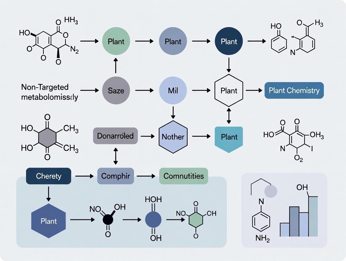
Non-Targeted Metabolomics in Plant Chemistry: A Comprehensive Guide from Discovery to Clinical Applications
This article provides a comprehensive overview of non-targeted metabolomics and its transformative role in plant chemical analysis.
Non-Targeted Metabolomics in Plant Chemistry: A Comprehensive Guide from Discovery to Clinical Applications
Abstract
This article provides a comprehensive overview of non-targeted metabolomics and its transformative role in plant chemical analysis. Aimed at researchers, scientists, and drug development professionals, it explores the foundational principles of untargeted approaches for hypothesis-free discovery of novel plant metabolites. The scope covers advanced LC-MS and NMR methodologies, practical applications in crop improvement and stress response studies, and critical troubleshooting for data accuracy and linearity challenges. It further examines validation strategies and comparative analyses with targeted methods, highlighting the direct pathway this technology provides for identifying bioactive plant compounds with therapeutic potential. The integration of these facets offers a complete resource for leveraging plant metabolomics in pharmaceutical and biomedical research.
Unlocking Plant Chemical Diversity: Foundations of Non-Targeted Metabolomics
Background: Non-targeted metabolomics is a powerful analytical strategy for the comprehensive analysis of small molecules in biological systems, enabling the discovery of novel compounds and biochemical pathways without a priori knowledge of sample composition. Aim of Review: This application note delineates the fundamental principles, standardized workflows, and practical protocols for implementing non-targeted metabolomics in plant chemistry research, with emphasis on its hypothesis-generating potential. Key Scientific Concepts: We elaborate the complete workflow from experimental design to data interpretation, highlighting feature-based molecular networking for chemical characterization, quality assurance measures for cross-laboratory reproducibility, and visualization strategies for effective data communication. This approach is particularly valuable for exploring the vast chemical diversity in plants, where much of the metabolome remains uncharacterized.
Non-targeted metabolomics represents a systematic approach for the simultaneous detection and relative quantification of a broad spectrum of metabolites within a biological system [1]. Unlike targeted analyses that focus on predefined compounds, non-targeted methods aim to capture as much of the metabolome as possible, serving as a powerful hypothesis-generating tool for discovering novel compounds, biomarkers, and biochemical pathways [2]. In plant chemistry research, this approach is particularly valuable for investigating the chemical diversity of both primary and specialized metabolites, which enables the comprehensive profiling of wild edible plants, understanding plant-environment interactions, and identifying bioactive compounds with potential pharmaceutical applications [3] [1].
The foundational principle of non-targeted metabolomics lies in its ability to provide a global overview of metabolic phenotypes without prior assumptions about which compounds are significant [2]. This methodology has revealed that our knowledge of food composition has traditionally focused on merely 35-160 molecular components, representing just a small fraction of the tens of thousands of molecules that constitute food, highlighting the vast potential for discovery in plant metabolomics [2]. The integration of high-resolution mass spectrometry with advanced computational analytics has positioned non-targeted metabolomics as an indispensable tool for expanding our understanding of plant chemical diversity and its applications in drug development and nutrition science.
Experimental Design and Workflow
The non-targeted metabolomics workflow encompasses multiple critical stages, from sample preparation to data interpretation, with rigorous quality control essential at each step to ensure reproducible and biologically meaningful results [1]. The workflow can be conceptually divided into wet laboratory and computational components, with visualizations playing a crucial role in data inspection, evaluation, and sharing throughout the process [4].
Table 1: Key Stages in Non-Targeted Metabolomics Workflow
| Stage | Key Activities | Output |
|---|---|---|
| Sample Collection & Preparation | Homogenization, metabolite extraction using appropriate solvents | Metabolite extract in solution |
| Chromatographic Separation | LC-MS (reversed-phase/HILIC) or GC-MS separation | Chromatograms with resolved peaks |
| Data Acquisition | High-resolution MS and MS/MS in data-dependent or data-independent modes | Raw spectral data files |
| Data Preprocessing | Peak detection, alignment, retention time correction, feature finding | Peak intensity table (feature matrix) |
| Statistical Analysis & Annotation | Multivariate analysis, molecular networking, database searching | Annotated metabolites, significantly altered features |
The following diagram illustrates the comprehensive workflow for non-targeted metabolomics in plant research:
Diagram 1: Non-targeted metabolomics workflow for plant chemistry.
Effective experimental design must account for biological replication, randomization, and incorporation of quality control samples throughout the analytical sequence [2]. Quality control (QC) samples, typically prepared from pooled aliquots of all study samples, are essential for monitoring instrument performance, evaluating technical variance, and correcting for systematic bias [1]. The data preprocessing stage involves noise reduction, retention time correction, peak detection and integration, and chromatographic alignment using specialized software platforms, after which data normalization is performed to reduce technical variation [1].
Core Methodologies and Protocols
Standardized Metabolomics Protocol for Cross-Laboratory Comparison
Recent advancements in non-targeted metabolomics have focused on standardizing protocols to enable data comparability across different laboratories and instrumentation platforms [2]. A validated approach for plant and food matrices involves solid phase extraction (SPE) reverse phase liquid chromatography (RPLC) positive mode electrospray (+ESI) high resolution mass spectrometry (HRMS), which balances broad metabolome coverage with practical implementation across different mass spectrometry platforms [2].
Table 2: Detailed Protocol for Non-Targeted Metabolomics of Plant Samples
| Step | Procedure | Parameters & Specifications |
|---|---|---|
| Sample Preparation | Homogenize lyophilized tissue to fine powder; weigh 50±1mg; add extraction solvent | Methanol:Water (80:20, v/v) with 0.1% formic acid; internal standards |
| Metabolite Extraction | Vortex, sonicate, centrifuge; transfer supernatant; repeat extraction; combine supernatants | 10 min vortex, 15 min sonication, 10 min centrifugation at 4°C |
| Sample Analysis | Inject onto LC-MS system; data acquisition in positive ESI mode with DDA | Reversed-phase C18 column; 35min gradient; MS1 (70,000 resolution), MS/MS (17,500) |
| Quality Control | Include pooled QC samples, solvent blanks, and internal standard mix throughout sequence | QC injection every 6-10 samples; monitor retention time stability and peak intensity |
This standardized approach has been demonstrated to effectively align small molecule data across different laboratories regardless of food type, establishing a foundational framework for generating high-quality, reproducible non-targeted metabolomics data [2]. The method incorporates a rationally-designed internal retention time standard (IRTS) mixture to correct for retention time shifts across different instruments and laboratories, significantly improving feature alignment and compound identification accuracy [2].
Data Processing and Molecular Networking
Following data acquisition, raw mass spectrometry files undergo preprocessing using specialized software such as XCMS, MZmine, or MS-DIAL for peak detection, retention time alignment, and feature quantification [3] [1]. The resulting feature tables are then subjected to feature-based molecular networking (FBMN) using the Global Natural Products Social Molecular Networking (GNPS) platform, which groups MS/MS spectra based on similarity to visualize the chemical relationships within samples [3].
The molecular networking approach enables the organization of complex metabolomic data into molecular families, facilitating the annotation of both known and novel compounds [3]. In plant metabolomics studies, this technique has successfully characterized diverse biochemical classes, with research on Rumex sanguineus demonstrating that approximately 60% of detected metabolites belonged to polyphenol and anthraquinone classes, while also enabling the quantification of potentially toxic compounds like emodin across different plant tissues [3].
Essential Research Reagents and Materials
Successful implementation of non-targeted metabolomics requires careful selection of research reagents and materials to ensure comprehensive metabolite coverage and analytical reproducibility.
Table 3: Essential Research Reagent Solutions for Non-Targeted Metabolomics
| Reagent/Material | Function/Purpose | Specifications/Alternatives |
|---|---|---|
| LC-MS Grade Solvents | Mobile phase preparation; sample extraction and reconstitution | Methanol, acetonitrile, water, isopropanol; with 0.1% formic acid or ammonium formate |
| Internal Standards | Quality control; retention time alignment; quantification | Internal Retention Time Standard (IRTS) mixture; stable isotope-labeled compounds |
| Solid Phase Extraction Cartridges | Sample clean-up; metabolite fractionation | Reversed-phase C18; mixed-mode cation/anion exchange; according to target metabolome |
| Chromatography Columns | Metabolite separation prior to MS detection | Reversed-phase C18 (for non-polar); HILIC (for polar); 100×2.1mm, 1.7-1.8μm particles |
| Mass Spectrometry Calibration Solutions | Instrument calibration; mass accuracy maintenance | Sodium formate or proprietary calibration solutions specific to instrument manufacturer |
The selection of appropriate reagents is critical for method robustness, particularly when implementing cross-laboratory standardized protocols [2]. The use of high-purity solvents and well-characterized internal standards significantly reduces technical variation and enhances the detection of true biological differences in plant metabolomics studies.
Data Visualization and Interpretation
Effective data visualization is indispensable throughout the non-targeted metabolomics workflow, serving critical functions in data inspection, quality assessment, and insight communication [4]. Visualization strategies range from basic quality control plots to advanced molecular networks that enable chemical structural annotations and hypothesis generation.
The following diagram illustrates the process of molecular networking and metabolite annotation:
Diagram 2: Molecular networking for metabolite annotation.
Visualizations serve as a means to augment researchers' decision-making capabilities by summarizing data, extracting and highlighting patterns, and organizing relations within complex datasets [4]. In non-targeted metabolomics, effective visualizations include scatter plots with line graphs for data summary, cluster heatmaps for pattern extraction, and network visualizations for organizing and showcasing relationships between metabolites [4]. These visual tools are particularly valuable for communicating the complex results of plant metabolomics studies, where chemical diversity can be substantial and novel compounds are frequently encountered.
Applications in Plant Chemistry Research
Non-targeted metabolomics has demonstrated significant utility across diverse applications in plant chemistry research, particularly in the characterization of wild edible plants, investigation of plant-environment interactions, and discovery of bioactive compounds with pharmaceutical potential.
Research on Rumex sanguineus, a traditional medicinal plant from the Polygonaceae family, exemplifies the power of non-targeted metabolomics for comprehensive chemical characterization [3]. By applying UHPLC-HRMS analysis and feature-based molecular networking to different plant tissues (roots, stems, and leaves), researchers annotated 347 primary and specialized metabolites grouped into 8 biochemical classes, with the majority (60%) belonging to polyphenols and anthraquinones [3]. This approach also facilitated the quantification of emodin, a potentially toxic anthraquinone, revealing its higher accumulation in leaves compared to stems and roots—information critical for assessing safety in culinary and medicinal applications [3].
The non-targeted approach also enables the detection of unexpected compounds, including environmental contaminants such as pesticides and per- and poly-fluoroalkyl substances (PFAS) in food matrices, expanding its utility beyond endogenous metabolite profiling to comprehensive chemical safety assessment [2]. This capability is particularly valuable for evaluating wild edible plants where contamination profiles may be unknown.
The plant metabolome represents the final downstream product of cellular regulation and encompasses a staggering chemical diversity that is central to a plant's existence, defense, and interactions with the environment. This complex universe of metabolites is broadly categorized into primary metabolites and specialized metabolites. Primary metabolites include compounds such as carbohydrates, lipids, and amino acids, which are universally essential for fundamental processes like growth, development, reproduction, and energy storage [5] [6]. In contrast, specialized metabolites (historically termed secondary metabolites) are a vast array of compounds—including terpenoids, phenylpropanoids, polyketides, and alkaloids—that are not directly involved in primary growth processes but are crucial for the plant's survival and perpetuation [5] [6]. These specialized metabolites facilitate communication and interactions with other organisms and serve as an alternative defense mechanism, with over 200,000 distinct types identified across the plant kingdom [6].
The biosynthetic pathways for these compounds are sophisticated and energetically expensive. While the building blocks for specialized metabolites originate from highly conserved central metabolic pathways (e.g., glycolysis, shikimate, mevalonate), the later stages of biosynthesis are notably complex and diverse [6]. This diversity is influenced by factors such as cell type, developmental stage, and environmental cues, leading to the immense structural variety observed in plant specialized metabolites [6]. This chemical complexity, while a rich source of bioactive compounds for medicine and agriculture, also presents a significant analytical challenge, which non-targeted metabolomics is uniquely positioned to address.
Analytical Platforms for Non-Targeted Metabolomics
Non-targeted metabolomics aims to provide a comprehensive, unbiased analysis of all measurable metabolites in a biological sample. The two foremost analytical techniques employed in this field are Mass Spectrometry (MS) and Nuclear Magnetic Resonance (NMR) spectroscopy, each with distinct advantages and limitations, as detailed in the table below [5].
Table 1: Comparison of Primary Analytical Platforms in Plant Metabolomics
| Feature | Mass Spectrometry (MS) | Nuclear Magnetic Resonance (NMR) |
|---|---|---|
| Sensitivity | High (Low LOD/LOQ) | Low to Moderate (µM range) |
| Metabolite Coverage | Hundreds per sample | Dozens per sample |
| Sample Preparation | Minimal; may require derivatization | Minimal |
| Analysis Nature | Destructive | Non-destructive |
| Quantification | Often requires internal standards | Directly quantitative |
| Structural Elucidation | Putative; requires fragmentation/chromatography | Direct; definitive for novel compounds |
| Key Strength | Broad metabolite coverage, high sensitivity | Structural identification, isomer differentiation, isotope tracing |
| Common Hyphenation | LC-MS, GC-MS | Not applicable |
MS is typically hyphenated with separation techniques like liquid or gas chromatography (LC-MS or GC-MS) to enhance metabolite coverage and identification [7] [5]. Its primary strength lies in its high sensitivity, enabling the detection of a vast range of metabolites. However, identification is often only putative and can lead to misidentifications [5]. Conversely, NMR spectroscopy is a nondestructive technique that allows for the simultaneous identification and quantification of metabolites without the need for extensive separation or reference standards [5]. Its powerful capability for de novo structural elucidation and isomer differentiation makes it particularly valuable for investigating plants where new or rare metabolites are present, though its lower sensitivity means it detects fewer metabolites per sample compared to MS [5]. Given their complementary capabilities, these techniques are often used in combination to provide a more holistic view of the plant metabolome [5].
Detailed Experimental Protocols
The following sections provide detailed, practical protocols for conducting a non-targeted metabolomics study in plants, incorporating both MS and NMR methodologies.
Protocol 1: Non-Targeted Metabolomics via LC-MS for Stress Response Investigation
This protocol is adapted from studies investigating the effects of abiotic stress and herbicide exposure on plant metabolism [7] [8]. It outlines the procedure from sample collection to data acquisition using Liquid Chromatography coupled to a high-resolution Mass Spectrometer.
1. Sample Collection and Preparation:
- Plant Material & Treatment: Grow plants under controlled conditions. Apply the stressor (e.g., drought simulated by PEG-6000, or chemical stressor like atrazine) to the treatment group while maintaining a control group [8]. The flowering period is often the most sensitive stage for stress studies [8].
- Harvesting: Collect leaf or other tissue samples at multiple time points post-treatment (e.g., day 0, 3, 6, 9). Immediately freeze the samples in liquid nitrogen to halt metabolic activity [8].
- Lyophilization and Homogenization: Lyophilize the frozen samples for 72 hours. Grind the lyophilized material into a fine powder using a homogenizer like a TissueLyser [8].
- Metabolite Extraction: Weigh ~50 mg of the lyophilized powder. Add 1000 µL of a cold extraction solvent (e.g., methanol:acetonitrile:water in a 2:2:1 ratio, containing internal standards). Vortex vigorously and homogenize with ceramic beads [8]. Centrifuge (e.g., 13,000 g for 15 min at 4°C) and collect the supernatant for analysis [7] [8].
2. LC-MS Data Acquisition:
- Chromatography: Use a UHPLC or UPLC system with a C18 reversed-phase column. A common mobile phase consists of (A) water with 0.1% formic acid and (B) acetonitrile with 0.1% formic acid. Employ a gradient elution, for example: 0-2 min, 5% B; 2-15 min, 5-95% B; 15-17 min, 95% B; 17-17.1 min, 95-5% B; 17.1-20 min, 5% B [7].
- Mass Spectrometry: Analyze the samples using a high-resolution mass spectrometer, such as a Q-TOF or ion trap-triple quadrupole system. Acquire data in both positive and negative electrospray ionization (ESI) modes to maximize metabolite coverage. Data-Dependent Acquisition (DDA) is often used, where the top N most intense ions from a full MS scan are selected for MS/MS fragmentation [7].
3. Data Processing and Analysis:
- Peak Picking and Alignment: Process the raw data using software (e.g., MS-DIAL, XCMS) to detect features (ions characterized by m/z and retention time), align them across samples, and perform peak table construction.
- Metabolite Annotation: Annotate features by comparing their accurate mass and MS/MS fragmentation spectra against public databases such as MassBank, Metlin, and GNPS [3] [8]. Feature-Based Molecular Networking on the GNPS platform is a powerful tool for organizing metabolites based on spectral similarity and annotating within molecular families [3].
- Statistical Analysis: Import the peak table with normalized intensities into statistical software. Use multivariate statistics like Principal Component Analysis (PCA) and Partial Least Squares-Discriminant Analysis (PLS-DA) to identify metabolites that discriminate between treatment and control groups. Univariate statistics (e.g., t-tests, ANOVA) with correction for multiple testing (e.g., False Discovery Rate) are also applied.
Figure 1: LC-MS non-targeted metabolomics workflow for plant stress studies.
Protocol 2: NMR-Based Metabolomics for Comprehensive Metabolite Profiling
This protocol is based on established NMR methodologies for plant metabolomics, which are particularly valuable for definitive structural identification and studies where sample preservation is desired [5].
1. Sample Preparation for NMR:
- Extraction: Prepare a polar extract from plant tissue (e.g., 100 mg fresh weight) using a deuterated solvent mixture like CD₃OD:KH₂PO₄ buffer in D₂O (1:1). The use of D₂O provides a field-frequency lock for the NMR spectrometer [5].
- Internal Standard: Include an internal chemical shift standard, such as 0.1 mM TSP (trimethylsilylpropanoic acid) or DSS, which also serves as a reference for quantification [5].
- Centrifugation: Centrifuge the extract (e.g., 13,000 g for 10 min) to remove any particulate matter.
- Loading: Transfer a precise volume (e.g., 600 µL) of the clear supernatant into a standard 5 mm NMR tube.
2. NMR Data Acquisition:
- Instrument Setup: Conduct experiments on a high-field NMR spectrometer (e.g., 500 MHz or 600 MHz). Maintain a constant temperature (e.g., 298 K) during analysis.
- Key Pulse Sequences:
- 1D ¹H NMR: Begin with a standard one-dimensional pulse sequence with water suppression (e.g., presat or NOESY-presat). This is the primary experiment for profiling and quantification. Typical parameters: 64-128 transients, spectral width of 12-16 ppm, and a relaxation delay of 2-4 seconds [5].
- 2D NMR: For structural elucidation of unknown metabolites, acquire two-dimensional experiments. Essential 2D experiments include:
- ¹H-¹H COSY (Correlation Spectroscopy): Identifies scalar-coupled protons.
- ¹H-¹H TOCSY (Total Correlation Spectroscopy): Reveals correlations within a spin system.
- ¹H-¹³C HSQC (Heteronuclear Single Quantum Coherence): Identifies direct ¹H-¹³C couplings, providing a map of protonated carbons.
- ¹H-¹³C HMBC (Heteronuclear Multiple Bond Correlation): Detects long-range ¹H-¹³C couplings, crucial for establishing connectivity between quaternary carbons and protons [5].
3. NMR Data Processing and Analysis:
- Processing: Apply Fourier transformation to the Free Induction Decay (FID). Use phase correction and baseline correction. Reference the spectrum to the internal standard (e.g., TSP at 0.0 ppm).
- Spectral Bucketing: To facilitate statistical analysis, segment the ¹H NMR spectrum into small regions (buckets or bins) and integrate the area under each segment. This reduces the complexity of the data.
- Metabolite Identification and Quantification: Identify metabolites by comparing the chemical shifts, coupling constants, and spin-spin correlations from 1D and 2D spectra with reference spectra in databases (e.g., HMDB, BMRB, in-house libraries). The concentration of metabolites can be directly calculated by integrating their resolved peaks relative to the internal standard [5].
- Chemometric Analysis: Subject the bucketed data or quantified concentrations to multivariate statistical analysis (PCA, PLS-DA) to identify metabolic patterns and biomarkers.
The Scientist's Toolkit: Essential Research Reagents and Materials
Successful non-targeted metabolomics relies on a suite of essential reagents and materials. The following table details key solutions and their specific functions in a typical workflow.
Table 2: Key Research Reagent Solutions for Plant Non-Targeted Metabolomics
| Reagent/Material | Function & Application in Protocol |
|---|---|
| Methanol, Acetonitrile, Water (LC-MS Grade) | Used as extraction solvents and LC-MS mobile phases. High purity is critical to minimize background noise and ion suppression in MS [7] [8]. |
| Deuterated Solvents (e.g., CD₃OD, D₂O) | NMR solvent that provides a field-frequency lock and enables the accurate shimming of the magnetic field [5]. |
| Internal Standards (e.g., TSP, DSS) | Added to NMR samples for chemical shift referencing (calibration) and as a known concentration for quantitative analysis [5]. |
| Formic Acid (LC-MS Grade) | A mobile phase additive in LC-MS (0.1%) to improve chromatographic peak shape and enhance ionization efficiency in positive ESI mode [7]. |
| Polyethylene Glycol (PEG-6000) | Used to simulate osmotic (drought) stress in plant growth experiments by creating a negative water potential in the growth medium [8]. |
| Chemical Shift Reference Databases (e.g., HMDB, BMRB) | Electronic libraries of known metabolite NMR spectra used for the definitive identification of compounds in complex plant extracts [5]. |
| MS/MS Spectral Libraries (e.g., GNPS, MassBank) | Public repositories of mass spectral fragmentation data used for the annotation of metabolites in LC-MS/MS studies [3] [8]. |
Integration with Other Omics and Visualization of Metabolic Pathways
Non-targeted metabolomics is most powerful when integrated with other omics technologies in a multi-omics approach. The combination of metabolomics with transcriptomics is particularly effective for identifying genes involved in specialized metabolic pathways [9] [6]. This integration operates on the hypothesis that the expression of genes encoding enzymes in a biosynthetic pathway will be co-regulated and will correlate with the accumulation of the pathway's end product [9]. For instance, this approach has been successfully used to discover pathways for compounds like kavalactones in kava and podophyllotoxin in mayapple [9].
Specialized metabolite biosynthesis begins with primary metabolic pathways, which provide the essential precursors. The diagram below illustrates the major branching points from primary to specialized metabolism.
Figure 2: Biosynthetic origins of major specialized metabolite classes from primary metabolism.
This multi-omics framework, supported by the detailed protocols for MS and NMR, provides researchers with a comprehensive strategy to move from simply observing metabolic changes to understanding their genetic and enzymatic basis, ultimately enabling the engineering of pathways for sustainable production of valuable plant metabolites [9] [6].
Non-targeted metabolomics has emerged as a powerful analytical strategy in plant chemistry research, enabling the comprehensive investigation of low-molecular-weight metabolites without prior hypothesis. This approach captures the metabolic phenotype of plants, reflecting interactions between genetics, development, and environmental influences [5]. The field primarily distinguishes between metabolic fingerprinting, which provides a rapid, high-throughput overview of sample classification, and comprehensive profiling, which aims to identify and quantify a broader range of metabolites for detailed biochemical interpretation [10] [11].
In plant sciences, these techniques are particularly valuable because plants produce a vast array of specialized metabolites—estimated at over a million across the plant kingdom—that play crucial roles in survival, defense, and communication [11]. These compounds also have significant applications in drug development, agriculture, and food science. However, the tremendous structural diversity of plant metabolites presents substantial analytical challenges, with current technologies able to identify only a fraction of the metabolites detected in typical plant extracts [11].
This application note outlines the core principles, methodologies, and practical applications of non-targeted metabolomics in plant research, providing detailed protocols for researchers and scientists seeking to implement these approaches in their workflows.
Key Concepts and Analytical Approaches
Defining Metabolic Fingerprinting and Profiling
Metabolic fingerprinting is a non-targeted approach focused on rapid sample classification and pattern recognition without necessarily identifying all metabolites. It generates global spectral signatures that can be used to discriminate between sample groups based on their biological origin or treatment condition [5]. This approach is particularly useful for quality control, phenotyping, and detecting metabolic responses to environmental stressors or genetic modifications.
Comprehensive metabolic profiling extends beyond fingerprinting by aiming to identify and quantify a wide range of metabolites, providing deeper biochemical insights. While still non-targeted in nature, profiling seeks to put names to the discriminating features, enabling biological interpretation at the pathway level [7] [12]. This approach is more resource-intensive but offers greater mechanistic understanding of plant metabolic processes.
Analytical Techniques in Non-Targeted Metabolomics
Two principal analytical platforms dominate non-targeted metabolomics, each with distinct advantages and limitations:
Table 1: Comparison of Major Analytical Platforms in Plant Metabolomics
| Platform | Sensitivity | Metabolite Coverage | Key Strengths | Primary Limitations |
|---|---|---|---|---|
| Mass Spectrometry (MS) | High (low LOD/LOQ) | Hundreds to thousands of features | High sensitivity, broad dynamic range, structural information via MS/MS | Destructive analysis, typically requires chromatography, putative identification only |
| Nuclear Magnetic Resonance (NMR) | Moderate (µM-mM range) | Dozens to hundreds of metabolites | Non-destructive, quantitative, structural elucidation power, high reproducibility | Lower sensitivity, spectral overlap challenges |
Mass Spectrometry platforms, particularly when coupled with separation techniques like liquid chromatography (LC-MS) or gas chromatography (GC-MS), offer high sensitivity and can detect thousands of metabolite features in a single sample [7] [11]. LC-MS is preferred for thermally labile compounds such as alkaloids, phenolic compounds, and most secondary metabolites, while GC-MS is suitable for volatile compounds and those made amenable to analysis through derivatization (e.g., organic acids, sugars) [10]. The workflow typically involves sample extraction, chromatographic separation, ionization (commonly electrospray ionization), and mass analysis using high-resolution instruments such as time-of-flight (TOF) or Orbitrap mass analyzers [12].
Nuclear Magnetic Resonance spectroscopy provides a complementary approach, with the key advantage of being non-destructive and inherently quantitative without requiring reference standards [5]. Although less sensitive than MS, NMR excels at structural elucidation of unknown compounds and isomer differentiation, making it particularly valuable for investigating novel plant metabolites [5]. Proton (¹H) NMR is most commonly used due to the high natural abundance of hydrogen and relatively short experiment times.
Diagram: Decision Workflow for Selecting Analytical Platforms in Plant Metabolomics
Experimental Design and Workflow
Sample Preparation and Extraction
Proper sample preparation is critical for generating reliable and reproducible metabolomic data. The general workflow begins with immediate quenching of metabolism, typically using liquid nitrogen, to preserve the metabolic state at the time of collection [5]. For plant tissues, this is followed by homogenization (often with liquid nitrogen), and then metabolite extraction.
A common effective extraction protocol for comprehensive plant metabolomics involves:
- Tissue Processing: Grind 30-50 mg of lyophilized leaf material under liquid nitrogen to a fine powder [12].
- Biphasic Extraction: Use a methyl-tert-butyl-ether:methanol (MTBE:MeOH, 3:1 v:v) solvent system to extract both polar and non-polar metabolites simultaneously [12].
- Internal Standards: Add stable isotope-labeled internal standards (e.g., U-¹³C-sorbitol, L-Alanine-d4) for quality control and potential normalization [12].
- Extraction Procedure: Vortex, incubate on an orbital shaker (40 rpm, 45 min, 4°C), sonicate in a water bath (15 min, 4°C), and centrifuge (10,000×g, 10 min, 4°C) [12].
- Sample Preparation for Analysis: Transfer aliquots of the soluble fraction to new vials and dry under a stream of nitrogen or in a speed vacuum concentrator.
For NMR-based approaches, samples are typically reconstituted in deuterated solvents (e.g., D₂O, CD₃OD) containing a reference standard such as trimethylsilylpropanoic acid (TSP) for chemical shift calibration [5].
Data Acquisition and Metabolite Identification
The data acquisition strategy depends on the analytical platform selected. For LC-MS-based non-targeted metabolomics, reverse-phase chromatography with C18 columns is commonly used, with gradients typically employing water and acetonitrile or methanol, both modified with 0.1% formic acid to enhance ionization [7]. Data-dependent acquisition (DDA) is frequently employed, where the top N most intense ions from the full MS scan are selected for MS/MS fragmentation to generate structural information.
For GC-MS analyses, samples typically require derivatization to increase volatility and stability. A common approach follows the protocol of Erban et al. (2007), using methoxyamine hydrochloride in pyridine followed by N-trimethylsilyl-N-methyl trifluoroacetamide (MSTFA) [12]. Separation is achieved using DB-35MS or similar columns with temperature ramping from 85°C to 360°C.
Metabolite identification remains a significant challenge in non-targeted plant metabolomics, with typically only 2-15% of detected peaks confidently annotated through spectral library matching [11]. The Metabolomics Standards Initiative (MSI) has established confidence levels for metabolite identification:
Table 2: Metabolite Identification Confidence Levels
| Confidence Level | Identification Evidence | Typical Approaches |
|---|---|---|
| Level 1: Identified | Matching to authentic standard using two orthogonal properties (e.g., RT + MS/MS) | Commercial standards, in-house libraries |
| Level 2: Putatively Annotated | Spectral similarity to reference library without RT match | GNPS, MassBank, METLIN, RefMetaPlant |
| Level 3: Putative Class | Characteristic chemical class features | CANOPUS, NPClassifier, rule-based fragmentation |
| Level 4: Unknown | Distinguished only by m/z and RT | De novo characterization needed |
Several databases and software tools have been developed to facilitate metabolite annotation in plants:
- RefMetaPlant: A reference metabolome database specifically for plants [11]
- Plant Metabolome Hub (PMhub): Consolidates MS/MS data for nearly 189,000 plant metabolites [11]
- GOLM Metabolome Database: Particularly useful for GC-MS based metabolomics [12]
- KEGG and HMDB: Provide pathway information and metabolite annotations [12]
- GNPS (Global Natural Products Social Molecular Networking): Enables community-wide sharing of MS/MS spectra [11]
Advanced computational approaches, including machine learning tools like CSI-FingerID, CANOPUS, and Mass2SMILES, are increasingly being employed to improve annotation rates and predict compound classes from MS/MS data without authentic standards [11].
Applications in Plant Chemistry Research
Investigating Environmental Stress Responses
Non-targeted metabolomics has proven valuable for understanding how plants respond to abiotic and biotic stressors. A recent study investigated the hidden effects of the herbicide atrazine and its degradation products on Japanese radish (Raphanus sativus var. longipinnatus) metabolism [7]. Using LC-MS-based non-targeted metabolomics, researchers discovered that both atrazine and its metabolites (DEA, DIA, DEDIA) significantly altered amino acid profiles in the plants, despite the absence of visible stress symptoms. This demonstrates the sensitivity of metabolomics in detecting subtle biochemical changes before morphological symptoms appear.
The study employed chemometric tools for data analysis, including partial least squares-discriminant analysis (PLS-DA), to identify metabolic patterns distinguishing treatment groups. Key findings included disruptions in branched-chain amino acid metabolism, highlighting the potential impact of environmental contaminants on plant nutritional quality [7].
Cultivar Differentiation and Precision Breeding
Non-targeted metabolomics enables the identification of metabolic fingerprints that distinguish plant cultivars with different genetic backgrounds. Research on five Coffea arabica cultivars grown in field conditions demonstrated distinct metabolic signatures among cultivars, with 41 metabolites identified as key discriminators [12]. The non-targeted GC-MS approach detected 463 metabolic features, with major classes including sugars, amino acids, lipids, phenylpropanoids, and phenolic compounds.
PLS-DA analysis revealed that ferulic acid, theobromine, octopamine, rosmarinic acid, and gibberellin were particularly important for cultivar discrimination [12]. This metabolic fingerprinting approach provides valuable tools for coffee breeding programs, allowing selection of cultivars with desirable traits such as stress resistance or cup quality based on their metabolic profiles.
Diagram: Integrated Workflow for Non-Targeted Plant Metabolomics
Essential Research Reagent Solutions
Successful implementation of non-targeted metabolomics requires careful selection of reagents and materials. The following table outlines key solutions for plant metabolomics research:
Table 3: Essential Research Reagent Solutions for Plant Metabolomics
| Reagent/Material | Function/Purpose | Application Notes |
|---|---|---|
| MTBE:MeOH (3:1, v:v) | Biphasic extraction solvent | Simultaneously extracts polar and non-polar metabolites; enables comprehensive metabolite coverage [12] |
| Deuterated Solvents (D₂O, CD₃OD) | NMR sample preparation | Provides locking signal for NMR stability; enables quantitative analysis without internal standards [5] |
| Methoxyamine hydrochloride | GC-MS derivatization agent | Protects carbonyl groups and reduces tautomerization; improves metabolite stability and separation [12] |
| MSTFA | GC-MS silylation reagent | Increases volatility of metabolites; essential for GC-MS analysis of non-volatile compounds [12] |
| Stable Isotope-Labeled Standards | Quality control and normalization | Corrects for instrument variation; validates analytical performance [12] |
| C18 LC Columns | Reverse-phase chromatography | Separates metabolites by hydrophobicity; workhorse for LC-MS metabolomics [7] |
| DB-35MS GC Columns | GC-MS separation | Mid-polarity stationary phase; suitable for diverse metabolite classes [12] |
Data Analysis and Interpretation Strategies
Statistical and Bioinformatics Approaches
The analysis of non-targeted metabolomics data requires specialized statistical approaches to extract meaningful biological information from complex multivariate datasets. Common strategies include:
- Unsupervised methods such as Principal Component Analysis (PCA) for exploratory data analysis and outlier detection [7] [12]
- Supervised methods including Partial Least Squares-Discriminant Analysis (PLS-DA) and Orthogonal PLS-DA (OPLS-DA) for identifying metabolites that discriminate between predefined sample groups [7] [12]
- Molecular networking based on MS/MS similarity to visualize chemical relationships and identify structurally related metabolites without requiring prior identification [11]
- Information theory-based metrics to assess metabolic diversity and complexity without complete metabolite identification [11]
For studies where metabolite identification remains challenging, several identification-free approaches have been developed. These include analyzing spectral features directly, comparing fold-changes of unknown features, and employing database-independent visualization tools that cluster metabolites based on fragmentation patterns or chromatographic behavior [11].
Data Visualization and Reporting
Effective visualization of metabolomics data is essential for interpretation and communication of results. The complexity of metabolomic datasets often requires multiple visualization strategies:
- Volcano plots to display both statistical significance (p-values) and magnitude of change (fold-change) simultaneously
- Heatmaps with hierarchical clustering to visualize patterns in metabolite abundance across sample groups
- Pathway maps to display metabolic perturbations in the context of biochemical networks
- Box and whisker plots for comparing distributions of individual metabolites across experimental groups [13]
When preparing metabolomics data for publication, it is essential to follow journal guidelines regarding data presentation. Key considerations include providing clear, self-explanatory titles for all tables and figures, defining all abbreviations in footnotes, and ensuring consistency in formatting across all visual elements [13]. Most journals now require raw metabolomics data to be deposited in public repositories such as MetaboLights or the Metabolomics Workbench.
Non-targeted metabolomics provides powerful approaches for investigating plant chemistry, from initial metabolic fingerprinting for sample classification to comprehensive profiling for detailed biochemical interpretation. The integration of advanced analytical platforms, particularly high-resolution mass spectrometry and NMR spectroscopy, with sophisticated bioinformatics tools has dramatically enhanced our ability to characterize the complex metabolomes of plants.
Despite significant advances, challenges remain in metabolite identification, data integration, and biological interpretation. Ongoing developments in computational approaches, including machine learning and artificial intelligence, are promising strategies to address these limitations. As the field continues to evolve, non-targeted metabolomics will play an increasingly important role in plant research, from fundamental studies of metabolic diversity to applied applications in crop improvement, natural product discovery, and environmental monitoring.
For researchers implementing these approaches, careful attention to experimental design, sample preparation, quality control, and data analysis is essential for generating robust and biologically meaningful results. The protocols and applications outlined in this article provide a foundation for developing effective metabolomics strategies in plant chemistry research.
In the field of plant chemistry research, a significant challenge persists: the vast majority of metabolites detected through modern analytical techniques remain unidentified. This unexplored chemical space, often termed "metabolic dark matter," represents a critical knowledge gap in understanding plant physiology, stress responses, and biosynthetic potential [11]. Current untargeted liquid chromatography-tandem mass spectrometry (LC-MS/MS) analyses typically detect thousands of metabolic features from plant extracts, yet studies consistently report that >85% of these peaks cannot be annotated with confidence using standard approaches [11]. This identification bottleneck limits our ability to fully decipher the chemical diversity that plants employ for defense, communication, and adaptation.
The plant metabolome is estimated to contain over a million metabolites, yet comprehensive databases contain only a fraction of these compounds. For instance, the KNApSAcK plant metabolite database lists approximately 63,723 compounds as of its 2024 update, highlighting the immense disparity between known and unknown chemical space in plants [11]. This review outlines integrated experimental and computational strategies to illuminate this dark matter, with particular emphasis on approaches relevant to plant specialized metabolism, natural product discovery, and crop improvement research.
Experimental Workflows for Functional Group Characterization
Multiplexed Chemical Labeling (MCheM)
Principle: MCheM introduces chemical reactivity as an additional dimension to LC-MS/MS analyses by using selective derivatization reagents that target specific functional groups, thereby revealing structural information through predictable mass shifts [14].
Protocol:
- Post-column Derivatization Setup: Integrate a microfluidic reactor after the analytical column but prior to MS ionization.
- Reagent Selection: Employ multiple, parallel derivatization reagents targeting complementary functional groups (e.g., hydroxyls, amines, carboxylic acids).
- Reaction Optimization: Adjust flow rates, reaction temperature, and solvent compatibility to maintain chromatographic integrity while ensuring complete derivatization.
- Data Acquisition: Acquire MS/MS data in real-time during reagent introduction.
- Data Analysis: Identify mass shifts corresponding to specific functional group additions across different reagent channels.
Applications in Plant Research: This approach proved particularly valuable for characterizing unknown compounds in complex plant extracts, where it helped identify a Michael system in previously unannotated metabolites, dramatically narrowing plausible substructures [14].
Feature-Based Molecular Networking (FBMN)
Principle: FBMN groups metabolites based on similarity of their MS/MS fragmentation patterns, creating visual networks where structurally related compounds cluster together [15] [3].
Protocol:
- LC-MS/MS Data Acquisition: Perform untargeted LC-MS/MS analysis using data-dependent acquisition (DDA) or data-independent acquisition (DIA).
- Feature Detection: Process raw data using tools like MZmine or OpenMS to extract chromatographic features, alignment, and MS/MS spectral decomposition.
- Spectral Networking: Upload processed data to GNPS platform to construct molecular networks based on MS/MS similarity.
- Statistical Analysis: Apply multivariate statistics to identify significant features across experimental conditions using provided R or Python scripts [15].
- Annotation Propagation: Leverage network neighborhoods to propagate annotations from known to unknown metabolites.
Implementation Example: In a study of Rumex sanguineus, FBMN enabled the comprehensive annotation of 347 primary and specialized metabolites, with 60% belonging to polyphenols and anthraquinones classes, demonstrating the power of this approach for characterizing chemically complex plant extracts [3].
Table 1: Comparison of Experimental Approaches for Metabolite Annotation
| Method | Key Principle | Structural Information Gained | Limitations | Suitable for Plant Sample Types |
|---|---|---|---|---|
| MCheM [14] | Selective post-column derivatization | Functional group presence (hydroxyls, amines, carboxylic acids) | Requires optimization of reaction conditions; commercially available reagents | Complex plant extracts; natural product mixtures |
| FBMN [15] [3] | MS/MS spectral similarity networking | Structural similarity; compound classes | Limited for novel scaffolds without reference spectra | Wild edible plants; medicinal plants; stress-responsive tissues |
| KGMN [16] | Multi-layer network integration | Biochemical relationships; putative identities | Depends on quality of initial seed annotations | Plant tissues with well-annotated core metabolomes |
Computational Frameworks for Systematic Annotation
Knowledge-Guided Multi-Layer Network (KGMN)
Principle: KGMN integrates three complementary networks to enable annotation propagation from knowns to unknowns [16]:
- Knowledge-based Metabolic Reaction Network (KMRN): Biochemical transformations from databases (KEGG) expanded with in silico enzymatic reactions
- Knowledge-guided MS/MS Similarity Network: Structural relationships constrained by biochemical plausibility
- Global Peak Correlation Network: Different ion forms (adducts, fragments) of the same metabolite
Workflow Implementation:
- Seed Annotation: Confidently identify a subset of metabolites using standard MS/MS library matching
- Network Expansion: Recursively propagate annotations to reaction-paired neighbors using mass differences, retention time predictions, and MS/MS similarity
- Unknown Metabolite Prediction: Generate putative structures for unannotated features using in silico enzymatic transformations from known seed metabolites
- Validation: Corroborate predictions using in silico MS/MS tools and repository mining
Plant Research Applications: KGMN has demonstrated capability to annotate ~100-300 putative unknowns in individual datasets, with >80% corroboration rate by in silico MS/MS tools, making it particularly valuable for exploring plant specialized metabolism [16].
ATLASx for Biochemical Space Expansion
Principle: ATLASx predicts hypothetical biochemical transformations using 489 generalized enzymatic reaction rules applied to a unified database of 1.5 million biological compounds [17].
Protocol for Plant Natural Product Discovery:
- Query Compound Input: Input a plant natural product of interest or use the database exploration tools
- Pathway Prediction: Identify potential biosynthetic pathways or transformation products
- Reaction Rule Application: Apply expert-curated reaction rules from BNICE.ch to predict novel derivatives
- Structural Evaluation: Assess predicted compounds for novelty and biological relevance
- Experimental Prioritization: Select high-priority targets for isolation or synthesis based on predicted properties
This approach has been successfully used to predict over 5 million reactions and integrate nearly 2 million compounds into biochemical space, significantly expanding the framework for identifying plant natural products [17].
Integrated Multi-Omics Approaches for Plant Metabolism
The integration of metabolomics with genomics and transcriptomics provides a powerful strategy for linking metabolites to their biosynthetic origins. This is particularly relevant for plant natural products, where many biosynthetic gene clusters (BGCs) remain uncharacterized [18].
Protocol for Integrated Omics in Plant Research:
- Genome Sequencing and Assembly: Use long-read technologies (PacBio, Oxford Nanopore) to obtain contiguous assemblies capable of capturing complete BGCs
- BGC Identification: Annotate BGCs using antiSMASH with plant-specific rules [18]
- Metabolite Profiling: Perform untargeted LC-MS/MS under various growth conditions and tissues
- Correlation Analysis: Integrate gene expression and metabolite abundance data to identify candidate genes for specific metabolites
- Functional Validation: Use heterologous expression or gene silencing to verify gene-metabolite relationships
This integrated approach has accelerated the discovery of novel plant natural products by providing a direct link between genetic capacity and metabolic output [18].
Table 2: Computational Tools for Metabolite Annotation in Plant Research
| Tool/Platform | Primary Function | Data Input Requirements | Strengths for Plant Metabolomics | Integration Capabilities |
|---|---|---|---|---|
| GNPS/FBMN [15] | Molecular networking & annotation propagation | LC-MS/MS raw data or feature tables | Extensive plant-relevant spectral libraries; user-friendly web interface | Cytoscape visualization; statistical analysis tools |
| SIRIUS/CANOPUS [11] | In silico fragmentation & compound class prediction | MS/MS spectra | Predicts structural classes using NPClassifier ontology; requires no reference spectra | Standalone tool; can process output from various pre-processing pipelines |
| KGMN [16] | Multi-layer network annotation | LC-MS/MS data with minimal seed annotations | Excellent for annotating unknown plant metabolites using biochemical context | Compatible with MS data pre-processed by common tools |
| ATLASx [17] | Biochemical reaction prediction | Compound structures or queries | Expands known biochemical space; predicts novel transformations | Web interface; connects with biochemical databases |
Table 3: Key Research Reagent Solutions for Plant Metabolite Discovery
| Reagent/Resource | Function | Application Example in Plant Research | Considerations |
|---|---|---|---|
| Derivatization Reagents (e.g., hydroxyl-, amine-targeting) [14] | Reveal specific functional groups through predictable mass shifts | Characterizing reactive groups in unknown plant specialized metabolites | Commercial availability; compatibility with LC mobile phases |
| Authentic Standards | Confident metabolite identification (MSI Level 1) | Quantification of emodin in Rumex sanguineus tissues [3] | Cost; availability of rare plant compounds |
| Stable Isotope-Labeled Precursors (e.g., ^13C, ^15N) | Tracing metabolic pathways and confirming formula assignments | Elucidating biosynthetic pathways of plant natural products | Incorporation efficiency; cost for multiple labeling |
| Specialized LC Columns (HILIC, reversed-phase) | Separation of diverse metabolite classes | Comprehensive coverage of polar and non-polar plant metabolites | Method development time; column longevity with crude extracts |
| Enzyme Inhibitors/Activators | Probing metabolic pathways in vivo | Investigating flux through competing biosynthetic routes | Specificity; potential pleiotropic effects |
Visualizing the Integrated Workflow
The following diagram illustrates the integrated experimental and computational pipeline for advancing from unknown metabolic features to annotated metabolites in plant research:
Diagram 1: Integrated metabolite discovery workflow for plant research.
The challenge of metabolic dark matter in plant chemistry research is being addressed through innovative experimental and computational strategies that create additional layers of information beyond traditional MS/MS matching. The integration of chemical derivatization, molecular networking, knowledge-guided algorithms, and multi-omics approaches provides a powerful framework for systematically annotating previously unknown metabolites. As these technologies continue to mature and become more accessible to the research community, we anticipate significant advances in our understanding of plant chemical diversity, biosynthetic pathways, and ecological functions. The protocols and resources outlined herein provide a roadmap for researchers seeking to illuminate the dark corners of plant metabolism and unlock the full potential of plant-derived compounds for pharmaceutical applications, crop improvement, and fundamental biological discovery.
Non-targeted metabolomics has emerged as a powerful analytical strategy for comprehensively characterizing the small molecule composition of biological systems without prior hypothesis. In the context of biodiversity screening and novel compound discovery, this approach enables researchers to capture the vast chemical diversity present in plants, marine organisms, and other biological resources, much of which remains unexplored [11]. The technological advancement of liquid chromatography-mass spectrometry (LC-MS) platforms now allows researchers to detect thousands of metabolite features from single organ extracts, providing unprecedented access to nature's chemical treasury [11]. This capability is particularly valuable given that current plant metabolite databases document only a fraction of the estimated over one million metabolites existing in the plant kingdom [11]. The application of non-targeted metabolomics within biodiversity research thus addresses a critical bottleneck in natural product discovery, enabling the systematic mapping of chemical diversity across species and ecosystems while facilitating the identification of novel compounds with potential applications in pharmaceuticals, nutraceuticals, and agriculture.
Standardized Experimental Protocol for Cross-Laboratory Metabolomics
The reproducibility of non-targeted metabolomics data across different laboratories and instrumentation platforms remains a significant challenge in biodiversity research. To address this limitation, a standardized protocol has been developed specifically for cross-laboratory comparison of biological samples, focusing on solid phase extraction (SPE) reverse phase liquid chromatography (RPLC) positive mode electrospray (+ESI) high resolution mass spectrometry (HRMS) analysis [2]. This protocol serves as a foundational framework for generating high-quality, reproducible nontargeted metabolomics data that enables alignment of small molecule data across different laboratories, regardless of biological source [2].
Sample Preparation and Processing
Consistent practices of sample collection, handling, storage, and transportation are maintained from the point of collection through preparation and processing. Biological materials are collected in a "ready-to-analyze" manner from their natural environment or cultivated sources. For field-collected specimens, a random selection of individuals is recommended to account for biological variation. Samples undergo pre-processing steps that include lyophilization followed by homogenization into a fine powder to normalize variation in water content and create a consistent analytical matrix [2].
The extraction process employs a standardized solid phase extraction protocol using 96-well SPE plates, which provides a balance between broad metabolome coverage and practical implementation across different mass spectrometry instrument platforms. This approach demonstrates robustness to matrix variation across diverse biological samples [2].
Data Acquisition Parameters
The analytical workflow employs reverse phase liquid chromatography separation coupled to high-resolution mass spectrometry detection. A key innovation in this standardized protocol is the implementation of a rationally-designed internal retention time standard (IRTS) mixture, which enables retention time alignment across different laboratories and instrumentation platforms [2]. This IRTS mixture is spiked into every sample prior to analysis, facilitating cross-laboratory data comparison.
Mass spectrometry analysis is performed in data-dependent acquisition (DDA) mode, which collects both precursor (MS1) and fragmentation (MS/MS) spectra. The MS settings include:
- Resolution: 120,000 for MS1 and 30,000 for MS/MS
- Scan range: m/z 100-1500
- Collision energies: Stepped normalized collision energy (NCE) of 20, 40, and 60 eV
- Dynamic exclusion: 10 seconds after fragmentation [2]
Data Processing and Analysis
Raw data files are processed using feature detection software (e.g., Progenesis QI, XCMS, or MS-DIAL) with consistent parameter settings across all participating laboratories. The processing includes retention time alignment using the internal standard mixture, peak picking, deconvolution, and adduct identification [2].
Feature-based molecular networking through the Global Natural Products Social Molecular Networking (GNPS) platform is employed for metabolite annotation and comparison across samples [3]. This computational approach groups related metabolite features based on similarity of their MS/MS fragmentation patterns, enabling the organization of complex metabolomic data into molecular families and facilitating the identification of novel compounds through structural relationships to known metabolites [3] [11].
Table 1: Key Steps in Standardized Non-Targeted Metabolomics Protocol
| Protocol Step | Key Parameters | Purpose |
|---|---|---|
| Sample Preparation | Lyophilization, homogenization, SPE extraction | Normalize matrix variation, broad metabolome coverage |
| Chromatography | Reverse phase LC, C18 column, 30°C column temperature | Separate complex metabolite mixtures |
| Mass Spectrometry | +ESI, 120,000 resolution MS1, DDA MS/MS | High-quality spectral data for compound identification |
| Quality Control | Internal RT standards, pooled QC samples | Monitor system performance, enable cross-lab alignment |
| Data Processing | Feature detection, RT alignment, molecular networking | Annotate metabolites, identify novel compounds |
Essential Research Reagents and Materials
The successful implementation of non-targeted metabolomics for biodiversity screening requires specific research reagents and analytical tools that enable comprehensive metabolite profiling and accurate compound identification.
Table 2: Essential Research Reagents and Materials for Biodiversity Metabolomics
| Reagent/Material | Function/Application | Examples/Specifications |
|---|---|---|
| Solid Phase Extraction (SPE) Plates | Metabolite extraction and cleanup | 96-well format for high-throughput processing |
| Internal Retention Time Standards (IRTS) | Retention time alignment across platforms | Rationally-designed mixture spiked in all samples [2] |
| LC-MS Grade Solvents | Mobile phase preparation | Methanol, acetonitrile, water with 0.1% formic acid |
| Analytical Standards | Metabolite identification and quantification | Pure compounds for confirmation (e.g., emodin) [3] |
| HILIC & RPLC Columns | Complementary separation mechanisms | RPLC for non-polar, HILIC for polar metabolites [2] |
| Mass Spectral Libraries | Metabolite annotation | GNPS, METLIN, MassBank, RefMetaPlant [11] |
| Bioassay Kits | Bioactivity screening | Anti-inflammatory, antimicrobial, anticancer assays [19] |
The selection of appropriate reagents and materials must consider the specific biological matrix being analyzed. For plant materials rich in polyphenols and anthraquinones (such as Rumex sanguineus), specific analytical standards like emodin are essential for quantitative analysis and toxicity assessment [3]. The integration of bioassay screening materials enables simultaneous chemical characterization and biological activity assessment, creating a direct path from compound discovery to functional validation [19].
Data Analysis and Computational Workflows
The analysis of non-targeted metabolomics data generated from biodiversity screening involves multiple computational steps that transform raw spectral data into biologically meaningful information about novel compounds.
Molecular Networking and Metabolite Annotation
Feature-based molecular networking (FBMN) has become a cornerstone technique for organizing and annotating the complex metabolomic data generated from biological samples. This approach, implemented through platforms like GNPS, groups metabolite features based on the similarity of their MS/MS fragmentation patterns, creating visual networks where structurally related compounds cluster together [3]. This technique is particularly valuable for biodiversity screening as it enables the identification of novel compounds through their structural relationships to known metabolites, effectively mapping the chemical diversity within biological samples [3] [11].
Statistical analysis techniques including partial least squares-discriminant analysis (PLS-DA) and volcano plot analysis are employed to identify metabolites that differentiate sample groups, such as resistant versus susceptible plant accessions or infected versus control samples [20]. These approaches help prioritize novel compounds with potential biological significance for further investigation.
Compound Identification and Classification
Metabolite annotation in non-targeted metabolomics follows the confidence levels established by the Metabolomics Standards Initiative (MSI). Computational tools such as CSI-FingerID and CANOPUS enable the prediction of compound structures and classification into chemical classes based solely on MS/MS fragmentation data, significantly expanding annotation coverage beyond library-based approaches [11]. These tools are particularly valuable for novel compound discovery as they can propose structural classifications for previously uncharacterized metabolites.
For biodiversity applications, specialized compound class annotation tools can detect characteristic fragmentation patterns associated with specific metabolite families, such as flavonoids, resin glycosides, and acylsugars, enabling class-level annotation even when exact structures are unknown [11].
Diagram 1: Data analysis workflow for novel compound discovery.
Application Case Studies in Biodiversity Research
Chemical Characterization of Medicinal Plants
Non-targeted metabolomics has proven particularly valuable for the comprehensive chemical characterization of medicinal plants with historical traditional use. In a study of Rumex sanguineus, a traditional medicinal plant from the Polygonaceae family, non-targeted metabolomics based on UHPLC-HRMS and feature-based molecular networking enabled the annotation of 347 primary and specialized metabolites grouped into 8 biochemical classes [3]. The analysis revealed that most detected metabolites (60%) belonged to polyphenols and anthraquinones classes, providing a scientific basis for understanding both the potential beneficial and harmful compounds in this species [3]. Importantly, the quantification of emodin across different plant tissues (leaves, stems, and roots) demonstrated higher accumulation in leaves, highlighting the importance of thorough metabolomic studies for safety assessment of plants transitioning from traditional medicinal use to modern culinary applications [3].
Uncovering Biochemical Resistance Mechanisms in Wild Plant Species
The application of non-targeted metabolomics to study plant-insect interactions has revealed sophisticated chemical defense systems in wild plant species. Research on wild tomato accessions (Solanum cheesmaniae and Solanum galapagense) subjected to herbivory by whitefly (Bemisia tabaci) and tomato leafminer (Phthorimaea absoluta) employed LC-HRMS-based non-targeted metabolomics to identify resistance-related metabolites [20]. The study revealed distinct sets of resistance-related constitutive (RRC) and induced (RRI) metabolites, with key compounds involved in fatty acid and associated biosynthesis pathways, including triacontane, di-hexanoic acid, dodecanoic acid, and 12-hydroxyjasternic acid [20].
Volcano plot analysis demonstrated a higher number of significantly upregulated metabolites in wild accessions following herbivory, indicating precise metabolic reprogramming in response to insect attack [20]. This application exemplifies how non-targeted metabolomics can uncover biochemical mechanisms governing economically valuable traits in wild species, providing both candidate metabolites for breeding programs and potential novel compounds for agrochemical development.
Table 3: Quantitative Metabolite Findings from Biodiversity Case Studies
| Study | Biological System | Total Metabolites Detected | Key Compound Classes Identified | Significant Findings |
|---|---|---|---|---|
| Rumex sanguineus Analysis [3] | Medicinal plant (Polygonaceae) | 347 metabolites | Polyphenols (60%), Anthraquinones | Emodin accumulation highest in leaves |
| Wild Tomato Insect Resistance [20] | Solanum accessions under herbivory | 7,884 consistent peaks at 6 hpi | Fatty acids, Galactolipids, Sphinganine | 503 induced metabolites post-herbivory |
| Convolvulaceae Resin Glycosides [11] | 30 Convolvulaceae species | Thousands of features | Resin glycosides | Expanded known resin glycosides from 300 to thousands |
Integration with Biodiversity Conservation and Bioprospecting
The application of non-targeted metabolomics in biodiversity screening aligns with growing efforts in biodiversity conservation and sustainable bioprospecting. Modern approaches emphasize sustainable sourcing methods to avoid environmental concerns, including the use of in vitro cultivation and biotechnological production to reduce pressure on wild resources [21]. The Marbio platform in Norway exemplifies this integrated approach, combining marine biology, chemistry, and biomedical applications while adhering to ethical collection practices that avoid overharvesting and focus on Red List species protection [19].
The development of comprehensive reference databases and digital resources represents another critical integration point for non-targeted metabolomics in biodiversity research. Initiatives such as the Reference Metabolome Database for Plants (RefMetaPlant) and the Plant Metabolome Hub (PMhub) consolidate standard MS/MS and in silico MS/MS spectral data for hundreds of thousands of metabolites across various plant species, significantly enhancing annotation capabilities [11]. These resources, coupled with the application of artificial intelligence and machine learning tools, are transforming how researchers explore chemical diversity in nature and accelerating the discovery of novel compounds with potential applications across pharmaceuticals, nutraceuticals, and agriculture [11] [22].
Diagram 2: Integrated bioprospecting and conservation workflow.
From Lab to Lead: Methodologies and Real-World Applications in Plant Metabolomics
Non-targeted metabolomics has emerged as a powerful approach for comprehensively characterizing the complex chemical profiles of plant systems. This methodology provides a holistic snapshot of the metabolome, capturing dynamic metabolic changes in response to genetics, environment, and stress conditions [23]. The analytical platforms of Liquid Chromatography-High Resolution Mass Spectrometry (LC-HRMS), Gas Chromatography-Mass Spectrometry (GC-MS), and Nuclear Magnetic Resonance (NMR) spectroscopy form the technological foundation for these investigations, each offering complementary strengths in metabolite separation, detection, and identification.
Each platform possesses distinct capabilities regarding sensitivity, metabolite coverage, and analytical output, making their selection and application crucial for answering specific biological questions in plant chemistry research. This article presents detailed application notes and experimental protocols for these core analytical platforms, providing researchers and drug development professionals with practical frameworks for implementing non-targeted metabolomics in their investigations of plant chemical diversity.
Platform Fundamentals and Comparative Analysis
The selection of an appropriate analytical platform is dictated by the specific research objectives, the chemical properties of target metabolites, and the required depth of metabolome coverage. The following table summarizes the core characteristics, advantages, and limitations of each major platform.
Table 1: Comparative Analysis of Major Analytical Platforms in Non-Targeted Plant Metabolomics
| Platform | Metabolite Coverage | Key Strengths | Key Limitations | Typical Applications in Plant Research |
|---|---|---|---|---|
| LC-HRMS | Broad range of semi-polar and non-volatile compounds (e.g., phenolics, saponins, lipids) | High sensitivity and resolution; does not require derivatization; capable of detecting thousands of features | Difficulties in identifying unknown compounds; matrix effects can suppress ionization; requires specialized expertise in data processing | Chemical fingerprinting for authentication [24]; discovery of novel natural products [25]; studying plant-insect interactions [20] |
| GC-MS | Volatile and thermally stable compounds; derivatization expands coverage to polar metabolites (e.g., sugars, amino acids, organic acids) | Highly reproducible; robust compound identification using standardized spectral libraries; high sensitivity | Requires derivatization for many metabolites; limited to smaller, volatile, or derivatizable molecules; analysis can be destructive | Profiling primary metabolism [26]; analysis of fruit volatile aromas [27]; seed composition studies [26] |
| NMR | Wide range of metabolites, provided they are present in sufficient concentration | Highly quantitative and reproducible; non-destructive; requires minimal sample preparation; provides structural information | Lower sensitivity compared to MS techniques; limited dynamic range; spectral overlap can complicate analysis | Authenticity and origin verification [28]; metabolic fingerprinting [12]; in vivo analysis of intact tissues via HR-MAS [29] |
Detailed Experimental Protocols
Protocol for LC-HRMS Analysis of Plant Metabolites
LC-HRMS is ideal for characterizing a wide range of semi-polar secondary metabolites in plant tissues, such as phenolics, alkaloids, and terpenes [24] [25].
1. Sample Preparation and Extraction:
- Homogenization: Fresh or frozen plant tissue (e.g., leaf, root) is flash-frozen in liquid nitrogen and ground to a fine powder using a mortar and pestle or a tissue lyser [20] [25].
- Extraction: Transfer approximately 30-50 mg of the homogenized powder into a microcentrifuge tube.
- Add 1.0-1.5 mL of a pre-chilled extraction solvent. A commonly used solvent for comprehensive metabolite extraction is Methyl-tert-butyl-ether:methanol:water in a ratio such as 3:1:1 (v/v/v) [25]. The solvent mixture should include internal standards (e.g., U-13C sorbitol, L-Alanine-d4) for quality control and potential normalization [12].
- Vortex vigorously, then incubate on an orbital shaker (40 rpm) for 45 minutes at 4°C.
- Sonicate the samples in a cold water bath (4°C) for 15 minutes.
- Centrifuge at 10,000-14,000 × g for 10-15 minutes at 4°C to pellet insoluble material [20] [12] [25].
- Analysis Ready: Transfer the supernatant (the metabolite-containing layer) to a new vial. For LC-HRMS, dry the extract under a nitrogen stream or speed vacuum and reconstitute it in a suitable LC-compatible solvent (e.g., methanol/water, 1:1). Filter the reconstituted sample before injection [25].
2. Instrumental Analysis:
- Chromatography: Utilize a UHPLC system with a C18 reversed-phase column (e.g., 2.1 mm × 100 mm, 1.7-1.8 µm). A typical mobile phase consists of:
- Mass Spectrometry: Operate the HRMS instrument (e.g., Q-Exactive Orbitrap) in both positive and negative electrospray ionization (ESI) modes to maximize metabolite coverage. Data should be acquired in full-scan mode with a mass range of m/z 100-1500 at a high resolution (e.g., >70,000). Data-Dependent Acquisition (DDA) MS/MS is performed to obtain fragmentation data for metabolite annotation [24] [25].
3. Data Processing: Process raw data using software (e.g., Compound Discoverer, XCMS) for peak picking, alignment, and normalization. Annotate metabolites by matching accurate mass and MS/MS spectra against databases like mzCloud, GNPS, and in-house libraries, reporting confidence levels per the Metabolomics Standards Initiative (MSI) [24].
Protocol for GC-MS Analysis of Plant Metabolites
GC-MS is highly effective for profiling primary metabolites like sugars, amino acids, and organic acids, which are crucial for understanding plant physiology [27] [26].
1. Sample Preparation and Derivatization:
- Extraction: Weigh 50 mg of ground plant material. Add 0.5 mL of a methanol:chloroform (3:1, v/v) mixture and a known amount of an internal standard (e.g., ribitol). Homogenize using a bead beater, then centrifuge. Collect the polar (upper) phase and dry it completely in a centrifugal concentrator [26].
- Derivatization (Critical Step):
- Methoximation: Add 60 µL of methoxyamine hydrochloride (20 mg/mL in pyridine) to the dried extract and incubate at 80°C for 30 minutes. This step protects carbonyl groups and reduces ring formation in sugars.
- Trimethylsilylation: Add 70 µL of N-Methyl-N-(trimethylsilyl)trifluoroacetamide (MSTFA) with 1% TMCS as a catalyst. Incubate at 70°C for 1.5 hours. This replaces active hydrogens with a trimethylsilyl group, making metabolites volatile and thermally stable [12] [26].
2. Instrumental Analysis:
- Gas Chromatography: Use an Agilent 7890 GC system equipped with a non-polar DB-5MS capillary column (30 m × 0.25 mm × 0.25 µm). Helium is the carrier gas at a constant flow of 1.0-1.5 mL/min. The temperature program is:
- Mass Spectrometry: Operate the MS with an electron impact (EI) ion source at 70 eV. Acquire data in full-scan mode over a mass range of m/z 50-600. The ion source temperature is typically set to 250°C [27] [26].
3. Data Processing: Use instrument software (e.g., ChromaTOF) and the LECO-Fiehn Rtx5 library or NIST database for peak deconvolution and metabolite identification based on retention index and mass spectral matching [12] [26].
Protocol for NMR Analysis of Plant Metabolites
NMR spectroscopy offers a highly reproducible and quantitative profile of major metabolites in plant samples with minimal sample preparation [28] [30] [29].
1. Sample Preparation for Liquid NMR:
- Extraction: Prepare a hydroalcoholic extract as described in the LC-HRMS protocol. Alternatively, for a more targeted polar metabolite profile, extract ~50 mg of ground tissue with 1 mL of deuterated phosphate buffer (e.g., 100 mM, pD 7.0) in D2O containing 0.5 mM of a chemical shift reference standard like TSP (trimethylsilylpropanoic acid) or DSS (4,4-dimethyl-4-silapentane-1-sulfonic acid) [28] [29].
- Centrifuge at high speed and transfer 600 µL of the supernatant into a standard 5 mm NMR tube.
2. Sample Preparation for HR-MAS NMR (Intact Tissue):
- This method eliminates extraction and allows in vivo-like analysis. Place ~30 mg of fresh or frozen plant tissue directly into a zirconium oxide HR-MAS rotor.
- Add 10-20 µL of D2O for field-frequency locking and a reference compound like TSP [29].
3. Data Acquisition:
- Experiment Type: For a standard metabolic profile, a 1D proton NMR experiment with water suppression (e.g., 1D NOESY-presat or CPMG pulse sequence) is used. The CPMG sequence adds a T2 filter to suppress broad signals from macromolecules like proteins [29].
- Acquisition Parameters: Acquire data at a controlled temperature (e.g., 298 K). Typical parameters include a spectral width of 12-20 ppm, relaxation delay of 4 seconds, and 64-128 transients to achieve a good signal-to-noise ratio [30] [29].
4. Data Processing and Analysis:
- Process FIDs (Free Induction Decays) by applying exponential multiplication (line broadening of 0.3-1.0 Hz), followed by Fourier Transform. Manually phase and baseline correct the spectra. Reference the spectrum to the internal standard peak (e.g., TSP at 0.0 ppm).
- For multivariate analysis, segment the spectrum into consecutive "buckets" (integral regions), normalize the data (e.g., constant sum or probabilistic quotient normalization), and then apply PCA or PLS-DA to identify metabolic differences between sample groups [29].
The Scientist's Toolkit: Essential Research Reagents and Materials
Successful execution of non-targeted metabolomics requires carefully selected reagents and materials. The following table details key solutions used across the protocols.
Table 2: Essential Research Reagent Solutions for Plant Non-Targeted Metabolomics
| Reagent/Material | Function/Application | Example Usage in Protocol |
|---|---|---|
| MTBE:MeOH:Water Solvent System | Comprehensive extraction of a wide range of polar and non-polar metabolites from plant tissue. | Used in the initial biphasic extraction to separate metabolites from the solid matrix [12] [25]. |
| Deuterated Solvents (e.g., D2O, CD3OD) & NMR Reference Standards (TSP, DSS) | Provides a locking signal for the NMR spectrometer and an internal chemical shift reference for quantitative and reproducible NMR spectroscopy. | Added to the NMR sample to ensure all spectra are accurately aligned and metabolites can be quantified [28] [29]. |
| Derivatization Reagents: Methoxyamine Hydrochloride & MSTFA | Chemically modify metabolites to make them volatile and thermally stable for GC-MS analysis. | Sequentially added to dried polar extracts for methoximation and trimethylsilylation [12] [26]. |
| Stable Isotope-Labeled Internal Standards (e.g., 13C-Sorbitol, D4-Alanine) | Monitors and corrects for variability during sample preparation and instrument analysis; can aid in quantification. | Added at the very beginning of the extraction process to account for technical losses [12] [25]. |
| UHPLC Reversed-Phase C18 Column | Separates a complex mixture of metabolites based on hydrophobicity prior to introduction into the mass spectrometer. | The core component of the LC system, enabling the chromatographic separation of metabolites [24] [25]. |
Workflow and Data Interpretation Visualizations
The following diagrams illustrate the generalized workflow for non-targeted plant metabolomics and a specific example of a data processing and interpretation pathway.
Diagram 1: Non-targeted plant metabolomics workflow. The process begins with experimental design and proceeds sequentially through sample preparation, analysis on one or more platforms, data processing, and finally biological interpretation. PCA: Principal Component Analysis; PLS-DA: Partial Least Squares - Discriminant Analysis.
Diagram 2: Data processing and interpretation logic flow. This chart outlines the sequence from raw data to biological insight, highlighting how statistical analysis pinpoints significant features for annotation. The example shows how this pipeline can lead to discoveries, such as the role of fatty acids and the jasmonic acid pathway in insect resistance in wild tomatoes [20].
Concluding Remarks
The integration of LC-HRMS, GC-MS, and NMR spectroscopy provides a powerful, complementary framework for non-targeted plant metabolomics. LC-HRMS excels in broad metabolite discovery, GC-MS offers robust quantification of primary metabolites, and NMR delivers highly reproducible, quantitative profiles with minimal sample workup. The choice of platform(s) should be guided by the specific research question, whether it is the discovery of novel bioactive compounds [25], understanding plant stress responses [20], or verifying geographical origin and authenticity [28] [24].
As the field advances, emphasis on standardized reporting [30], improved metabolite annotation strategies like molecular networking [25], and the integration of metabolomic data with other omics layers will be crucial for deepening our understanding of plant chemistry and its application in drug development and agriculture.
Molecular Networking and Feature-Based Metabolite Annotation
Feature-Based Molecular Networking (FBMN) has emerged as a powerful computational strategy within untargeted metabolomics, addressing critical limitations of traditional molecular networking by integrating chromatographic separation data with mass spectral similarity [31]. This approach provides a framework for organizing complex metabolomic data, facilitating the discovery and annotation of novel natural products, particularly in plant chemistry research where chemical diversity presents significant analytical challenges [31] [11].
In conventional untargeted LC-MS/MS-based metabolomics, a major bottleneck persists: on average, less than 10% of detected features are confidently annotated, leaving the vast majority of the metabolome as "dark matter" [32] [11]. FBMN addresses this by leveraging both structural mass spectrometry data and the chromatographic behavior of metabolites, enabling effective distinction between positional and stereoisomers that exhibit similar mass spectra but different retention times [31]. This capability is particularly valuable for plant metabolomics, where organisms produce a tremendous number of metabolites—diversified in structure and abundance—as survival strategies in response to internal and external stimuli [11].
The integration of FBMN into plant chemistry research provides a systematic approach to navigate complex metabolite mixtures, guide the isolation of novel bioactive compounds, and uncover metabolic patterns underlying biological phenomena, thereby accelerating natural product discovery and functional characterization [31] [11] [33].
Key Principles and Advantages of FBMN
Core Technological Principles
Feature-Based Molecular Networking builds upon chromatographic feature detection and comparison tools, creating an interactive online-centric approach to metabolomic data management and analysis [31]. Unlike traditional molecular networking that relies primarily on MS/MS spectral similarity, FBMN incorporates retention time and ion abundance data as critical dimensions, transforming how researchers can interpret complex metabolite relationships [31].
The fundamental advance of FBMN lies in its ability to differentiate between isomeric compounds that would be collapsed into single nodes in conventional molecular networks. As noted in recent literature, "FBMN can differentiate between the spectra of positional and stereoisomers in MN that exhibit similar MS but have different retention times" [31]. This capability is essential for accurate metabolite annotation, particularly in plant systems where structural diversity abounds.
Comparative Advantages in Plant Metabolomics
Table 1: Advantages of Feature-Based Molecular Networking in Plant Research
| Advantage | Technical Basis | Impact on Plant Metabolomics |
|---|---|---|
| Isomer Distinction | Integration of retention time data with MS/MS spectra | Enables separation of stereoisomers and positional isomers common in plant specialized metabolism |
| Trace Compound Discovery | High sensitivity combined with chromatographic alignment | Facilitates detection of low-abundance bioactive compounds that may be missed in conventional approaches |
| Semi-Quantitative Analysis | Incorporation of ion abundance data from chromatographic features | Allows for relative quantification and comparison of metabolite levels across different plant samples or treatments |
| Open-Source Platform | Built on GNPS platform with multiple software integration | Provides accessible, cost-effective solution compared to expensive commercial databases, broadening research opportunities |
| Enhanced Annotation Confidence | Combination of multiple lines of evidence (retention time, fragmentation, abundance) | Increases confidence in metabolite annotations, reducing false positives in compound identification |
The value of FBMN for plant chemistry research is further enhanced through its implementation on the Global Natural Products Social Molecular Network (GNPS) platform, which "provides more diverse and accessible applications compared to expensive commercial mass spectrometry databases, thereby broadening opportunities for the research community" [31]. This open-access nature is particularly beneficial for comprehensive exploration of plant metabolomes, where chemical space vastly exceeds current library coverage.
Experimental Protocols and Workflows
Comprehensive FBMN Workflow
The successful application of Feature-Based Molecular Networking requires careful attention to three critical points: sample processing, optimization of acquisition conditions, and analysis of acquired MS/MS data [31]. Both sample processing and condition optimization significantly impact the successful acquisition of MS/MS data and the accurate identification of chemical information from test samples.
Detailed Sample Processing Protocol
Key natural products or metabolites in plant samples are often present in micro or trace amounts, making them extremely susceptible to loss during sample processing. Ideal sample processing should be as straightforward as possible to minimize alterations to sample composition due to human intervention [31].
Plant Material Extraction Procedure:
- Tissue Harvesting and Preservation: Rapidly freeze plant tissue in liquid nitrogen immediately after collection to preserve metabolic profiles. Store at -80°C until extraction.
- Sample Homogenization: Grind frozen tissue to fine powder under liquid nitrogen using pre-chilled mortar and pestle or mechanical homogenizer.
- Metabolite Extraction: Weigh 100 mg of homogenized tissue into pre-chilled microcentrifuge tubes. Add 1 mL of extraction solvent (typically 90% methanol in water for comprehensive polar metabolite coverage) containing internal standards [34].
- Vortex and Mix: Vigorously vortex samples for 30 seconds, then shake at 30 Hz in a mixer mill for 2 minutes to ensure complete tissue disruption and metabolite extraction.
- Precipitation Incubation: Store samples at -20°C for 2 hours to precipitate proteins and insoluble material.
- Centrifugation: Centrifuge at 14,000 × g for 10 minutes at 4°C to pellet debris.
- Supernatant Collection: Transfer 200 μL of supernatant to LC-MS vials.
- Sample Concentration: Evaporate to dryness in a SpeedVac concentrator and store at -80°C until LC-MS analysis [34].
- Reconstitution: Prior to analysis, reconstitute dried extracts in appropriate initial mobile phase compatible with LC separation.
Modern extraction techniques are typically utilized to enhance the extraction rate of the target product through pressurization and other auxiliary means. These methods offer advantages such as reduced solvent usage, shortened extraction times, high selectivity, and improved retention of trace compounds [31].
LC-MS/MS Acquisition Parameters
Chromatographic Separation:
- System: Ultra-High Performance Liquid Chromatography (UHPLC) system
- Column Selection: Choose appropriate stationary phase (C18 for general metabolomics, HILIC for polar compounds)
- Mobile Phase: Optimize based on compound characteristics (typically water/acetonitrile with formic acid or ammonium acetate modifiers)
- Gradient Elution: Implement optimized gradient settings (typically 5-100% organic modifier over 10-30 minutes)
- Column Temperature: Maintain constant temperature (usually 40-50°C)
- Flow Rate: Standard flow rates of 0.3-0.6 mL/min for analytical-scale separation [31]
With ongoing demand for higher resolution in separation systems, innovative techniques such as capillary liquid chromatography, two-dimensional liquid chromatography, and ion mobility spectrometry have gradually been adopted for enhanced compound separation [31].
Mass Spectrometry Detection:
- Ionization: Electrospray ionization (ESI) in both positive and negative ionization modes
- Mass Analyzer: High-resolution mass spectrometer (Orbitrap or TOF instruments preferred)
- Scan Modes: Full scan MS (m/z 50-1500) at high resolution (≥60,000) followed by data-dependent MS/MS acquisition
- Fragmentation: Collision-induced dissociation (CID) or higher-energy collisional dissociation (HCD) with stepped collision energies
- Dynamic Exclusion: Enable to improve coverage of lower abundance ions [31] [11]
Data Processing and FBMN Construction
FBMN is built on chromatographic feature detection and comparison tools, supporting multiple software programs for feature detection and alignment processing [31]. The typical workflow involves:
- Feature Detection: Use software such as MZmine or OpenMS to detect chromatographic peaks, align features across samples, and adduct/isotope grouping.
- Parameter Optimization: Utilize auto-optimization modules to fine-tune parameters, which is particularly valuable when using command-line interface tools.
- MS/MS Data Export: Export aligned feature tables with associated MS/MS spectra in appropriate formats (.mgf or .mzML).
- GNPS Upload and Processing: Upload data to GNPS platform, set appropriate networking parameters (cosine score threshold, minimum matched peaks, etc.).
- Network Analysis: Interpret resulting molecular networks through Cytoscape or GNPS web interface.
- Metabolite Annotation: Integrate spectral library matching, in silico fragmentation tools (SIRIUS, CSI:FingerID), and retention time prediction for comprehensive annotation [31] [11].
Studies have reported that different software and parameter settings can significantly impact FBMN results. For instance, "only three different positional isomers could be observed in FBMN using OpenMS. In contrast, MZmine successfully distinguished seven isomers in FBMN for the same sample, suggesting that varying treatments and/or parameters may yield different results in FBMN" [31].
Advanced Applications in Plant Chemistry Research
Discovery of Novel Natural Products
FBMN plays a crucial role not only in the targeted separation of novel compounds but also in the identification of isomers, enabling discovery of various natural products featuring new backbones and significant biological activities [31]. Recent applications demonstrate its power in plant natural product research:
In a study of Smallanthus sonchifolius extracts, researchers utilized FBMN to characterize the structural diversity of caffeic acid esters and to selectively separate novel trace caffeic acid esters from different plant organs. The FBMN approach enabled visualization of semi-quantitative differences through node sizes and organ-specific distribution through node colors, leading to identification of three new compounds, one with very low isolation yield that would likely have been missed using conventional approaches [31].
Similarly, investigation of Melicope pteleifolia using FBMN led to discovery of anti-inflammatory chromene dimers. Compared to previous studies, FBMN enabled identification of a rare family of trace chromene dimers demonstrating anti-inflammatory effects with IC₅₀ values up to 5.1 μmol/L [31].
A comprehensive study of eight Egyptian Centaurea species applied FBMN to explore metabolome diversity in relation to cytotoxic activity. The constructed molecular network consisted of 977 nodes grouped in 77 clusters, revealing diverse chemical classes including cinnamic acids, sesquiterpene lactones, flavonoids, and lignans. By linking the recorded metabolome to previously reported cytotoxicity, sesquiterpene lactones were identified as major contributors to bioactivity. Bioassay-guided fractionation of C. lipii led to isolation of the sesquiterpene lactone cynaropicrin with an IC₅₀ of 1.817 μM against leukemia cell lines, validating the FBMN predictions [33].
Table 2: Representative Novel Natural Products Discovered via FBMN in Plant Systems
| Plant Source | Compound Class | Bioactivity | FBMN Contribution |
|---|---|---|---|
| Smallanthus sonchifolius | Caffeic acid esters | Not specified | Identified three novel trace compounds; visualized organ-specific distribution |
| Melicope pteleifolia | Chromene dimers | Anti-inflammatory (IC₅₀ up to 5.1 μmol/L) | Discovered rare family of trace compounds missed in previous studies |
| Rosa roxburghii Tratt. | Ascorbic acid derivatives | Functional nutrients | Revealed 17 novel ascorbic acid derivatives coupled with organic acids, flavonoids, or glucuronides |
| Ajuga spectabilis | Ecdysteroids | Potential anti-aging agents | Isolated two new ecdysteroids influencing 11β-hydroxysteroid dehydrogenase type 1 expression |
| Centaurea lipii | Sesquiterpene lactones | Cytotoxic (IC₅₀ 1.817 μM) | Identified sesquiterpene lactones as cytotoxic principles; guided isolation of cynaropicrin |
Metabolite Profiling in Plant Physiology
FBMN serves as a powerful tool for annotating micro or even trace amounts of metabolites in both physiological and pathological conditions, enabling comprehensive understanding of plant metabolic responses [31]. A recent investigation of distant hybrid incompatibility between Paeonia sect. Moutan and P. lactiflora employed non-target metabolomics to identify key metabolites involved in cross-incompatibility [35].
The study analyzed metabolites in the stigma 12 hours after pollination using UPLC-MS, identifying 1242 differential metabolites with 433 up-regulated and 809 down-regulated. Most differential metabolites were down-regulated in hybrid stigmas, potentially affecting pollen germination and pollen tube growth. Cross-pollinated stigma exhibited lower levels of high-energy nutrients (such as amino acids, nucleotides, and tricarboxylic acid cycle metabolites) compared to self-pollinated stigma, suggesting that energy deficiency contributes to crossing barriers [35].
Additionally, hormone profiling revealed that contents of zeatin riboside (ZR) and indole-3-acetic acid (IAA) in hybrid stigmas were significantly lower than controls, while abscisic acid (ABA), brassinosteroid (BR), methyl jasmonate (MeJA), and melatonin (MT) were significantly higher. These metabolic changes provided insights into the physiological mechanisms underlying hybridization barriers in peony breeding [35].
Integration with Network Pharmacology
Integrating FBMN with network pharmacology addresses limitations of genomic technology in traditional Chinese medicine research [31]. Since FBMN provides relative quantitative information for each feature, it allows construction of correlated features and biological parameters derived from MS/MS quantification [31].
A study investigating hepatotoxic components and mechanisms of intrinsic hepatotoxicity of Epimedii Folium employed an integrated strategy combining network toxicology and FBMN. The results indicated that this combination could enhance understanding of the mechanisms of action of medicinal plants and aid in discovery of bioactive components [31].
Emerging Methodologies and Complementary Technologies
Multiplexed Chemical Metabolomics (MCheM)
A groundbreaking advancement in metabolite annotation is Multiplexed Chemical Metabolomics (MCheM), which employs orthogonal post-column derivatization reactions integrated into a unified mass spectrometry data framework [32]. MCheM generates orthogonal structural information that substantially improves metabolite annotation through in silico spectrum matching and open-modification searches [32].
The MCheM workflow utilizes selective post-column derivatization to reveal the presence of specific functional groups by triggering predictable mass shifts during LC-MS/MS acquisition. Multiple reagents are introduced in parallel, each targeting different chemical functionalities [14]. This approach adds a reactivity-based data layer that can be directly linked to chemical structure and combined with conventional mass spectrometry signals [14].
Experimental validation using 359 structurally diverse natural product standards demonstrated that MCheM significantly improves annotation rankings. When combined with CSI:FingerID, MCheM improved rankings for 49% of spectra, with 20% promoted into the top 3 and 6% reranked to the top 1 position [32]. For open modification searches, the average Tanimoto similarity score improved from 0.36 to 0.44 for top 1 matches [32].
Advanced Visualization Strategies
Effective data visualization is crucial for interpreting complex metabolomics data, with visual strategies employed throughout the untargeted metabolomics workflow for data inspection, evaluation, and sharing [4]. Recent advances include:
- Datasaurus Dataset Visualization: Illustrates how misleading summary statistics can be, and how powerful visualization can be at showing actual differences behind apparently similar overview statistics [4].
- Spatial Metabolomics Tools: Platforms like SMAnalyst provide integrated web-based solutions for spatial metabolomic data analysis, offering multi-dimensional data quality assessment, comprehensive metabolite annotation scoring systems, and dual-dimension spatial pattern discovery [36].
- Interactive Molecular Networks: Enhanced visualization capabilities in GNPS and Cytoscape enable interactive exploration of molecular families, facilitating hypothesis generation about biosynthetic relationships and structural features.
Identification-Free Analysis Approaches
Given that over 85% of LC-MS peaks remain unidentified in typical plant metabolomics studies, identification-free approaches provide complementary strategies for analyzing complex metabolomics data [11]. These methods bypass the need for metabolite identification while still enabling interpretation of global metabolic patterns and identification of key metabolite signals [11].
Key identification-free approaches include:
- Molecular Networking: Organizes MS/MS spectra based on spectral similarity, creating molecular families without requiring initial identification.
- Distance-Based Approaches: Utilize multivariate statistical analysis to compare metabolic profiles based on spectral features rather than identified compounds.
- Information Theory-Based Metrics: Apply entropy and other information theory concepts to quantify metabolic diversity and complexity.
- Discriminant Analysis: Identify features that discriminate between sample groups without prior identification.
These approaches enhance researchers' ability to uncover new insights into plant metabolism while acknowledging and working within the current limitations of metabolite identification [11].
The Scientist's Toolkit: Key Research Reagent Solutions
Table 3: Essential Research Reagents and Platforms for FBMN Implementation
| Tool/Resource | Function | Application Notes |
|---|---|---|
| GNPS Platform | Web-based mass spectrometry ecosystem for data sharing and analysis | Core platform for FBMN construction; provides spectral library matching and molecular networking capabilities |
| MZmine | Open-source software for mass spectrometry data processing | Primary tool for chromatographic feature detection; excels at isomer separation in FBMN workflows |
| SIRIUS | Computational framework for MS/MS data interpretation | Provides CSI:FingerID for structure prediction and CANOPUS for compound class prediction |
| Multiplexed Chemical Derivatization Reagents | Functional group-specific reagents for structural characterization | Includes L-cysteine (electrophiles), AQC (amines/phenols), hydroxylamine (aldehydes/ketones) |
| LC-MS Grade Solvents | High purity solvents for chromatographic separation | Essential for maintaining system performance and minimizing background interference |
| Authentic Standards | Chemical reference materials for validation | Crucial for confirming identifications and building in-house retention time libraries |
| Spatial Metabolomics Software (SMAnalyst) | Integrated web-based spatial metabolomics analysis | Provides quality control, preprocessing, annotation, and pattern discovery for MSI data |
Computational Platforms and Databases
Successful FBMN implementation relies on integrated computational ecosystems and comprehensive databases:
Global Natural Products Social Molecular Networking (GNPS):
- Live repository of MS/MS spectral data with community-contributed reference libraries
- Provides FBMN workflows, feature-based networking, and library search capabilities
- Enables collaborative data analysis and knowledge sharing [31] [11]
MetaboAnalyst:
- Web-based platform for comprehensive metabolomics data analysis and interpretation
- Offers statistical analysis, pathway analysis, and functional interpretation modules
- Version 6.0 includes tandem MS spectral processing and compound annotation capabilities [37]
Specialized Plant Metabolite Databases:
- RefMetaPlant: Phyla-specific Reference Metabolome Database for Plants
- Plant Metabolome Hub (PMhub): Consolidated 348,153 standard MS/MS and 1,130,197 in silico MS/MS spectral data of 188,837 metabolites across various plant species
- KNApSAcK: Comprehensive species-metabolite relationship database [11]
Concluding Remarks
Feature-Based Molecular Networking represents a significant advancement in plant metabolomics, effectively addressing the critical challenge of metabolite annotation in complex biological samples. By integrating chromatographic separation data with mass spectral similarity, FBMN provides a powerful framework for visualizing metabolic relationships, distinguishing isomeric compounds, and guiding the discovery of novel bioactive natural products.
The continuing evolution of FBMN, through integration with complementary approaches like multiplexed chemical derivatization, advanced visualization strategies, and identification-free analysis methods, promises to further enhance our ability to explore the vast chemical diversity of plant metabolomes. As these technologies become more accessible and computational tools more sophisticated, FBMN is poised to become an indispensable component of plant chemistry research, accelerating natural product discovery and deepening our understanding of plant metabolic systems.
For researchers implementing these approaches, success depends on careful attention to sample preparation, method optimization, and appropriate selection of computational tools. The open-source nature of many FBMN resources lowers barriers to adoption, while the growing community of practice ensures continuous refinement of methods and interpretation frameworks. Through strategic application of FBMN and related technologies, plant scientists can look forward to illuminating much of the "dark matter" of plant metabolomes, revealing new insights into plant chemistry, ecology, and potential therapeutic applications.
Non-targeted metabolomics has emerged as a powerful analytical approach for comprehensively characterizing the small molecule composition of plant systems. This methodology enables the simultaneous analysis of hundreds to thousands of metabolites without prior selection, facilitating discoveries in plant breeding, stress response, and bioactive compound identification [38] [25]. The complex chemical diversity within plant matrices—spanning primary metabolites involved in growth and development to specialized secondary metabolites with therapeutic potential—presents unique analytical challenges that require optimized workflows [2] [25]. This application note provides a detailed protocol for non-targeted metabolomics in plant research, encompassing sample preparation, data acquisition, and data processing, with a specific application to the analysis of Rumex sanguineus as a case study [25].
The fundamental workflow involves sample collection and stabilization, metabolite extraction, chromatographic separation, high-resolution mass spectrometric detection, and computational data processing. Recent advancements have addressed key challenges in plant metabolomics, including the vast chemical diversity of plant metabolites, their wide concentration range, and the presence of isomeric compounds [2]. Furthermore, spatial metabolomics techniques now enable the resolution of metabolite localization at tissue-specific and even subcellular levels, providing unprecedented insights into plant metabolic organization and function [39]. This protocol emphasizes standardized approaches that balance comprehensive metabolome coverage with practical implementation across different laboratory settings and instrumentation platforms.
Experimental Protocols
Sample Preparation and Metabolite Extraction
Proper sample preparation is critical for maintaining metabolite integrity and ensuring analytical reproducibility. For plant tissues, immediate stabilization after collection is essential to prevent metabolic changes.
Materials and Reagents:
- Liquid nitrogen
- Lyophilizer
- Homogenizer (e.g., bead mill or mortar and pestle)
- Solid Phase Extraction (SPE) cartridges (e.g., Oasis HLB, Strata WAX/WCX) [40]
- Extraction solvent (e.g., methanol, methyl tert-butyl ether, water mixtures) [25]
- Internal standards (e.g., L-tryptophan-d5) [25]
Protocol:
- Sample Collection and Stabilization: Collect plant tissues (leaves, stems, roots) and immediately flash-freeze using liquid nitrogen to quench metabolic activity [25].
- Lyophilization: Transfer frozen samples to a lyophilizer for 72 hours to remove water content completely while preserving labile metabolites [2] [25].
- Homogenization: Grind lyophilized tissues into a homogeneous fine powder using a bead mill or mortar and pestle cooled with liquid nitrogen [2].
- Metabolite Extraction:
- Weigh 25 mg of homogenized tissue into extraction tubes [25].
- Add 1650 µL of extraction solvent (e.g., water:methanol:methyl tert-butyl ether mixture) [25].
- Vortex vigorously for 30-60 seconds and centrifuge at 14,000 × g for 10 minutes at 4°C.
- Transfer 600 µL of supernatant and dry under a gentle nitrogen stream [25].
- Reconstitute dried extracts in 300 µL UPLC-grade methanol/water (1:1, v/v) [25].
- Quality Control (QC) Preparation: Create pooled QC samples by combining equal aliquots (10 µL) from all sample extracts. These QCs are injected at the beginning of the analytical run and at regular intervals throughout the sequence to monitor system stability and performance [25].
Table 1: Troubleshooting Guide for Sample Preparation
| Issue | Potential Cause | Solution |
|---|---|---|
| Incomplete homogenization | Insufficient grinding time | Extend homogenization time; ensure proper tissue drying |
| Poor metabolite recovery | Suboptimal solvent composition | Test different solvent ratios (e.g., methanol:water:chloroform) |
| High background noise | Matrix interference | Implement SPE clean-up (e.g., Oasis HLB cartridges) [40] |
| Inconsistent extraction | Variable tissue particle size | Standardize homogenization protocol and particle size distribution |
Data Acquisition via LC-HRMS
Liquid chromatography coupled to high-resolution mass spectrometry (LC-HRMS) provides the separation power and detection sensitivity necessary for comprehensive plant metabolome analysis.
Instrumentation and Materials:
- UHPLC system (e.g., Agilent 1290)
- High-resolution mass spectrometer (e.g., Q-Exactive Orbitrap, Orbitrap Astral)
- Chromatographic columns: C18 reversed-phase for mid-to-non-polar compounds; HILIC for polar metabolites [2]
- Mobile phase additives: formic acid, ammonium acetate
Protocol:
- Chromatographic Separation:
- Column: C18 reversed-phase column (e.g., 1.7 µm, 2.1 × 100 mm)
- Temperature: Maintain at 40°C
- Mobile Phase: A: Water with 0.1% formic acid; B: Acetonitrile with 0.1% formic acid
- Gradient: 5-100% B over 20-30 minutes
- Flow Rate: 0.3-0.4 mL/min
- Injection Volume: 1-5 µL
Mass Spectrometric Detection:
- Ionization Mode: Electrospray ionization (ESI) in both positive and negative polarity modes
- Resolution: ≥70,000 full width at half maximum (FWHM)
- Mass Range: m/z 50-1500
- Data Acquisition: Data-dependent acquisition (DDA) or data-independent acquisition (DIA)
- Collision Energies: Stepped collision energy (e.g., 20, 40, 60 eV) for fragmentation data
Quality Assurance:
Table 2: LC-HRMS Acquisition Parameters for Plant Metabolomics
| Parameter | Setting 1 | Setting 2 | Notes |
|---|---|---|---|
| Chromatography | Reversed-Phase C18 | HILIC | Polarity switching recommended [2] |
| MS Resolution | >70,000 FWHM | >35,000 FWHM | Higher resolution improves annotation |
| Mass Accuracy | <5 ppm | <3 ppm | Internal calibration recommended |
| Fragmentation | DDA | DIA | DIA provides more comprehensive MS/MS data |
| Polarity | Positive ESI | Negative ESI | Run separately or with fast switching |
Data Processing and Analysis
Computational processing of LC-HRMS data transforms raw instrument data into biologically interpretable information through a series of specialized algorithms.
Software Tools:
Protocol:
- Raw Data Conversion and Preprocessing:
Peak Detection and Componentization:
- Apply peak picking algorithms (e.g., MassCube's signal clustering with Gaussian-filter assisted edge detection) [41].
- Group related features (adducts, isotopes, in-source fragments) into molecular entities.
- Align retention times across samples using internal standards.
Metabolite Annotation and Identification:
- Level 1: Confirm identity with authentic standards using retention time and fragmentation data.
- Level 2: Annotate based on MS/MS spectral similarity to databases (GNPS, MassBank).
- Level 3: Putative characterization based on chemical class (e.g., molecular networking).
- Level 4: Differential analysis of m/z features without annotation.
Advanced Data Analysis:
- Perform multivariate statistical analysis (PCA, PLS-DA) to identify group differences.
- Apply feature-based molecular networking (FBMN) to visualize structural relationships [25].
- Integrate with other omics data (genomics, transcriptomics) for systems biology insights.
Table 3: Comparison of Data Processing Software for Plant Metabolomics
| Software | Strengths | Limitations | Best Use Cases |
|---|---|---|---|
| MassCube | High accuracy (96.4%), fast processing, 100% signal coverage [41] | Python knowledge beneficial | Large-scale studies, benchmarking |
| MS-DIAL | Comprehensive workflow, MS/MS focused | Slower with large datasets | Untargeted discovery with DDA |
| MZmine | Modular, flexible algorithms | Requires parameter optimization | Customized workflows |
| XCMS | Widely adopted, R-based | High false positive rate [41] | Statistical analysis integration |
Case Study: Chemical Characterization ofRumex sanguineus
Application of the Workflow
The non-targeted metabolomics workflow was applied to characterize the chemical profile of Rumex sanguineus, a wild edible plant with medicinal properties. This case study demonstrates the practical implementation of the protocol and its value in plant chemistry research [25].
Experimental Design:
- Plant Material: Rumex sanguineus tissues (roots, stems, young leaves, adult leaves) were collected with 4 biological replicates each.
- Extraction: Metabolites were extracted using the protocol described in section 2.1.
- Analysis: UHPLC-HRMS analysis was performed in both positive and negative ionization modes.
- Data Processing: Feature-based molecular networking (FBMN) was employed to enhance metabolite annotation.
Results:
- A total of 347 primary and specialized metabolites were annotated, with 60% belonging to polyphenols and anthraquinones classes [25].
- Molecular networking enabled the visualization of structural relationships and improved annotation rates.
- Emodin, a potentially toxic anthraquinone, was quantified and found to be higher in leaves compared to stems and roots, informing safety assessments for culinary use [25].
This case study highlights how non-targeted metabolomics provides comprehensive chemical characterization of plant species, enabling both the discovery of beneficial bioactive compounds and the identification of potentially harmful constituents.
The Scientist's Toolkit
Table 4: Essential Research Reagents and Materials for Plant Non-Targeted Metabolomics
| Item | Function | Examples/Alternatives |
|---|---|---|
| Solid Phase Extraction Cartridges | Metabolite enrichment and clean-up | Oasis HLB, Strata WAX, Strata WCX [40] |
| LC-MS Grade Solvents | Mobile phase preparation and extraction | Methanol, acetonitrile, water, methyl tert-butyl ether [25] |
| Internal Standards | Quality control and quantification | L-tryptophan-d5, isotopically labeled compounds [25] |
| UHPLC Columns | Chromatographic separation of metabolites | C18 reversed-phase, HILIC for polar compounds [2] |
| Mass Spectrometry Quality Control | System performance monitoring | Pooled QC samples, reference standards [25] |
| Chemical Derivatization Reagents | Enhanced detection of specific functional groups | Multiplexed Chemical Metabolomics (MCheM) reagents [14] |
This workflow breakdown provides a comprehensive protocol for implementing non-targeted metabolomics in plant chemistry research. From sample preparation to data processing, each step has been optimized to address the unique challenges presented by complex plant matrices. The standardized approach enables cross-laboratory comparability while maintaining the flexibility to adapt to specific research questions [2].
The integration of advanced computational tools, particularly feature-based molecular networking and machine learning algorithms, continues to expand our ability to annotate and interpret the complex chemical landscapes of plants [40] [25]. As the field advances, spatial metabolomics technologies promise to add another dimension to our understanding by resolving metabolite localization within tissues [39]. These developments position non-targeted metabolomics as an indispensable tool for unlocking the chemical diversity of plants and harnessing their potential for agricultural, nutritional, and pharmaceutical applications.
Within the context of a broader thesis on non-targeted metabolomics for plant chemistry research, this application note details a comprehensive methodology for investigating the biochemical foundations of insect resistance in wild tomatoes. Non-targeted metabolomics has emerged as a powerful discovery tool, enabling the systematic profiling of a plant's complete set of small-molecule metabolites in response to biotic stress [20]. This approach is particularly valuable for uncovering resistance-related metabolites and the associated pathways that cultivated crops may have lost during domestication [43]. This protocol outlines a complete workflow, from experimental design and sample preparation through data acquisition, statistical analysis, and biological interpretation, providing researchers with a robust framework for plant-insect interaction studies.
Experimental Design and Workflow
The following diagram summarizes the core experimental workflow and data analysis pipeline for a non-targeted metabolomics study of plant-insect interactions.
Plant and Insect Material
- Plant Genotypes: The study should include both resistant wild tomato accessions (e.g., Solanum cheesmaniae and Solanum galapagense) and susceptible cultivated controls (e.g., Solanum lycopersicum 'Moneymaker') [20] [43]. This comparative design is critical for distinguishing resistance-specific metabolic responses.
- Insect Herbivores: To investigate feeding-guild-specific responses, include insects with different modes of feeding, such as:
- Experimental Groups and Time Points:
- Infested Groups: Expose plants to insect herbivory.
- Mock-treated Controls: Use untreated or sham-treated plants.
- Time-Course Sampling: Collect leaf tissue at multiple time points post-infestation (e.g., 6 hours and 12 hours) to capture both early and later metabolic changes [20]. A minimum of 5-6 biological replicates per group is recommended for statistical robustness.
Detailed Protocols
Sample Preparation and Metabolite Extraction
Objective: To rapidly quench metabolism and efficiently extract a broad range of polar and semi-polar metabolites from plant leaf tissue.
Procedure:
- Harvesting: Snap-freeze leaf tissue using liquid nitrogen. Grind the tissue to a fine powder under continuous liquid nitrogen cooling using a mortar and pestle or a homogenizer.
- Weighing: Accurately weigh approximately 100 mg of the frozen powder into a pre-chilled 2 mL microcentrifuge tube.
- Extraction: Add 1 mL of a pre-chilled extraction solvent, such as a mixture of methanol:water (80:20, v/v) or acetonitrile:methanol:water (2:2:1, v/v/v). Vortex vigorously for 1 minute.
- Sonication: Sonicate the samples in an ice-water bath for 15 minutes to aid in metabolite extraction.
- Centrifugation: Centrifuge at 14,000 × g for 15 minutes at 4°C to pellet insoluble debris.
- Collection: Carefully transfer the supernatant (the metabolite extract) to a new LC-MS vial.
- Storage: Store the extracts at -80°C until LC-HRMS analysis. It is advisable to prepare and analyze a pooled Quality Control (QC) sample from all extracts to monitor instrument performance.
LC-HRMS Data Acquisition
Objective: To achieve chromatographic separation and high-resolution mass spectrometric detection of a wide array of metabolites.
Instrument Setup:
- Liquid Chromatography (LC): Utilize a reversed-phase C18 column (e.g., 2.1 x 100 mm, 1.8 µm) maintained at 40°C.
- Mobile Phase:
- A: Water with 0.1% formic acid.
- B: Acetonitrile or Methanol with 0.1% formic acid.
- Gradient:
- 5% B to 95% B over 15-20 minutes.
- Hold at 95% B for 3 minutes.
- Re-equilibrate at 5% B for 5 minutes.
- Flow Rate: 0.3 mL/min.
- Injection Volume: 5 µL.
- High-Resolution Mass Spectrometry (HRMS):
- Operate the mass spectrometer in both positive and negative electrospray ionization (ESI) modes to maximize metabolite coverage.
- Set the mass acquisition range to m/z 50-1200.
- Use a resolution of at least 70,000 (at m/z 200).
- Include data-dependent acquisition (DDA) or data-independent acquisition (DIA) modes to fragment precursor ions and obtain MS/MS spectra for metabolite identification.
Data Preprocessing and Statistical Analysis
Objective: To process raw LC-HRMS data, identify features that differ significantly between experimental groups, and annotate key metabolites.
Procedure:
- Preprocessing: Convert raw data files into a data matrix (features × samples) using software like XCMS, MS-DIAL, or Progenesis QI. This step includes peak picking, retention time alignment, and integration [44].
- Data Cleaning: Address missing values (e.g., replace with 1/5 of the minimum positive value for the corresponding variable) and normalize the data to correct for systematic bias [45]. Methods like probabilistic quotient normalization (PQN) or sample-specific factors are commonly used.
- Multivariate Analysis:
- Partial Least Squares-Discriminant Analysis (PLS-DA): A supervised method used to maximize the separation between predefined groups (e.g., resistant vs. susceptible, infested vs. control) and identify features contributing most to this separation [20].
- Sparse PLS-DA (sPLS-DA): A variant that incorporates feature selection to produce more robust and interpretable models, effectively reducing the number of variables in high-dimensional data [45].
- Univariate Analysis:
- Volcano Plot: Combines statistical significance (p-value from t-tests, often corrected for False Discovery Rate (FDR)) and magnitude of change (Fold Change, FC) to visually identify features that are both statistically significant and biologically relevant [20] [45]. A typical threshold is |FC| > 2 and p-value (or FDR) < 0.05.
- Metabolite Identification:
- Query accurate mass (< 5 ppm error) and MS/MS spectra against public databases such as HMDB, MassBank, and GNPS.
- Confirm identities using authentic chemical standards where available, reporting the level of confidence as per the COSMOS Metabolomics Standards Initiative.
Resistance-Related Metabolites in Wild Tomatoes
Non-targeted metabolomics of wild and cultivated tomato accessions following herbivory reveals distinct sets of resistance-related constitutive (RRC) and induced (RRI) metabolites [20]. The following table summarizes key metabolite classes and examples identified in resistant wild tomato accessions.
Table 1: Key resistance-related metabolites and their potential roles in wild tomato defense against insect herbivores.
| Metabolite Class | Example Metabolites | FC in Resistant vs. Susceptible | Proposed Role in Defense |
|---|---|---|---|
| Fatty Acids & Derivatives | Dodecanoic acid, N-Hexadecanoic acid, 12-Hydroxyjasmonic acid, Monogalactosyldiacylglycerols [20] | Significantly upregulated [20] | Jasmonic acid precursor; membrane-derived signaling; direct toxicity [20] [46] |
| Alkaloids | Tomatine [43] | Higher in wild species [43] | Direct toxicity; bitter taste deters herbivory [43] |
| Phenolamides | N-trans-Feruloyl-3-methoxytyramine, N-trans-Feruloyltyramine [46] | Significantly induced by herbivory (e.g., >16-fold) [46] | Direct anti-insect activity; inhibits detoxification enzymes in insects [46] |
| Hydrocarbons | Triacontane, Pentacosane [20] | Significantly upregulated [20] | Cuticular components; physical barrier; volatile signaling |
Differential Metabolite Accumulation
Statistical analysis reveals a significant reprogramming of the metabolome in wild tomatoes following herbivore attack.
Table 2: Summary of differential metabolite accumulation in response to herbivory in resistant wild tomato accessions.
| Herbivore | Time Post-Infestation (hpi) | Total Consistent Peaks Detected | Differentially Accumulated Metabolites (RRI) | Key Observations |
|---|---|---|---|---|
| Bemisia tabaci (Whitefly) | 6 hpi | 7884 | 503 induced metabolites [20] | Wild accessions showed a higher number of significantly upregulated metabolites post-herbivory [20] |
| Bemisia tabaci (Whitefly) | 12 hpi | 4786 | 161 constitutive metabolites [20] | PLS-DA showed clear clustering of resistant accessions separate from susceptible [20] |
| Phthorimaea absoluta (Leafminer) | 6 hpi | 2851 | 135 constitutive metabolites [20] | Metabolic profiles of resistant accessions clustered together [20] |
| Phthorimaea absoluta (Leafminer) | 12 hpi | 2284 | 155 constitutive metabolites [20] | Species-specific metabolic responses to the two feeding guilds were observed [20] |
Biochemical Pathways of Resistance
The integration of identified metabolites into biochemical pathways provides a systems-level understanding of resistance mechanisms. The following diagram illustrates the key defense-related pathways activated in wild tomatoes upon herbivory.
Pathway analysis (e.g., KEGG enrichment) of up-regulated metabolites in resistant plants often reveals significant enrichment in:
- Linoleic acid and α-Linolenic acid metabolism: These pathways are central to the production of oxylipins, including jasmonic acid, a master regulator of plant defense responses to herbivory [20] [46].
- Terpene, piperidine, and pyridine alkaloid biosynthesis: Alkaloids like tomatine are potent defense compounds that are often found in higher concentrations in wild tomato species [46] [43].
- Biosynthesis of various phenolamides: These compounds are synthesized from phenylpropanoid and polyamine pathways and have demonstrated direct anti-insect activity [46].
The Scientist's Toolkit
Table 3: Essential research reagents and solutions for non-targeted metabolomics of plant-insect interactions.
| Item | Function / Role | Example / Specification |
|---|---|---|
| Wild Tomato Seeds | Source of genetic resistance traits. | Solanum galapagense, S. cheesmaniae accessions (e.g., V3, V7, V10) [20]. |
| Insect Cultures | To provide consistent herbivory pressure. | Bemisia tabaci (Asia II 7), Phthorimaea absoluta [20]. |
| Extraction Solvent | Quenches metabolism and extracts metabolites. | Methanol:Water (80:20, v/v) or Acetonitrile:Methanol:Water (2:2:1, v/v/v), pre-chilled [20]. |
| LC-HRMS System | High-resolution separation and detection of metabolites. | UHPLC coupled to Q-Exactive Orbitrap or similar, with ESI source [20] [44]. |
| Chromatography Column | Separates metabolites prior to MS detection. | Reversed-phase C18 column (e.g., 2.1 x 100 mm, 1.8 µm) [20]. |
| Data Processing Software | Converts raw data into a feature matrix. | XCMS, MS-DIAL, Progenesis QI [44]. |
| Statistical Analysis Platform | Performs multivariate and univariate statistics. | MetaboAnalystR, R packages (ropls, mixOmics) [45]. |
| Metabolite Databases | For annotation and identification of metabolites. | HMDB, MassBank, GNPS [44]. |
Rumex sanguineus, commonly known as Bloody Dock or Red-Veined Dock, is a perennial plant valued for both its ornamental appeal and its historical use in traditional medicine [47]. This case study employs a non-targeted metabolomics approach to characterize the complex mixture of phytochemicals in R. sanguineus, providing a comprehensive chemical profile that links to its documented bioactivities. Non-targeted metabolomics is a powerful discovery tool that enables the simultaneous analysis of a vast array of small molecules, offering a holistic view of the plant's chemical constitution without prior bias [48] [49]. The workflow detailed herein—from sample preparation and LC-MS analysis to data processing and compound identification—exemplifies a robust protocol for plant chemistry research, aligning with the broader objective of validating traditional plant uses and discovering novel bioactive compounds for drug development.
Background and Medicinal Significance ofRumex sanguineus
Rumex sanguineus is a member of the Polygonaceae family, characterized by its lanceolate green leaves with striking red to purple venation [47]. It thrives in waste ground, grassy areas, and woodlands, and is a hardy perennial that can reach up to one meter in height [50].
- Traditional Medicinal Uses: Historically, various parts of the plant have been used for their therapeutic properties.
- The root is recognized for its astringent qualities. An infusion of the root has been used traditionally to treat bleeding and circulatory issues [47] [51].
- A decoction of the leaves is used externally to address a range of skin ailments, including wounds, burns, rashes, boils, and hemorrhoids [47] [51].
- Edible Uses: The young, tender leaves of R. sanguineus are edible, either raw in salads or cooked as a spinach substitute, to which they impart a mildly acidic, citrus-lemon flavor [47] [50]. It is crucial to note that the leaves become increasingly bitter and tough with age.
- Safety Considerations: A primary safety consideration for R. sanguineus, as with many members of the Rumex genus, is its content of oxalic acid [50] [51]. While safe for most people in small quantities, consuming large amounts can lead to mineral deficiencies (particularly calcium) by locking up nutrients. Individuals with conditions such as rheumatism, arthritis, gout, kidney stones, or hyperacidity are advised to exercise particular caution, as oxalic acid can exacerbate these conditions [50]. Cooking the leaves can reduce the oxalic acid content [51].
Table 1: Traditional and Potential Medicinal Uses of Rumex sanguineus
| Plant Part | Traditional Use | Reported Pharmacological Actions | Application Method |
|---|---|---|---|
| Root | Treatment of bleeding, circulatory diseases [47] | Astringent [47] [51] | Infusion [51] |
| Leaves | Healing wounds, burns, rashes, insect bites, boils, hemorrhoids, and other skin diseases [47] | Antiseptic, Astringent [47] | Decoction for external preparation [47], Salve [47] |
| Leaves | General health supplement | Rich in Vitamins A & C, Magnesium, Iron [47] | Culinary use in small amounts (young leaves) [47] [50] |
Experimental Protocol for Non-Targeted Metabolomics
Sample Preparation and Extraction
A critical first step in non-targeted metabolomics is a simple and comprehensive extraction protocol to capture the widest possible range of metabolites [48].
Protocol: Tissue Harvesting and Metabolite Extraction
- Plant Material Collection: Harvest young leaves of R. sanguineus during the active growth phase (e.g., late spring). Immediately freeze the material in liquid nitrogen to quench metabolic activity and preserve chemical integrity.
- Homogenization: Using a homogenizer (e.g., Bead Ruptor 24), grind the frozen leaf tissue to a fine powder in pre-chilled microtubes [48].
- Metabolite Extraction:
- Weigh an aliquot of the homogenized tissue (e.g., 50 mg).
- Add a cold extraction solvent (e.g., 300 µL of 80% methanol) at a ratio of 1:6 (tissue:solvent) [48].
- Vortex the mixture vigorously for 10-15 seconds and shake on a multi-platform shaker for 10 minutes at 4°C.
- Centrifuge at high speed (e.g., 14,000-16,000 x g) for 15 minutes at 4°C to pellet insoluble debris.
- Sample Filtration and Preparation: Transfer the supernatant to a filter plate or pass it through a syringe filter (e.g., 0.2 µm PALL syringe filter) to remove any remaining particulates [48].
- Quality Control (QC) Pool: Create a pooled QC sample by combining equal aliquots from all individual sample extracts. This QC pool is analyzed repeatedly throughout the LC-MS sequence to monitor instrument performance and stability [48].
Table 2: Essential Research Reagent Solutions for Metabolite Extraction and LC-MS Analysis
| Category | Item / Reagent | Function / Application |
|---|---|---|
| Homogenization | Homogenizer microtubes (e.g., OMNI International) | Contain tissue and grinding beads for efficient mechanical lysis [48]. |
| Extraction Solvents | Methanol (MeOH), LC-MS grade | Primary organic solvent for metabolite extraction; used in 80% aqueous solution for polar/semi-polar compounds [48]. |
| Acetonitrile (ACN), LC-MS grade | Alternative organic solvent for protein precipitation and broad-spectrum extraction [48]. | |
| Ultra-pure Water (Class 1) | Aqueous component of extraction solvents and mobile phases [48]. | |
| Mobile Phase Additives | Formic Acid, LC-MS grade | Acidic additive to mobile phase to promote protonation of analytes in positive electrospray ionization (ESI+) mode [48]. |
| Ammonium Formate, LC-MS grade | Volatile buffer salt for mobile phases to help control pH and improve ionization consistency [48]. | |
| Sample Filtration | Syringe Filters (e.g., 0.2 µm PALL) | Removal of particulate matter from sample extracts to prevent clogging of LC system and column [48]. |
| Chromatography | Reversed-Phase (RP) C18 Column (e.g., Zorbax Eclipse) | Separates metabolites based on hydrophobicity; ideal for semi-polar to non-polar compounds [48]. |
| HILIC Column (e.g., Acquity UPLC BEH Amide) | Separates metabolites based on hydrophilicity; ideal for polar compounds that do not retain well on RP columns [48]. |
LC-MS Analysis and Data Acquisition
The analysis utilizes liquid chromatography coupled to a high-resolution mass spectrometer (LC-HRMS), the workhorse of non-targeted metabolomics due to its high sensitivity and ability to handle complex mixtures [48].
Instrumentation and Data Acquisition Protocol:
- Liquid Chromatography (LC) System: 1290 Infinity Binary UPLC system (Agilent Technologies) or equivalent [48].
- Mass Spectrometer: High-accuracy quadrupole-time-of-flight (qTOF) mass spectrometer (e.g., Agilent 6540 UHD qTOF) with an electrospray ionization (ESI) source, capable of operating in both positive and negative ionization modes [48].
- Data-Dependent Acquisition (DDA): The MS method should be set to perform full MS scans (e.g., m/z 50-1700) to record all detectable ions, followed by MS/MS scans on the most abundant ions from the initial survey scan. This generates fragmentation data essential for compound identification [37].
- Quality Assurance: The analytical sequence should be interspersed with blank samples (pure solvent) and the pooled QC samples to assess background contamination and instrument stability, respectively [48].
Data Processing and Statistical Analysis
The raw LC-MS data undergoes extensive computational processing to extract meaningful biological information. This workflow can be implemented using platforms like MetaboAnalyst and the notame R package [48] [37].
Data Processing Protocol:
- Peak Picking and Alignment: Use software tools (e.g., MS-DIAL, XCMS, or MetaboAnalyst's built-in module) to detect chromatographic peaks, deisotope, and align features across all samples based on their m/z and retention time [48] [37].
- Data Matrix Creation: Generate a peak intensity table (data matrix) where rows represent features (m/z @ RT), columns represent samples, and values represent peak intensities [48].
- Data Preprocessing:
- Imputation: Estimate and fill in low abundance values that are missing at random (e.g., using QRILC or MissForest methods) [37].
- Normalization: Correct for systematic technical variance using methods like probabilistic quotient normalization (PQN) or variance stabilizing normalization (VSN) [37].
- Data Scaling: Apply Pareto or unit variance scaling to make features more comparable before multivariate analysis [48].
- Statistical Analysis and Biomarker Discovery:
- Multivariate Analysis: Perform Principal Component Analysis (PCA) to visualize overall sample grouping and identify outliers. Use Partial Least Squares-Discriminant Analysis (PLS-DA) to maximize the separation between predefined groups (e.g., different plant parts or treatments) and identify features most responsible for the discrimination [37].
- Univariate Analysis: Apply statistical tests (e.g., t-tests, ANOVA with post-hoc analysis) to find individual features that are significantly different between groups. Generate volcano plots to visualize both statistical significance (p-value) and magnitude of change (fold-change) simultaneously [37].
- Biomarker Evaluation: Use Receiver Operating Characteristic (ROC) curve analysis to evaluate the sensitivity and specificity of potential biomarker features [37].
Metabolite Identification and Pathway Analysis
This is often the most challenging step in non-targeted metabolomics, requiring a combination of automated algorithms and manual validation [48].
Identification and Functional Interpretation Protocol:
- MS/MS Spectral Matching: For features of interest, compare the acquired MS/MS fragmentation spectra against reference spectral libraries in databases such as GNPS, HMDB, or MassBank. The MetaboAnalyst MS/MS Peak Annotation module can facilitate this process [37].
- In-Silico Fragmentation Prediction: For unknown compounds not found in libraries, use software like MS-FINDER to predict fragmentation patterns from candidate structures and rank the matches [48].
- Database Queries: Use the exact mass of a feature (with high mass accuracy < 5 ppm) to search putative compound databases (e.g., KEGG, PubChem, PlantCyc). The Metabolomics Workbench and its RefMet resource provide tools for standardizing metabolite nomenclature and calculating possible ion adducts [52].
- Pathway and Enrichment Analysis: Input the list of confidently or putatively identified metabolites into MetaboAnalyst's Pathway Analysis module. This will determine which metabolic pathways (e.g., flavonoid biosynthesis, TCA cycle) are significantly enriched in the dataset, providing a functional context for the findings [37]. For untargeted peak lists, the "MS Peaks to Pathways" module using the mummichog algorithm can predict pathway activity without full metabolite identification [37].
Table 3: Key Bioinformatics Resources for Metabolite Identification
| Resource Name | Primary Function | Application in This Study |
|---|---|---|
| MetaboAnalyst 6.0 | Comprehensive web-based platform for metabolomics data analysis, visualization, and interpretation [37]. | Statistical analysis, pathway enrichment, MS/MS spectral processing, and functional meta-analysis. |
| GNPS / FBMN | Global Natural Products Social Molecular Networking for MS/MS spectral similarity networking and annotation [49]. | Identifying related compound families and annotating unknowns via molecular networking. |
| Metabolomics Workbench / RefMet | NIH data repository with tools for standardized nomenclature and data exploration [52]. | Converting identified compound names to a standard reference; searching public metabolomics data. |
| notame R Package | R package bundling data-analysis tools for non-targeted metabolic profiling [48]. | Implementing the data preprocessing and statistical analysis workflow within the R environment. |
| MS-DIAL | Open-source software for MS-based metabolomics data analysis [48]. | Performing peak picking, alignment, and deconvolution of LC-MS/MS data. |
Anticipated Results and Discussion
Applying the above protocol to R. sanguineus is expected to yield a comprehensive phytochemical profile that corroborates its traditional uses.
Identification of Bioactive Compounds: The non-targeted approach is anticipated to detect and putatively identify a range of compound classes. Key among these are flavonoids (potentially responsible for antioxidant and anti-inflammatory effects), tannins (contributing to the well-documented astringent property), and various phenolic acids [47]. The characteristic red-veined pigmentation of the leaves strongly suggests the presence of anthocyanins, a subclass of flavonoids, which could be a key differentiator from other Rumex species. The presence of oxalic acid and its salts will also be confirmed, aligning with known safety considerations [50].
Correlation of Metabolites with Bioactivity: The statistical and bioinformatic analysis will allow researchers to hypothesize which specific metabolites or metabolic pathways are linked to the plant's traditional applications. For instance, features significantly abundant in the leaf extract could be correlated with its external use for skin diseases, while unique compounds in the root could be investigated further for their astringent and circulatory effects [47] [51].
Data Repositories and Future Research: To ensure reproducibility and contribute to the broader scientific community, the raw and processed data generated from this study should be deposited in a public repository such as the Metabolomics Workbench [52]. This facilitates meta-analysis and comparison with future studies, ultimately accelerating the discovery of novel plant-based therapeutics.
Integrating metabolomics with genomics and transcriptomics represents a powerful approach in plant systems biology. This multi-omics strategy enables researchers to move beyond simple correlation to establish causal relationships between genotype and phenotype, uncovering the functional mechanisms underlying complex plant traits [53]. By systematically connecting variation at the genomic level with transcript abundance and metabolite accumulation, scientists can construct comprehensive regulatory networks that reveal how plants respond to environmental stimuli, develop specialized metabolic pathways, and express key agricultural traits [23] [54].
The integration of these molecular layers is particularly valuable for non-targeted metabolomics in plant chemistry research, where unexpected metabolites often emerge from comprehensive profiling. When these metabolic discoveries are contextualized with genomic and transcriptomic data, researchers can identify biosynthetic gene clusters, regulatory hotspots, and key enzymatic steps in specialized metabolite pathways [3]. This holistic perspective accelerates the discovery of novel bioactive compounds and provides insights into their regulation and ecological functions.
Multi-Omics Integration Frameworks and Approaches
Systematic Integration Workflow
Table: Levels of Multi-Omics Integration in Plant Research
| Integration Level | Description | Key Methods | Applications |
|---|---|---|---|
| Element-Based (Level 1) | Unbiased statistical integration without prior knowledge | Correlation analysis, clustering, multivariate statistics | Identify coordinated changes across molecular layers |
| Pathway-Based (Level 2) | Knowledge-guided integration using pathway databases | Co-expression analysis, pathway mapping, enrichment analysis | Contextualize findings within established biological pathways |
| Mathematical (Level 3) | Quantitative modeling of system-wide relationships | Genome-scale metabolic models, network inference | Predictive modeling and hypothesis testing |
The systematic integration of multi-omics data can be conceptualized through three progressive levels of analysis [53]. Element-based integration employs statistical approaches to identify coordinated changes across molecular layers without incorporating prior biological knowledge. This unbiased approach can reveal novel relationships but may lack biological context. Pathway-based integration maps multi-omics data onto established biological pathways, leveraging curated knowledgebases to interpret results within known metabolic and regulatory networks. Mathematical integration represents the most sophisticated approach, using quantitative models to simulate system behavior and generate testable predictions [53].
Visualization and Analysis Platforms
Several specialized computational platforms facilitate multi-omics integration. The Omics Dashboard provides a hierarchical visualization system that enables researchers to survey the state of cellular systems across multiple omics datasets simultaneously [55]. This tool organizes data into panels representing major cellular functions, allowing scientists to quickly identify systems of interest and drill down into successive levels of functional detail. The dashboard can accommodate metabolomics, transcriptomics, proteomics, and reaction-flux data, displaying all data modalities for a given system side by side [55].
Complementary to this approach, the Cellular Overview within Pathway Tools enables simultaneous visualization of up to four omics data types on organism-scale metabolic network diagrams [56]. This system maps different omics datasets to distinct visual channels—for example, displaying transcriptomics data as reaction arrow colors, proteomics data as arrow thickness, and metabolomics data as metabolite node colors. This coordinated visualization helps researchers identify patterns and relationships across molecular layers within the context of complete metabolic networks [56].
Standardized Experimental Protocols
Cross-Laboratory Metabolomics Profiling
Table: Key Research Reagent Solutions for Multi-Omics Integration
| Reagent/Resource | Function | Application Notes |
|---|---|---|
| Solid Phase Extraction (SPE) Cartridges | Metabolite purification and concentration | Balances broad coverage with practical implementation across labs |
| Internal Retention Time Standard (IRTS) Mixture | Chromatographic alignment | Enables cross-laboratory data comparison and retention time correction |
| Lyophilization Equipment | Sample preservation and homogenization | Maintains metabolite stability; enables powder homogenization |
| Reversed-Phase Liquid Chromatography (RPLC) | Metabolite separation | Ideal for non-polar compounds; paired with positive mode ESI |
| High-Resolution Mass Spectrometer (Orbitrap/TOF) | Metabolite detection and quantification | Provides accurate mass measurements for compound identification |
A standardized non-targeted metabolomics method has been developed specifically to enable cross-laboratory comparison of molecular profiles, which is essential for reproducible multi-omics research [2]. This protocol employs solid phase extraction (SPE) for sample preparation, followed by reversed-phase liquid chromatography (RPLC) with positive mode electrospray ionization (+ESI) coupled to high-resolution mass spectrometry (HRMS). The method balances broad metabolome coverage with robustness to matrix variation, making it applicable to diverse plant tissues [2].
The protocol incorporates a rationally-designed internal retention time standard (IRTS) mixture that serves as a critical tool for aligning chromatographic data across different instruments and laboratories. This standardization enables the creation of comparable datasets that can be aggregated across research groups, addressing a major challenge in metabolomics research [2]. When this metabolomic data is integrated with genomic and transcriptomic profiles, researchers can construct unified molecular inventories that capture the complex interactions between different regulatory layers in plant systems.
Integrated Sample Collection and Preparation
For robust multi-omics integration, careful experimental design must coordinate the collection, processing, and analysis of samples across all molecular layers. Plant tissues should be collected in a "ready-to-extract" state, with immediate freezing in liquid nitrogen to preserve metabolic profiles and prevent degradation of RNA and proteins [2]. For cross-laboratory studies, lyophilization followed by homogenization into a fine powder effectively normalizes variation in water content and creates homogeneous samples suitable for multiple extraction protocols [2].
The extraction process must be optimized to yield high-quality material for each omics platform. For integrated transcriptomics and metabolomics, methods that enable sequential extraction of RNA and metabolites from the same tissue sample are preferred, as they minimize biological variation between analyses. The standardized SPE-based extraction effectively captures a broad range of metabolites while maintaining compatibility with downstream transcriptomic analysis, providing a practical foundation for multi-omics integration [2].
Data Analysis and Integration Workflow
Metabolomics Data Acquisition and Preprocessing
Modern plant metabolomics relies primarily on mass spectrometry coupled with separation techniques such as liquid chromatography (LC-MS) or gas chromatography (GC-MS) [23]. LC-MS is particularly valuable for analyzing non-volatile and thermally labile compounds prevalent in plant extracts, while GC-MS offers robust analysis of volatile and thermally stable metabolites [23]. High-resolution mass analyzers including Orbitrap and time-of-flight (TOF) instruments provide the accurate mass measurements necessary for compound identification and differentiation [23].
Data preprocessing converts raw instrument data into a structured format suitable for integration. This includes peak detection, retention time alignment, and compound annotation using mass spectral libraries [2]. For cross-study comparisons, the use of internal standards and quality control samples is essential to normalize technical variation and ensure data quality [2]. The resulting feature tables contain quantified abundances for hundreds to thousands of metabolites across all experimental samples.
Integration with Genomic and Transcriptomic Data
Integration methods span from simple correlation analyses to sophisticated machine learning approaches. Correlation analysis identifies statistical associations between transcript levels and metabolite abundances, revealing potential regulatory relationships [53]. However, these correlations are often weak due to post-transcriptional regulation and complex metabolic networks, highlighting the importance of incorporating genomic data to establish causal links [53].
Machine learning models provide powerful tools for predictive integration. Studies in Arabidopsis have demonstrated that models integrating genomic, transcriptomic, and methylomic data outperform single-omics approaches for predicting complex traits such as flowering time [54]. These integrated models not only achieve higher prediction accuracy but also reveal feature interactions that extend knowledge about existing regulatory networks [54]. The interpretation of these models using techniques such as SHapley Additive exPlanations (SHAP) values helps identify the most influential features across omics layers [54].
Advanced computational approaches include graph machine learning, which represents multi-omics data as heterogeneous networks with different node types (genes, transcripts, metabolites) and edges representing their relationships [57]. Graph neural networks can then process these structured representations to discern complex patterns suitable for predictive modeling and biomarker discovery [57]. These methods effectively capture the complex relational dependencies between different molecular modalities that are often missed by conventional approaches.
Application Case Study: Chemical Characterization of Rumex sanguineus
A recent investigation of Rumex sanguineus, a traditional medicinal plant, demonstrates the power of integrated multi-omics approaches for comprehensive chemical characterization [3]. Researchers employed UHPLC-HRMS analysis followed by feature-based molecular networking to annotate 347 primary and specialized metabolites grouped into eight biochemical classes [3]. This non-targeted metabolomics approach revealed that most detected metabolites (60%) belonged to polyphenols and anthraquinones classes, highlighting the plant's chemical richness.
Integration of metabolomic data with genomic and transcriptomic resources enabled the researchers to investigate potential toxicity concerns associated with anthraquinones, particularly emodin [3]. By quantifying emodin accumulation across different plant tissues and contextualizing these measurements with expression data for biosynthetic genes, they determined that leaves contained significantly higher levels than stems and roots [3]. This finding illustrates how multi-omics integration provides crucial insights for safety assessment of medicinal plants, particularly those transitioning from traditional use to modern culinary applications.
The study demonstrates a practical workflow for connecting metabolomic fingerprints with genetic underpinnings: non-targeted metabolite profiling identified features of interest, molecular networking grouped structurally related compounds, and integration with transcriptomic data helped prioritize key biosynthetic genes for further functional characterization [3]. This systematic approach enables comprehensive understanding of both beneficial and potentially harmful compounds in plant species.
Future Perspectives and Challenges
As multi-omics technologies continue to advance, several emerging trends are shaping the future of integrated analyses in plant chemistry research. Single-cell omics approaches are beginning to reveal the cellular heterogeneity of metabolite production in plant tissues, moving beyond bulk measurements that average across cell types [23]. Similarly, spatial metabolomics techniques such as mass spectrometry imaging enable the precise localization of metabolite distributions within plant tissues, providing critical context for understanding their biological functions [23].
The field continues to face significant challenges in data standardization and method harmonization. Despite efforts to develop cross-laboratory protocols, seemingly minor changes in experimental variables can significantly alter qualitative and quantitative findings [2]. Addressing these challenges requires community-wide adoption of standardized practices, sharing of reference materials, and development of improved data integration algorithms.
Looking forward, the integration of metabolomics with genomics and transcriptomics will play an increasingly central role in plant breeding and biotechnology. By connecting metabolic traits to their genetic determinants, researchers can accelerate the development of crop varieties with enhanced nutritional quality, stress resistance, and desirable chemical profiles [23] [54]. These applications highlight the transformative potential of multi-omics integration for advancing both fundamental plant science and agricultural innovation.
Navigating Analytical Challenges: Troubleshooting and Optimizing Your Metabolomics Workflow
Overcoming the Metabolite Identification Bottleneck and 'Dark Matter'
In the field of plant chemistry research, non-targeted metabolomics has emerged as a powerful tool for comprehensively studying the vast array of small molecules produced by plants. These metabolites, which number over an estimated million in the plant kingdom, play crucial roles in plant survival, communication, and adaptation [11]. However, a significant challenge persists: the majority of metabolites detected in liquid chromatography–mass spectrometry (LC–MS/MS) experiments remain unidentified, creating what is often referred to as the "dark matter" of the metabolome [11] [58]. Current studies indicate that 85% or more of metabolite features detected in untargeted LC–MS/MS analyses of plant extracts lack confident annotations, severely limiting biological interpretation [11]. This application note details integrated experimental and computational protocols designed to address this bottleneck, enabling researchers to transition from unknown metabolic features to biologically significant discoveries in plant chemistry.
The Scale of the Challenge in Plant Metabolomics
Plant metabolomes present unique challenges due to their tremendous structural diversity, which arises as a survival strategy in response to internal and external stimuli [11]. Unlike human metabolomics, where database coverage is more extensive, plant metabolite annotation suffers from limited spectral library coverage and an enormous chemical space that remains unexplored.
Table 1: Metabolite Annotation Rates in Typical Plant Metabolomics Studies
| Plant Species/Sample Type | Total LC–MS Features Detected | Confidently Identified (MSI Level 1) | Putatively Annotated (MSI Level 2-3) | Unknown ("Dark Matter") |
|---|---|---|---|---|
| Malpighiaceae (39 genera) | Not specified | Not specified | ~25% at Superclass level [11] | ~75% |
| Convolvulaceae species | Thousands of resin glycosides | ~300 previously known | Thousands via rule-based fragmentation [11] | Significant proportion |
| General plant extracts | Thousands of peaks | 2-15% [11] | Varies | 85%+ [11] |
The Metabolomics Standards Initiative (MSI) has established confidence levels for metabolite identification, with Level 1 representing the highest confidence (identified compounds) and Level 4 representing complete unknowns [1]. Most plant metabolomics studies struggle to move beyond Level 3-4 annotations for the majority of detected features, creating a critical bottleneck in data interpretation [11].
Integrated Workflow for Addressing Metabolite Dark Matter
The following integrated workflow combines experimental and computational approaches to tackle the metabolite identification challenge in plant research, from sample preparation to structural annotation.
Diagram 1: Integrated workflow for metabolite annotation
Sample Preparation and LC-HRMS/MS Acquisition Protocol
Protocol 3.1.1: Comprehensive Plant Metabolite Extraction
- Materials: Liquid nitrogen, mortar and pestle, extraction solvents (methanol, water, chloroform), centrifuge, solid phase extraction (SPE) cartridges (Oasis HLB, ISOLUTE ENV+), nitrogen evaporator [40].
- Procedure:
- Flash-freeze 100 mg of plant tissue in liquid nitrogen and homogenize to fine powder using mortar and pestle.
- Add 1 mL of optimized methanol-water-chloroform (2:1:1, v/v/v) extraction solvent to simultaneously extract hydrophilic and hydrophobic compounds [10].
- Vortex vigorously for 1 minute, then sonicate in ice water bath for 15 minutes.
- Centrifuge at 14,000 × g for 15 minutes at 4°C to separate phases.
- Collect both upper (aqueous) and lower (organic) layers for analysis.
- For complex matrices, employ multi-sorbent SPE cleanup using combinations of Oasis HLB with ISOLUTE ENV+, Strata WAX, and WCX cartridges [40].
- Concentrate extracts under nitrogen stream and reconstitute in initial mobile phase for LC-MS analysis.
Protocol 3.1.2: LC-HRMS/MS Data Acquisition
- Instrumentation: UHPLC system coupled to high-resolution mass spectrometer (Q-TOF or Orbitrap) [10] [1].
- Chromatography:
- Column: Reversed-phase C18 (e.g., 2.1 × 100 mm, 1.7-1.8 μm) for non-polar metabolites; HILIC column for polar metabolites [10].
- Mobile Phase: (A) Water with 0.1% formic acid; (B) Acetonitrile with 0.1% formic acid.
- Gradient: 5-100% B over 15-20 minutes, flow rate 0.4 mL/min.
- Injection Volume: 5 μL.
- Mass Spectrometry:
- Ionization: ESI positive and negative modes.
- Mass Resolution: >50,000 FWHM.
- Mass Range: m/z 50-1500.
- Fragmentation: Data-dependent acquisition (DDA) of top 10-20 ions per cycle; stepped collision energies (10-40 eV).
Computational Annotation Protocols
Protocol 3.2.1: Knowledge-Guided Multi-Layer Network (KGMN) Analysis
The KGMN approach integrates multiple networks to propagate annotations from knowns to unknowns [16].
- Software Tools: KGMN implementation, MetDNA, SIRIUS, GNPS [16].
- Procedure:
- Initial Seed Annotation: Identify known metabolites by matching MS1 m/z, retention time, and MS/MS spectra against standard spectral libraries (MassBank, GNPS, RefMetaPlant) [11].
- Knowledge-Based Metabolic Reaction Network (KMRN) Construction: Map seed metabolites to reaction networks from KEGG, then expand with in silico enzymatic reactions to generate possible unknown metabolites [16].
- Knowledge-Guided MS2 Similarity Network: Link reaction-paired neighbor metabolites from KMRN to experimental data using four constraints: MS1 m/z, predicted RT, MS/MS similarity, and metabolic biotransformation type [16].
- Global Peak Correlation Network: Annotate different ion forms (adducts, isotopes, in-source fragments) using chromatographic co-elution correlation [16].
- Recursive Annotation: Use newly annotated metabolites as seeds for further annotation cycles until no new metabolites can be annotated.
Table 2: Key Resources for Multi-Layer Network Analysis
| Resource Type | Specific Tools/Databases | Application in Workflow |
|---|---|---|
| Spectral Libraries | MassBank, GNPS, METLIN, RefMetaPlant, PMhub [11] | Initial seed annotation, spectral matching |
| In Silico Tools | SIRIUS, CSI-FingerID, CANOPUS, MetFrag, CFM-ID [11] [16] | Structure prediction, compound class annotation |
| Reaction Databases | KEGG, MetaCyc, Model SEED [10] [16] | Knowledge-based network construction |
| Analysis Platforms | KGMN, GNPS, MZmine3, XCMS [1] [16] | Data processing, network analysis, visualization |
Protocol 3.2.2: Multiplexed Chemical Labeling for Functional Group Detection
Multiplexed Chemical Metabolomics (MCheM) uses selective derivatization to reveal functional groups, providing an additional data layer for structural annotation [14].
- Materials: Post-column derivatization system, microfluidic flow controller, derivatization reagents (targeting hydroxyls, amines, carboxylic acids) [14].
- Procedure:
- Set up post-column derivatization apparatus with multiple reagent channels.
- Optimize reaction conditions for each functional group-specific reagent.
- Acquire LC-MS/MS data with parallel derivatization channels.
- Monitor predictable mass shifts in each channel to identify specific functional groups.
- Integrate functional group information with conventional MS data in MZmine, SIRIUS, or GNPS for enhanced annotation [14].
Diagram 2: KGMN annotation propagation workflow
Advanced Data Analysis and Visualization Protocols
Protocol 3.3.1: Identification-Free Data Analysis Strategies
When metabolite identification remains challenging, identification-free approaches can extract biological insights from unknown features [11].
- Molecular Networking: Create MS/MS similarity networks using GNPS to cluster structurally related metabolites without requiring identification [11] [16].
- Distance-Based Approaches: Calculate chemical similarity matrices between samples based on metabolic profiles.
- Information Theory Metrics: Apply entropy measures and other information theory concepts to quantify metabolic diversity.
- Discriminant Analysis: Use statistical models to identify features that discriminate between sample groups regardless of identity.
Protocol 3.3.2: Advanced Visualization for Metabolite Annotation
Effective visualization is crucial for interpreting complex metabolomics data and validating annotation quality [4].
- MS2 Similarity Networks: Visualize spectral relationships using force-directed layouts in Cytoscape or GNPS.
- Van Krevelen Diagrams: Plot H/C vs O/C ratios to visualize chemical space and compound class distributions.
- Motif Visualization: Use MS2LDA to extract and visualize conserved fragmentation motifs [16].
- Annotation Propagation Maps: Create subnetworks showing how annotations propagate from known seeds to unknown features.
Research Reagent Solutions
Table 3: Essential Research Reagents and Platforms for Metabolite Annotation
| Reagent/Platform | Function | Application Notes |
|---|---|---|
| Multi-sorbent SPE cartridges (Oasis HLB, ISOLUTE ENV+, Strata WAX/WCX) [40] | Comprehensive metabolite extraction | Combine multiple sorbents to broaden metabolite coverage; essential for capturing diverse plant metabolites |
| Post-column derivatization reagents [14] | Functional group detection | Target specific functionalities (hydroxyls, amines, carboxylic acids); commercially available and relatively inexpensive |
| Liquid chromatography columns (C18, HILIC) [10] | Metabolic separation | Employ orthogonal separation mechanisms to increase metabolite coverage; C18 for non-polar, HILIC for polar metabolites |
| SIRIUS computational platform [11] [14] | In silico structure annotation | Predicts compound structures and classes from MS/MS data; integrates CSI:FingerID and CANOPUS |
| KGMN platform [16] | Multi-layer network analysis | Integrates reaction, spectral similarity, and correlation networks; enables annotation propagation from knowns to unknowns |
| Reference compound libraries | Seed identification | Critical for establishing initial known metabolites; can be compiled from commercial sources or isolated natural products |
Concluding Remarks
The integration of experimental and computational strategies outlined in this application note provides a comprehensive framework for addressing the critical challenge of metabolite identification in plant chemistry research. By implementing multi-layer network analysis, functional group detection through chemical labeling, and advanced visualization techniques, researchers can significantly reduce the "dark matter" in their metabolomics studies. The protocols detailed here enable the propagation of annotations from known metabolites to structurally related unknowns, transforming uncharacterized spectral features into biologically meaningful discoveries. As these approaches continue to evolve and integrate with emerging technologies such as machine learning and repository-scale mining, they promise to dramatically expand our understanding of plant chemical diversity and its biological significance.
High-Resolution Mass Spectrometry (HRMS) has emerged as a cornerstone technology in modern analytical chemistry, particularly within non-targeted metabolomics approaches for plant chemistry research. The integration of accurate mass measurement with superior resolution enables simultaneous targeted quantification and untargeted compound discovery [59]. For researchers investigating complex plant metabolomes, the technical challenges of maintaining linear dynamic range and analytical accuracy across diverse metabolite concentrations remain significant hurdles. These challenges are particularly acute in plant systems containing specialized metabolites with vast concentration ranges and structural diversity, such as the polyphenols and anthraquinones found in Rumex sanguineus [3]. This application note examines the key parameters governing linearity and accuracy in HRMS quantification, provides validated experimental protocols, and demonstrates their application within plant chemistry research to generate publication-quality data.
Critical HRMS Performance Characteristics in Plant Metabolomics
Defining Linearity and Accuracy Parameters
For HRMS data to be scientifically valid, methods must demonstrate acceptable linearity and accuracy across the expected concentration range of target analytes. Linearity refers to the ability of the method to obtain test results proportional to analyte concentration, while accuracy represents the closeness of agreement between measured and reference values [59]. In plant metabolomics, where analyte concentrations may span several orders of magnitude, establishing a wide linear dynamic range is essential for comprehensive metabolite profiling.
The measurement range must be established during validation. Recent research demonstrates that HRMS methods can achieve linear ranges from 100 to 40,000 ng/mL for key plant metabolites including indoxyl sulfate and p-cresyl sulfate, with a lower limit of quantification (LLOQ) of 100 ng/mL [59]. This extensive range is sufficient to cover concentration variations commonly encountered in plant extracts.
Precision and accuracy should be determined using quality control samples at multiple concentrations. Validation studies show that HRMS can deliver high accuracy (99.5-104%) and precision (2-9%) comparable to traditional tandem mass spectrometry methods [60]. These performance characteristics make HRMS particularly valuable for quantifying both primary and specialized metabolites in plant extracts, where concentration variability can be substantial between different plant tissues and developmental stages.
Comparative Performance of HRMS Versus MS/MS
The analytical performance of HRMS has been rigorously compared to established tandem mass spectrometry (MS/MS) approaches. In a systematic evaluation of nerve agent metabolites (relevant to certain plant defense compounds), HRMS demonstrated comparable sensitivity with limits of detection overlapping (0.2-0.7 ng/mL) with MS/MS methods [60]. This performance confirms that HRMS can achieve the sensitivity required for detecting low-abundance specialized metabolites in complex plant matrices.
Table 1: Comparison of HRMS and MS/MS Performance Characteristics
| Performance Parameter | HRMS Performance | MS/MS Performance | Application in Plant Chemistry |
|---|---|---|---|
| Accuracy | 99.5-104% [60] | 99.5-104% [60] | Reliable quantification of plant metabolites across tissue types |
| Precision | 2-9% [60] | 2-9% [60] | Reproducible measurement of seasonal metabolite variation |
| Limit of Detection | 0.2-0.7 ng/mL [60] | 0.2-0.7 ng/mL [60] | Detection of low-abundance signaling molecules |
| Linear Range | 100-40,000 ng/mL [59] | Varies by method | Coverage of concentrated and dilute metabolites in extracts |
| Untargeted Capability | Full scan data available [59] | Limited to pre-selected transitions | Simultaneous targeted quantification and untargeted discovery |
A significant advantage of HRMS in plant chemistry research is its dual functionality. While providing quantitative data comparable to MS/MS, HRMS simultaneously acquires full-scan high-resolution data enabling untargeted compound identification [59]. This capability is particularly valuable for discovering novel plant metabolites or characterizing unexpected metabolic changes in response to environmental stimuli.
Validated Experimental Protocol for HRMS Quantification
Sample Preparation and Chromatography
The following protocol has been validated for plant metabolite quantification and can be adapted for various plant specialized metabolites.
Materials and Reagents:
- Plant tissue samples (leaves, roots, stems)
- Liquid nitrogen for flash freezing
- Methanol (HPLC grade)
- Water (HPLC grade)
- Formic acid (MS grade)
- Internal standards: Isotopically labeled compounds (e.g., IndS-13C6, pCS-d7) [59]
- Analytical standards for target metabolites
Sample Preparation:
- Tissue Homogenization: Flash-freeze plant tissue in liquid nitrogen and homogenize using a pre-cooled mortar and pestle or bead beater.
- Metabolite Extraction: Weigh 50 mg of homogenized tissue into a microcentrifuge tube. Add 500 µL of cold methanol containing appropriate internal standards.
- Protein Precipitation: Vortex vigorously for 30 seconds, then incubate at -20°C for 1 hour to precipitate proteins.
- Clarification: Centrifuge at 14,000 × g for 10 minutes at 4°C. Transfer supernatant to a new LC-MS vial.
- Concentration (optional): For low-abundance metabolites, evaporate under nitrogen gas and reconstitute in 50 µL of initial mobile phase.
Chromatographic Conditions:
- Column: HALO 90 Å C18 (100 × 0.3 mm, 2.7 µm) or equivalent [59]
- Mobile Phase A: Water with 0.1% formic acid
- Mobile Phase B: Methanol with 0.1% formic acid
- Flow Rate: 10 µL/min (micro-LC) [59]
- Gradient Program:
- 0-1 min: 5% B
- 1-10 min: 5-95% B
- 10-15 min: 95% B
- 15-16 min: 95-5% B
- 16-20 min: 5% B (column re-equilibration)
- Injection Volume: 5 µL
- Column Temperature: 40°C
HRMS Instrumentation and Data Acquisition
Mass Spectrometer Parameters:
- Instrument: High-resolution mass spectrometer (Orbitrap or Q-TOF)
- Ionization Mode: Negative electrospray ionization [59]
- Resolution: 50,000 or higher [60]
- Mass Accuracy: < 5 ppm
- Scan Range: m/z 50-1000
- Sheath Gas Flow: 25-35 arbitrary units
- Aux Gas Flow: 10-15 arbitrary units
- Spray Voltage: 3.0 kV
- Capillary Temperature: 320°C
- Data Acquisition: Full scan with parallel data-dependent MS/MS
Quantification Method:
- Calibration Curve: Prepare fresh calibration standards spanning the expected concentration range (e.g., 100-40,000 ng/mL) [59]. Include quality control samples at low, medium, and high concentrations.
- Data Processing: Use exact mass extraction with a narrow mass window (10 ppm) for quantification [60].
- Peak Integration: Manually review automated integration to ensure accuracy.
- Quality Assurance: Ensure calibration curve R² value >0.99 and QC samples within 15% of nominal values.
Figure 1: HRMS Quantitative Analysis Workflow
Application in Plant Chemistry Research
Case Study: Quantification of Specialized Metabolites in Rumex sanguineus
Non-targeted metabolomics using HRMS has enabled comprehensive chemical characterization of medicinal plants such as Rumex sanguineus (bloody dock). Recent research applied UHPLC-HRMS with feature-based molecular networking to annotate 347 primary and specialized metabolites grouped into eight biochemical classes [3]. The majority (60%) belonged to polyphenols and anthraquinones, highlighting the importance of accurate quantification for understanding plant chemistry.
A critical application involved quantifying the anthraquinone emodin across different plant tissues. HRMS analysis revealed higher accumulation in leaves compared to stems and roots [3]. This tissue-specific distribution has implications for both medicinal applications and safety assessment, demonstrating how targeted quantification within untargeted metabolomics workflows provides biologically actionable data.
Data Analysis and Metabolite Identification Strategies
The integration of quantitative data with untargeted discovery requires specialized bioinformatic approaches:
Meta-analysis of Untargeted Data: Software tools like metaXCMS enable efficient comparison of metabolic profiles across multiple sample groups, facilitating prioritization of interesting metabolite features before structural identification [61]. This approach is particularly valuable in plant chemistry for comparing different cultivars, tissue types, or treatment conditions.
Molecular Networking: Feature-based molecular networking groups metabolites by structural similarity, aiding in the annotation of unknown compounds within plant metabolomes [3]. This technique leverages the high-resolution MS/MS data acquired simultaneously with quantitative information.
Table 2: HRMS Quantitative Validation Data for Plant Metabolites
| Metabolite | Class | Linear Range (ng/mL) | LLOQ (ng/mL) | Precision (% RSD) | Accuracy (%) | Plant Application |
|---|---|---|---|---|---|---|
| Indoxyl Sulfate | Protein-bound uremic toxin | 100-40,000 [59] | 100 [59] | 2-9 [60] | 99.5-104 [60] | Model compound for method validation |
| p-Cresyl Sulfate | Protein-bound uremic toxin | 100-40,000 [59] | 100 [59] | 2-9 [60] | 99.5-104 [60] | Model compound for method validation |
| Emodin | Anthraquinone | Tissue-dependent [3] | Not specified | Not specified | Not specified | Quantification in Rumex sanguineus [3] |
| Nerve Agent Metabolites | Organophosphorus | 1-200 [60] | 0.2-0.7 [60] | 2-9 [60] | 99.5-104 [60] | Relevant to plant defense compounds |
Essential Research Reagent Solutions
Successful HRMS quantification in plant chemistry research requires carefully selected reagents and materials. The following table details key solutions for implementing robust HRMS methods.
Table 3: Essential Research Reagent Solutions for HRMS Quantification
| Reagent/Material | Function | Application Example | Considerations |
|---|---|---|---|
| Isotopically Labeled Internal Standards (e.g., IndS-13C6, pCS-d7) | Correction for matrix effects and recovery variations | Accurate quantification of target metabolites [59] | Select compounds with minimal isotopic contribution to target analytes |
| HPLC-grade Methanol with 0.1% Formic Acid | Protein precipitation and mobile phase component | Sample preparation and chromatographic separation [59] | Formic acid improves ionization efficiency in ESI-negative mode |
| Micro-LC Columns (C18, 100 × 0.3 mm, 2.7 µm) | Chromatographic separation of metabolites | Improved sensitivity with minimal mobile phase consumption [59] | Reduced matrix effects compared to conventional LC |
| Reference Standard Materials | Calibration curve preparation | Quantification of emodin in plant tissues [3] | Essential for method validation and accurate quantification |
| Solid Phase Extraction Cartridges | Sample clean-up and concentration | Removal of interfering matrix components [60] | Particularly important for complex plant extracts |
Analytical Workflow Integration
Figure 2: Integrated Targeted and Untargeted HRMS Workflow
The synergy between targeted quantification and untargeted discovery represents the most powerful application of HRMS in plant chemistry research. This integrated approach enables comprehensive metabolic characterization while generating precise quantitative data for key metabolites. As demonstrated in studies of Rumex sanguineus, this strategy can identify both expected and unexpected metabolic differences, providing deeper insights into plant biochemistry and supporting drug development from plant-derived compounds [3]. By maintaining rigorous validation of linearity and accuracy parameters, HRMS methods deliver the reliability required for publication-quality research in plant chemistry.
Mitigating Matrix Effects and Ion Suppression in Complex Plant Extracts
In mass spectrometry (MS)-based plant metabolomics, matrix effects present a significant challenge to quantitative accuracy and reproducibility. Matrix effects are defined as the combined influence of all sample components, other than the analyte, on its measurement [62]. In liquid chromatography-mass spectrometry (LC-MS), these effects occur when co-eluting compounds from complex plant extracts alter the ionization efficiency of target analytes in the ion source, leading to either ion suppression or enhancement [62] [63]. Ion suppression, the more common phenomenon, can dramatically decrease measurement accuracy, precision, and sensitivity, with documented cases of suppression exceeding 90% for some metabolites [64].
The complexity of plant matrices exacerbates these challenges. Plants produce a tremendous diversity of metabolites—estimated at over 200,000 across the plant kingdom—with any single species potentially containing 7,000-15,000 different compounds [65]. These metabolites encompass a wide structural variety including primary metabolites essential for growth and development, and secondary metabolites (such as alkaloids, flavonoids, and terpenes) crucial for environmental adaptation and defense [66] [65]. This phytochemical diversity, combined with varying concentrations across tissue types and environmental conditions, creates a challenging analytical environment where co-eluting compounds consistently compete for ionization, compromising data quality and reliability in non-targeted metabolomics studies [11] [67].
Mechanisms of Matrix Effects
Matrix effects in electrospray ionization (ESI) primarily occur through two mechanisms: competition for charge and perturbation of droplet desolvation [63]. When co-eluting compounds enter the ion source, they may compete with target analytes for available charges, reducing the ionization efficiency of the compounds of interest. Additionally, matrix components can alter the physical properties of electrospray droplets, affecting the efficiency of solvent evaporation and gas-phase ion release. The extent of ion suppression is influenced by multiple factors including ionization source type, mobile phase composition, gas temperature, and physicochemical properties of both analytes and matrix components [64]. Notably, matrix effects tend to be more pronounced in ESI than in atmospheric pressure chemical ionization (APCI) because ESI ionization occurs in the liquid phase before transfer to the gas phase, while APCI occurs primarily in the gas phase [62].
Assessment Methods for Matrix Effects
Robust assessment of matrix effects should be embedded throughout method development and validation. Three primary approaches provide complementary data for evaluating matrix effects:
Post-Column Infusion: This qualitative method involves injecting a blank sample extract while continuously infusing analyte standards post-column via a T-piece. The resulting chromatogram identifies retention time zones experiencing ion suppression or enhancement, providing a "map" of problematic regions [62]. This approach is particularly valuable during method development to assess sample preparation performance and optimize chromatographic separation to minimize co-elution of interferents.
Post-Extraction Spike Method: This quantitative approach compares the response of an analyte in a pure standard solution to its response when spiked into a blank matrix extract at the same concentration. The percentage difference between these responses quantifies the degree of ion suppression or enhancement [62]. This method requires access to a blank matrix, which can be challenging for plant studies where true blank matrices are seldom available.
Slope Ratio Analysis: A semi-quantitative extension of the post-extraction spike method, this approach evaluates matrix effects across a range of concentrations by comparing the calibration curve slopes of standards in solvent versus matrix extracts [62]. This provides a more comprehensive assessment of concentration-dependent matrix effects.
Table 1: Comparison of Matrix Effect Assessment Methods
| Method | Type of Data | Key Advantages | Limitations |
|---|---|---|---|
| Post-Column Infusion | Qualitative | Identifies suppression zones across chromatogram; guides separation optimization | Does not provide quantitative data; labor-intensive for multiple analytes |
| Post-Extraction Spike | Quantitative | Provides numerical matrix effect percentage; standardized approach | Requires blank matrix; single concentration assessment |
| Slope Ratio Analysis | Semi-quantitative | Assesses concentration-dependent effects; more comprehensive | Still requires blank matrix; more resource-intensive |
Established Mitigation Strategies
Sample Preparation and Chromatographic Approaches
Effective mitigation of matrix effects begins with strategic sample preparation designed to reduce the concentration of interfering compounds while maintaining target analyte recovery. Sample dilution represents the most straightforward approach, with studies demonstrating that reducing the relative enrichment factor (REF) can decrease median signal suppression from 67% to below 30% in complex environmental samples [68]. For plant matrices, comprehensive extraction protocols utilizing solvent combinations like methanol, acetonitrile, and ethyl acetate in varying ratios have shown efficacy in balancing extraction efficiency with matrix complexity reduction [67]. Solid-phase extraction (SPE) provides another valuable clean-up strategy, particularly for removing phospholipids and other interferents, with multilayer SPE approaches demonstrating effectiveness for challenging matrices [68].
Chromatographic optimization plays a crucial role in mitigating matrix effects by separating analytes from interfering compounds. Gradient elution methods superior to isocratic approaches for spreading matrix components across the chromatographic timeline [63]. Strategic use of divert valves to switch early-eluting salts and late-eluting lipids to waste prevents source contamination and reduces suppression in critical regions [62]. The selection of stationary phase should be matched to analyte properties, with reversed-phase (C18), hydrophilic interaction liquid chromatography (HILIC), and ion chromatography (IC) each offering distinct separation mechanisms suitable for different metabolite classes [64].
Internal Standard-Based Correction Methods
The use of internal standards represents the most widely employed strategy for compensating for residual matrix effects after sample preparation and chromatographic optimization. Several approaches provide varying levels of correction accuracy:
Stable Isotope-Labeled Internal Standards (SIL-IS): These chemically identical but isotopically distinct analogues experience nearly identical matrix effects as their target analytes, enabling accurate correction through response ratio calculation [62] [69]. The primary limitation lies in the availability and cost of SIL-IS for all potential metabolites of interest, particularly in non-targeted workflows.
Best-Matched Internal Standard (B-MIS) Normalization: For non-targeted analyses where SIL-IS are unavailable for all features, this approach uses a pool of internal standards to correct unknown features based on retention time proximity [68]. While practical, this method assumes similar matrix effects for closely eluting compounds, which may not always hold true due to structure-specific ionization effects.
Individual Sample-Matched Internal Standard (IS-MIS): This novel approach analyzes individual samples at multiple dilutions to establish feature-specific correction factors, consistently outperforming pooled sample approaches in heterogeneous sample sets like urban runoff, achieving <20% RSD for 80% of features compared to 70% with conventional methods [68]. Although requiring approximately 59% more analytical runs, this strategy provides superior accuracy for variable matrices.
Advanced Workflow: The IROA TruQuant Approach
Principles and Implementation
The IROA TruQuant workflow represents a significant advancement in addressing matrix effects through the use of a stable isotope-labeled internal standard (IROA-IS) library and companion algorithms [64] [70]. This approach utilizes internal standards with a distinctive isotopic pattern created by mixing chemically identical standards with natural (1% ¹³C) and enriched (95% ¹³C) carbon isotopes, generating a characteristic "ladder" pattern that distinguishes biological metabolites from artifacts [64]. The fundamental principle underlying this correction method is that while both the ¹²C (sample) and ¹³C (internal standard) isotopologs experience identical suppression, their ratio remains constant and unaffected by matrix effects [70].
The implementation protocol involves four key steps:
Sample Preparation: Spike experimental samples with the IROA-IS mixture during extraction. The IROA-IS should be added at a constant concentration across all samples to enable quantitative comparisons [64].
LC-MS Analysis: Analyze samples using optimized chromatographic conditions (IC, HILIC, or RPLC) in both positive and negative ionization modes. The IROA isotopic pattern enables differentiation of true metabolites from artifacts regardless of chromatographic system [64].
Data Processing with ClusterFinder Software: Use the companion algorithm to automatically identify metabolites based on their characteristic IROA isotopic patterns, calculate ion suppression factors, and perform correction [64] [70].
Dual MSTUS Normalization: Apply the normalized data for biological interpretation, with the assurance that ion suppression has been mathematically accounted for across all detected metabolites [64].
Performance and Applications
The IROA TruQuant workflow has been rigorously evaluated across multiple chromatographic systems (IC, HILIC, RPLC) in both positive and negative ionization modes, with both cleaned and unclean ion sources [64]. Across these diverse conditions, the method effectively corrected ion suppression ranging from 1% to >90%, with coefficients of variation ranging from 1% to 20% [64]. Specific examples demonstrate its efficacy: phenylalanine (M+H) exhibiting 8.3% ion suppression in RPLC positive mode was accurately corrected, while pyroglutamylglycine (M-H) with up to 97% suppression in ICMS negative mode was similarly restored to expected linearity [64].
In practical application, this workflow has enabled the identification and measurement of 539 different metabolites across sample sets, with an average of 422 metabolites observed per sample [64]. The approach has proven particularly valuable in studying metabolic responses to perturbations, such as ovarian cancer cell response to L-asparaginase, where IROA-normalized data revealed significant alterations in peptide metabolism that had not been previously reported [64]. This demonstrates how effective matrix effect correction can uncover biologically relevant insights that might otherwise remain obscured by analytical artifacts.
IROA Workflow for Ion Suppression Correction
Experimental Protocols
Post-Column Infusion for Matrix Effect Assessment
Purpose: To identify regions of ion suppression or enhancement throughout the chromatographic run time.
Materials and Equipment:
- LC-MS system with column and post-column T-piece accessory
- Syringe pump for continuous standard infusion
- Blank plant matrix extract
- Standard solutions of target analytes or representative compounds
- Mobile phase components
Procedure:
- Connect the syringe pump containing standard solution (typical concentration 1-5 μg/mL) to the post-column T-piece.
- Set the syringe pump to deliver a constant flow (typically 5-20 μL/min) throughout the chromatographic run.
- Inject blank matrix extract onto the LC column using standard chromatographic conditions.
- Perform LC-MS analysis with continuous post-column infusion.
- Record the extracted ion chromatograms for target analytes.
- Identify regions of signal deviation (suppression or enhancement) by comparing the baseline signal during neat solution infusion versus blank matrix elution.
Interpretation: Stable baseline indicates minimal matrix effects. Signal depression indicates ion suppression; signal elevation indicates ion enhancement. The retention time zones showing deviations guide further method optimization.
IROA TruQuant Protocol for Comprehensive Correction
Purpose: To measure and correct for ion suppression across all detected metabolites in plant extracts.
Materials and Equipment:
- IROA TruQuant Internal Standard mixture (IROA-IS)
- IROA Long-Term Reference Standard (IROA-LTRS)
- Extraction solvent (typically methanol or methanol:water mixture)
- LC-MS system with appropriate chromatographic column
- ClusterFinder software (IROA Technologies)
Procedure:
- Sample Preparation:
- Homogenize plant tissue (100 mg) in liquid nitrogen.
- Add IROA-IS (typically 10-100 μL, concentration optimized for metabolite class) to 1 mL extraction solvent.
- Extract metabolites using appropriate method (e.g., vortexing, sonication, or shaking).
- Centrifuge at 14,000 × g for 15 minutes at 4°C.
- Transfer supernatant to fresh vial and evaporate under nitrogen stream.
- Reconstitute in 100 μL initial mobile phase for LC-MS analysis.
LC-MS Analysis:
- Inject 5-10 μL onto LC-MS system.
- Use gradient elution with appropriate column (HILIC, RPLC, or IC based on metabolite polarity).
- Acquire data in high-resolution mode with positive and negative ionization switching.
- Ensure mass resolution sufficient to distinguish ¹²C and ¹³C isotopologs (typically >30,000).
Data Processing:
- Import raw data into ClusterFinder software.
- Automatic detection of IROA patterns and metabolite identification.
- Software calculation of ion suppression using the equation: [AUC{12C}^{corrected} = \frac{AUC{12C}^{observed} \times IS{13C}^{unsuppressed}}{AUC{13C}^{observed}}]
- Application of Dual MSTUS normalization.
Quality Control:
- Analyze IROA-LTRS regularly to monitor system performance.
- Track internal standard peak areas for retention time stability and signal intensity.
Table 2: Quantitative Performance of Mitigation Strategies Across Studies
| Mitigation Strategy | Matrix | Performance Metrics | Reference |
|---|---|---|---|
| Sample Dilution (REF 50) | Urban Runoff | Median suppression reduced to 0-67% | [68] |
| Sample Dilution (REF 100) | "Clean" Urban Runoff | Suppression below 30% | [68] |
| IS-MIS Normalization | Urban Runoff | <20% RSD for 80% of features | [68] |
| IROA TruQuant Workflow | Multiple Biological Matrices | Corrected suppression ranging from 1% to >90% | [64] |
| IROA TruQuant Workflow | Plasma Extracts | CVs of 1-20% after correction | [64] |
The Scientist's Toolkit: Essential Research Reagents and Materials
Table 3: Research Reagent Solutions for Matrix Effect Mitigation
| Reagent/Material | Function | Application Notes |
|---|---|---|
| IROA TruQuant Internal Standard | Isotopic ratio-based correction | Enables suppression correction across hundreds of metabolites; requires ClusterFinder software [64] [70] |
| Stable Isotope-Labeled Internal Standards | Analyte-specific correction | Ideal for targeted analyses; should be spiked early in extraction to correct for preparation losses [62] [69] |
| Mixed Internal Standard Kit (23 compounds) | Retention time-based correction | Covers wide polarity range for IS-MIS normalization; particularly effective for heterogeneous samples [68] |
| Multilayer SPE Cartridges | Matrix clean-up | Combination of ENVI-Carb, Oasis HLB, and Isolute ENV+ effective for complex environmental samples [68] |
| Artificial Urine/Matrix | Calibration standard preparation | Creates consistent matrix-matched standards for quantitative workflows; composition should mimic target matrix [69] |
| ClusterFinder Software | Data processing algorithm | Automated IROA pattern recognition, suppression calculation, and normalization [64] [70] |
Effective mitigation of matrix effects and ion suppression is prerequisite for generating reliable, reproducible data in non-targeted plant metabolomics. The strategies presented herein, ranging from fundamental chromatographic optimization to advanced isotopic correction workflows, provide researchers with a comprehensive toolkit for addressing these challenges. The IROA TruQuant approach represents a particularly significant advancement, demonstrating robust correction of ion suppression across diverse analytical conditions and biological matrices [64]. For plant-specific applications, where matrix complexity exceeds many other biological systems, implementation of these mitigation strategies will enhance data quality, facilitate cross-study comparisons, and ultimately strengthen biological conclusions drawn from metabolomic investigations.
As the field progresses toward increasingly comprehensive metabolomic profiling, the integration of effective matrix effect management with emerging data science approaches—including machine learning, network analysis, and statistical modeling—will further advance our understanding of plant metabolism in all its complexity [11] [66]. Through the application of rigorous, validated protocols for assessing and correcting matrix effects, plant metabolomics will continue to provide valuable insights into plant growth, development, environmental adaptation, and the discovery of valuable bioactive compounds.
This application note provides a detailed protocol for identification-free data analysis in plant chemistry research, leveraging molecular networking and discriminant analysis within non-targeted metabolomics. We outline computational workflows that enable researchers to compare metabolic profiles and pinpoint biologically significant compounds without initial metabolite identification, thereby accelerating the discovery of novel phytochemicals and their functional roles. The methodologies described are particularly valuable for functional genomics, drug development, and understanding plant responses to environmental stimuli.
Non-targeted metabolomics provides a comprehensive snapshot of the small molecules within a plant system. However, the immense chemical diversity of plant metabolomes—estimated at 200,000 to over 1 million metabolites—presents a significant challenge for comprehensive analysis [71]. Traditional workflows that rely on initial metabolite identification create a bottleneck, limiting the scope and speed of discovery.
Identification-free data analysis flips this paradigm. By focusing on the relative abundance of spectral features across sample groups, researchers can first identify features of biological interest based on statistical significance, postponing identification until the final stages. This approach is especially powerful in plant specialized metabolism, where a vast proportion of compounds remain uncharacterized [72]. Molecular networking, particularly Feature-Based Molecular Networking (FBMN), visualizes the chemical relationships between thousands of features based on the similarity of their MS/MS fragmentation patterns, grouping structurally related molecules without requiring their identities [73] [74]. When integrated with discriminant analysis, this allows for the unbiased discovery of metabolic patterns differentiating plant phenotypes, such as different cultivars, stress conditions, or developmental stages [75].
This protocol details the application of these computational strategies to plant chemistry, enabling researchers to link metabolic phenotypes to genetic or environmental factors efficiently.
Experimental Protocols
This section provides a detailed, step-by-step guide for conducting identification-free analysis from sample preparation to statistical interrogation.
Sample Collection and Metabolite Extraction
Proper sample handling is critical for capturing an accurate snapshot of the plant metabolome.
- Sample Collection: To minimize enzymatic degradation and metabolite alteration, collect plant tissue and immediately freeze-clamp bulky samples between pre-frozen metal blocks, then submerge in liquid nitrogen. This rapid quenching prevents artificial metabolite changes [72].
- Sample Preparation: Lyophilize (freeze-dry) the frozen samples to remove water, ensuring chemical and biological stability. Grind the lyophilized tissue into a fine powder using a pre-chilled mortar and pestle or a mixer mill to increase the surface area for subsequent solvent extraction [72].
- Metabolite Extraction: For broad coverage of specialized metabolites, use ethyl acetate as the extraction solvent via maceration. Add solvent to the powdered sample (e.g., a 10:1 solvent-to-sample ratio) and sonicate for 15 minutes to enhance extraction efficiency. Centrifuge the mixture, collect the supernatant, and evaporate it to dryness under a nitrogen stream. Reconstitute the dried extract in a solvent compatible with your LC-MS system (e.g., methanol) [72].
Table 1: Solvent Selection for Targeted Metabolite Classes
| Metabolite Class | Recommended Solvent |
|---|---|
| Hydrophilic compounds (e.g., sugars, amino acids) | Methanol/Water |
| Broad-range specialized metabolites | Ethyl Acetate |
| Lipids and non-polar compounds | Chloroform/Methanol |
Liquid Chromatography-Tandem Mass Spectrometry (LC-MS/MS) Analysis
High-resolution mass spectrometry (HRMS) is a prerequisite for the computational workflows described herein.
- Liquid Chromatography: Employ reverse-phase liquid chromatography using a C18 column for separating weakly polar and non-polar specialized metabolites. A typical mobile phase consists of (A) water with 0.1% formic acid and (B) acetonitrile with 0.1% formic acid. Use a linear gradient from 5% B to 100% B over 20-30 minutes [72].
- Mass Spectrometry: Acquire data in data-dependent acquisition (DDA) mode on a high-resolution mass spectrometer (e.g., Q-TOF). First, collect a full MS1 scan (e.g., m/z 100-1500). Then, select the most intense ions from the MS1 scan for fragmentation to produce MS/MS spectra. Use collision-induced dissociation (CID) as the fragmentation method [72]. High mass accuracy and resolution are critical for confident downstream annotation.
Computational Data Analysis Workflow
The core of the identification-free approach lies in the computational processing of the raw LC-MS/MS data.
- LC-MS/MS Data Pre-processing: Convert raw data into an open format (e.g., .mzML). Use software tools (e.g., MZmine, XCMS) for peak picking, feature detection, and alignment across all samples. The output is a feature table containing m/z, retention time (RT), and intensity for each feature in every sample, alongside a directory of associated MS/MS spectra [73] [74].
- Feature-Based Molecular Networking (FBMN): Upload the feature table and MS/MS spectra to the Global Natural Products Social Molecular Networking (GNPS) platform. FBMN groups features into molecular families based on MS/MS spectral similarity, creating a visual network where nodes represent features (parent ions) and edges represent significant spectral similarities. This clusters structurally related metabolites, such as different glycosides of the same aglycone [73] [74] [76].
- Statistical (Discriminant) Analysis: The feature table is the input for statistical analysis.
- Data Cleanup and Normalization: Log-transform the data to reduce heteroscedasticity. Apply normalization techniques (e.g., total ion count, probabilistic quotient normalization) to correct for systematic biases [73] [74].
- Multivariate Analysis: Perform Principal Component Analysis (PCA) to get an unsupervised overview of data clustering and identify outliers. Follow this with Orthogonal Projections to Latent Structures Discriminant Analysis (OPLS-DA), a supervised method designed to maximize the separation between predefined sample groups (e.g., treated vs. control) and identify the features most responsible for this discrimination [73] [75].
- Data Integration and Interpretation: Integrate the statistical results with the molecular network by visualizing the OPLS-DA results (e.g., VIP scores, p-values) directly on the FBMN in Cytoscape. This highlights which nodes in the network are statistically significant between groups, immediately directing attention to chemical families of biological interest [73] [74]. Annotation of these priority features can then be pursued using in silico tools (e.g., SIRIUS) and spectral library matching.
Workflow for identification-free data analysis in plant metabolomics.
Results and Data Presentation
This section demonstrates typical outcomes of the protocol, using simulated data based on published studies.
Key Quantitative Findings from Integrated Analysis
The integration of molecular networking and discriminant analysis enables the prioritization of features from thousands of detected signals.
Table 2: Summary of Key Metabolomic Features Discriminating Between Two Hypothetical Plant Groups
| Feature Index | m/z | Retention Time (min) | VIP Score | p-value | Fold Change | Putative Class |
|---|---|---|---|---|---|---|
| F_1256 | 479.082 | 8.5 | 2.5 | 0.001 | 25.8 | Flavonoid glycoside |
| F_0871 | 331.139 | 12.2 | 2.1 | 0.005 | 0.05 | Terpenoid |
| F_2045 | 609.145 | 6.7 | 1.9 | 0.008 | 12.5 | Flavonoid glycoside |
| F_3310 | 453.118 | 9.1 | 1.8 | 0.010 | 0.1 | Phenolic acid derivative |
Molecular Networking and Statistical Outputs
A molecular network constructed from a plant dataset typically contains hundreds to thousands of nodes (features), clustered into distinct chemical families.
Table 3: Molecular Network Topology and Statistical Summary for a Simulated 60-Sample Plant Study
| Parameter | Value |
|---|---|
| Total Number of Nodes (Features) | 1,950 |
| Total Number of Edges (Spectral Similarities) | 8,540 |
| Number of Connected Components | 185 |
| Features with VIP > 2.0 (Significant) | 45 |
| Significant Features in Network Clusters | 38 |
| Largest Cluster (Nodes) | 55 |
Molecular network with OPLS-DA results mapped onto nodes. Red nodes indicate features with high discriminatory power (VIP > 1.5).
The Scientist's Toolkit
A successful identification-free analysis relies on a suite of specialized software tools and databases.
Table 4: Essential Research Reagent Solutions and Computational Tools
| Tool/Resource | Type | Primary Function | Application in Protocol |
|---|---|---|---|
| GNPS | Web Platform | Molecular Networking, Spectral Library Matching | Core environment for creating FBMNs and community-wide data analysis [73] [74]. |
| MZmine / XCMS | Software Package | LC-MS Data Pre-processing | Detects, aligns, and quantifies features from raw LC-MS data to create the feature table [74]. |
| MetaboAnalyst 5.0 | Web Tool | Statistical Analysis & Integration | Performs data normalization, PCA, OPLS-DA, and visualization of results [77] [74]. |
| Cytoscape | Software | Network Visualization & Analysis | Visualizes molecular networks and allows integration of statistical data (e.g., coloring nodes by VIP score) [73] [77]. |
| SIRIUS | Software | In-silico Annotation | Predicts molecular formulas and structures for prioritized features using MS/MS and isotope pattern analysis [72]. |
This application note delineates a robust protocol for identification-free data analysis in plant non-targeted metabolomics. By integrating molecular networking with discriminant analysis, this workflow allows researchers to efficiently navigate complex plant metabolomes, pinpoint features of biological significance, and prioritize compounds for downstream identification. This approach accelerates the discovery of novel phytochemicals, facilitates the understanding of plant biochemistry, and supports drug development efforts by providing a clear path from raw spectral data to biologically relevant chemical insights.
Best Practices for Sample Preparation and Instrument Calibration
Non-targeted metabolomics has emerged as a powerful approach for comprehensively analyzing the complex chemical profiles of plants, providing unique insights into their biochemical composition, stress responses, and nutritional value. This methodology aims to capture the broadest possible range of metabolites—from highly polar to non-polar compounds—without prior selection of specific analytes [78]. The chemical diversity of plants is extraordinary, with estimates suggesting over 200,000 metabolites across the plant kingdom, and individual species potentially containing between 7,000–15,000 different compounds [23]. This complexity presents significant challenges for sample preparation and analysis, as no single analytical technique can comprehensively analyze the full range of metabolites present in plant tissues [78].
The success of non-targeted metabolomics studies critically depends on robust sample preparation protocols and precise instrument calibration. Variations in these preliminary steps can introduce substantial artifacts, compromising data quality and reproducibility [2]. This application note provides detailed protocols and best practices for sample preparation and instrument calibration specifically tailored for non-targeted metabolomics in plant chemistry research, framed within the context of improving reproducibility and reliability in pharmaceutical and agricultural development.
Experimental Design and Quality Control
Foundational Experimental Considerations
A well-defined research hypothesis is the cornerstone of a successful metabolomics study, as it directly informs the choice of analytical tools and sampling strategy [78]. When designing plant metabolomics experiments, researchers must carefully consider biological scale—metabolite concentrations can vary significantly between leaves on the same branch, different branches, or individual plants grown under identical conditions [78]. Consistent sampling across developmental stages and environmental conditions is paramount for maintaining data integrity.
True biological replication is essential for generating statistically valid results. Sampling different parts of the same plant or multiple samples from a single source constitutes pseudo-replication, which fails to capture genuine biological variation [78]. True replication requires independent experimental units, such as different plants, to provide meaningful biological insights [78]. Randomization of sample collection order or treatment application helps control potential biases, particularly when sample sizes are sufficient to distribute systematic effects evenly [78].
Statistical power analysis is particularly challenging in metabolomics due to the high dimensionality of data and multicollinearity between variables [78]. Tools such as MetSizeR and MetaboAnalyst offer practical methods for calculating appropriate sample sizes, addressing the challenges inherent in high-dimensional data [78]. For plant studies, ensuring adequate power often requires careful consideration of genetic heterogeneity, environmental influences, and developmental stages.
Quality Assurance and Control Frameworks
Comprehensive quality control (QC) protocols are indispensable for generating reliable and reproducible metabolomics data. Global initiatives such as the Metabolomics Standards Initiative (MSI), COordination of Standards in MetabOlomicS (COSMOS), and the Metabolomics Quality Assurance and Quality Control Consortium (mQACC) have established guidelines to promote consistency across laboratories [78]. These frameworks provide structured approaches for quality assurance throughout the metabolomics workflow.
The implementation of a robust QC system includes several key elements. Pooled QC samples, created by combining equal aliquots from all experimental samples, should be analyzed at regular intervals throughout the analytical sequence to monitor instrument stability, signal drift, and reproducibility [78]. Internal standards are critical for correcting retention time shifts and monitoring ionization efficiency; they should be spiked into all samples at consistent concentrations prior to extraction [2]. Standard reference materials with certified metabolite concentrations help validate analytical accuracy across different batches and instruments [79]. Process blanks (extraction solvents without biological material) must be included to identify contamination originating from solvents, tubes, or extraction procedures [2].
Recent surveys of metabolomics practices reveal that approximately 83% of laboratories use synthetic chemical standards for instrument qualification, while 78% employ them for calibration [79]. Matrix reference materials are primarily applied for quality control (52%) and method validation (44%) [79]. Despite these practices, there remains a strong demand for more standardized reference materials, particularly for metabolite identification and quantification, with cost being a significant barrier, especially for isotopically labelled standards and certified reference materials [79].
Table 1: Key Quality Control Components in Plant Metabolomics
| QC Component | Frequency | Purpose | Acceptance Criteria |
|---|---|---|---|
| Pooled QC Samples | Every 6-10 analytical samples | Monitor system stability & reproducibility | <15% RSD for peak area; <0.5% RSD for retention time [80] |
| Internal Standards | All samples | Correct retention time shifts; monitor ionization | Consistent peak areas across samples |
| Standard Reference Materials | Each analysis batch | Validate analytical accuracy | Quantification within certified ranges |
| Process Blanks | Each extraction batch | Identify contamination | Absence of biological metabolites |
Sample Collection and Preparation Protocols
Plant Harvesting and Stabilization
The initial steps of sample collection are critical for preserving the authentic metabolic state of plant tissues. Rapid quenching of metabolic activity is essential to prevent post-harvest alterations in metabolite profiles. For most plant tissues, immediate freezing in liquid nitrogen is the preferred method, as it effectively halts enzymatic activity and preserves labile metabolites [78]. The specific harvesting protocol should be tailored to the plant species, tissue type, and research objectives.
When designing collection protocols, researchers should consider several key factors. Developmental stage must be carefully documented and standardized across biological replicates, as metabolite profiles change significantly throughout growth [78]. Diurnal variation can substantially influence metabolite levels; therefore, consistent collection times should be maintained throughout the study [78]. Environmental conditions at the time of collection, including temperature, light intensity, and humidity, should be recorded as they may introduce systematic variations [23]. For spatial metabolomics, specific embedding protocols may be required—tissues with high water content (e.g., leaves, fruits) often benefit from embedding in carboxymethyl cellulose (CMC) or hydroxypropyl methylcellulose with polyvinylpyrrolidone (HPMC+PVP) prior to freezing to preserve tissue architecture [81].
Snap-freezing in liquid nitrogen or a dry ice-ethanol bath is strongly recommended over slower freezing methods at -80°C. Studies have demonstrated that snap-freezing (completed within 1-2 minutes) preserves tissue morphology and prevents metabolite displacement, whereas slower freezing processes (taking 15-20 minutes) cause ice crystal formation that disrupts tissue integrity and leads to metabolite leakage [81]. Once frozen, samples should be stored at -80°C and transported on dry ice to maintain metabolic stability.
Metabolite Extraction Strategies
Comprehensive metabolite extraction presents significant challenges due to the immense chemical diversity of plant metabolites, which vary widely in polarity, solubility, and stability. No single extraction method can efficiently recover all metabolite classes, necessitating strategic decisions based on research priorities [78].
Biphasic extraction systems (e.g., methanol-methyl tert-butyl ether-water) offer broad coverage by separating metabolites into polar and non-polar fractions, enabling comprehensive analysis of diverse compound classes including sugars, organic acids, phospholipids, and neutral lipids [25]. This approach is particularly valuable for untargeted discovery studies where the goal is maximal metabolite coverage. Monophasic methanol-water or acetonitrile-water mixtures provide efficient extraction of medium to high polarity metabolites with simpler protocols, making them suitable for studies focusing on central carbon metabolism [20]. Solid-phase extraction (SPE) can be employed for sample cleanup or fractionation, particularly when analyzing complex plant matrices, though it may introduce selective metabolite losses [2].
A standardized SPE reverse-phase liquid chromatography (RPLC) positive mode electrospray ionization (+ESI) high-resolution mass spectrometry (HRMS) non-targeted metabolomics protocol has been developed through coordination among expert laboratories to balance broad metabolome coverage, robustness to food matrix variation, and practical implementation across different instrument platforms [2]. This method, along with a rationally-designed internal retention time standard (IRTS) mixture, serves as a foundational element for standardizing non-targeted metabolomics across laboratories and instrumentation [2].
Table 2: Comparison of Metabolite Extraction Methods for Plant Tissues
| Extraction Method | Optimal For | Protocol Summary | Limitations |
|---|---|---|---|
| Biphasic (MeOH/MTBE/H₂O) | Broad metabolite coverage [25] | 1. Homogenize tissue in 3:1:1 MTBE:MeOH:H₂O2. Phase separation with H₂O addition3. Collect both phases | Requires processing of two fractions; more complex |
| Monophasic (MeOH/H₂O/ACN) | Polar & mid-polar metabolites [20] | 1. Homogenize in 2:1:1 MeOH:ACN:H₂O2. Centrifuge & collect supernatant3. Evaporate & reconstitute in MS-compatible solvent | Limited coverage of very non-polar compounds |
| Solid-Phase Extraction | Sample clean-up; fractionation [2] | 1. Pre-condition SPE cartridge2. Load sample3. Elute with solvents of increasing strength | Potential selective loss of metabolites; variable recovery |
Figure 1: Comprehensive workflow for plant metabolomics sample preparation, highlighting critical steps for maintaining metabolite integrity from harvesting to analysis.
Analytical Techniques and Instrument Calibration
Liquid Chromatography-Mass Spectrometry Platforms
Liquid chromatography coupled to high-resolution mass spectrometry (LC-HRMS) has become the cornerstone technique for non-targeted plant metabolomics due to its sensitivity, versatility, and ability to analyze a wide range of metabolites [23]. The selection of chromatographic separation and mass spectrometry parameters should be guided by the specific research questions and metabolite classes of interest.
Reversed-phase liquid chromatography (RPLC) employing C18 columns with water-acetonitrile or water-methanol mobile phases containing acidic modifiers (e.g., 0.1% formic acid) is ideal for separating medium to non-polar metabolites, including many secondary metabolites such as flavonoids, alkaloids, and terpenoids [2] [80]. Hydrophilic interaction liquid chromatography (HILIC) provides complementary coverage of polar metabolites that are poorly retained in RPLC, including sugars, organic acids, and amino acids [64]. Ion chromatography (IC) coupled to MS offers specialized separation for ionic compounds, such as organic acids, phosphorylated metabolites, and nucleotides, though it is less commonly used in comprehensive non-targeted workflows [64].
High-resolution mass analyzers, particularly Orbitrap and time-of-flight (TOF) instruments, are preferred for non-targeted metabolomics due to their high mass accuracy and resolution, which facilitate metabolite identification [23]. Data acquisition typically involves both MS1 (precursor ion) and MS2 (fragmentation) scanning. Data-dependent acquisition (DDA) selects the most abundant ions for fragmentation, providing valuable structural information, while data-independent acquisition (DIA) fragments all ions within selected m/z windows, ensuring comprehensive coverage of fragment ions [2].
Instrument Calibration and Advanced Correction Techniques
Rigorous instrument calibration is fundamental for generating high-quality metabolomics data. Mass accuracy calibration should be performed according to manufacturer specifications using standard calibration solutions, with verification continuing throughout the analysis sequence [2]. Retention time stability is critical for metabolite identification and alignment across multiple samples; internal retention time standards (IRTS) spiked into all samples enable correction of minor chromatographic shifts [2].
A significant advancement in quantitative accuracy is the IROA TruQuant Workflow, which uses a stable isotope-labeled internal standard (IROA-IS) library and companion algorithms to measure and correct for ion suppression while performing Dual MSTUS normalization of MS metabolomic data [64]. This approach addresses a major challenge in mass spectrometry-based metabolomics where ion suppression can dramatically decrease measurement accuracy, precision, and sensitivity [64].
The IROA workflow identifies each molecule based on a unique, formula-specific isotopolog ladder and uses a 1:1 mixture of chemically equivalent IROA standards at 95% 13C and 5% 13C to create a distinctive isotopic pattern that distinguishes real metabolites from artifacts [64]. Since metabolites in the internal standard are spiked into samples at constant concentrations, the loss of 13C signals due to ion suppression in each sample can be determined and used to correct for the loss of corresponding 12C signals [64]. This method has demonstrated effectiveness across ion chromatography (IC), hydrophilic interaction liquid chromatography (HILIC), and reversed-phase liquid chromatography (RPLC)-MS systems in both positive and negative ionization modes, with studies showing it can correct ion suppression ranging from 1% to over 90% [64].
Table 3: Key Research Reagent Solutions for Plant Metabolomics
| Reagent/Material | Function | Application Notes |
|---|---|---|
| Internal Retention Time Standards (IRTS) | Chromatographic alignment [2] | Enables correction of retention time shifts across samples |
| IROA Internal Standard (IROA-IS) | Ion suppression correction & normalization [64] | 95% 13C labeled metabolite library for quantitative accuracy |
| Stable Isotope-Labeled Standards | Metabolite identification & quantification [79] | Cost major barrier; essential for definitive identification |
| Matrix Reference Materials | Quality control & method validation [79] | Used by 52% of labs for QC; 44% for method validation |
| HPMC+PVP Hydrogel | Tissue embedding for spatial metabolomics [81] | Superior to OCT; preserves morphology & minimizes analyte displacement |
Data Processing and Normalization Strategies
Advanced Data Correction Algorithms
Effective data processing is essential for transforming raw instrument data into biologically meaningful information. The IROA TruQuant Workflow incorporates sophisticated algorithms that automatically calculate and correct for ion suppression using the formula [64]:
Where AUC-12Ccorrected represents the ion suppression-corrected peak area for the endogenous metabolite, AUC-12Cobserved is the measured endogenous metabolite peak area, AUC-13Cexpected is the theoretical internal standard peak area based on the spiked concentration, and AUC-13Cobserved is the measured internal standard peak area [64].
This correction enables analysts to inject larger sample volumes to ensure robust measurement of low-abundance analytes while simultaneously performing ion suppression correction to achieve more accurate results [64]. The workflow produces accurate concentration values for most analytes, even in highly concentrated samples where metabolites might experience up to 97% suppression [64].
Dual-MSTUS (MS Total Useful Signal) normalization further refines data quality by normalizing the sum of all peak areas in each sample, reducing technical variability while preserving biological differences [64]. This approach significantly improves quantitative accuracy and precision across diverse analytical conditions and biological matrices.
Metabolite Identification and Annotation
Metabolite identification remains a significant challenge in non-targeted metabolomics, with conventional approaches typically annotating only approximately 5% of detected features [25]. Advanced computational approaches have emerged to address this limitation. Feature-Based Molecular Networking (FBMN) groups features with similar MS/MS fragmentation patterns, organizing them into molecular families that facilitate annotation of unknown compounds through spectral similarity to known metabolites [25]. This approach can increase annotation rates to approximately 10% by leveraging structural relationships between compounds [25].
Multiplexed Chemical Metabolomics (MCheM) represents another innovative approach that introduces chemical reactivity as an additional information layer through selective post-column derivatization, triggering predictable mass shifts that reveal specific functional groups during LC-MS/MS acquisition [14]. This reactivity-based data can be directly linked to chemical structure and combined with conventional mass spectrometry signals to dramatically narrow the set of plausible substructures for unknown compounds [14].
These computational approaches, integrated with open-source platforms such as MZmine, GNPS, and SIRIUS, create powerful pipelines for structural annotation that extend beyond traditional database matching [14] [25].
Implementing standardized sample preparation and rigorous instrument calibration protocols is fundamental for advancing plant metabolomics research. The integration of stable isotope-based correction methods, such as the IROA TruQuant Workflow, represents a significant step toward achieving quantitative rigor in non-targeted studies [64]. Similarly, computational advances like Feature-Based Molecular Networking and Multiplexed Chemical Metabolomics are expanding our ability to characterize the vast chemical diversity present in plant systems [14] [25].
As the field continues to evolve, several emerging trends promise to further enhance plant metabolomics. Spatial metabolomics techniques enable the precise localization of metabolite distributions within plant tissues, providing insights into compartmentalized metabolic processes [23] [81]. Single-cell metabolomics approaches offer the potential to resolve metabolic heterogeneity at cellular resolution, revealing metabolic specializations within seemingly uniform tissues [78]. Integration with other omics technologies (genomics, transcriptomics, proteomics) provides systems-level understanding of plant metabolism and its regulation [23].
For researchers in pharmaceutical development and natural products discovery, these advances in non-targeted metabolomics present unprecedented opportunities to comprehensively characterize the chemical composition of medicinal plants, identify novel bioactive compounds, and understand metabolic responses to biotic and abiotic stresses. By adopting the standardized protocols and best practices outlined in this application note, researchers can generate more reproducible, reliable, and biologically meaningful metabolomic data that accelerates discovery in plant chemistry research.
Ensuring Biological Relevance: Validation, Comparative Analysis, and Biomedical Translation
Plant metabolomics has emerged as a cornerstone of systems biology, providing a direct readout of cellular physiological status by comprehensively analyzing the small molecule metabolites within a biological system [77] [82]. While untargeted metabolomics excels at global biomarker discovery and hypothesis generation, it often faces challenges in quantification accuracy and compound identification, with over 85% of detected peaks typically remaining unannotated [11]. Widely-targeted metabolomics has evolved as a powerful hybrid approach that bridges the gap between discovery and validation, combining the high-throughput capability of untargeted methods with the precision and sensitivity of targeted analyses [83] [84]. This Application Note delineates the strategic implementation of widely-targeted metabolomics within plant chemistry research, providing detailed protocols and analytical frameworks to effectively navigate the discovery-validation continuum.
Widely-targeted metabolomics represents an innovative metabolite profiling methodology that synergistically integrates the broad coverage of untargeted screening with the quantitative rigor of targeted analysis [84]. This approach utilizes high-resolution mass spectrometry platforms (e.g., QTOF) for unbiased data acquisition and metabolite identification, then applies multiple reaction monitoring (MRM) on triple quadrupole instruments (QQQ) for precise quantification of hundreds to thousands of predefined metabolites across extensive sample sets [83] [84]. The core strength of this methodology lies in its utilization of curated metabolite databases that facilitate accurate compound annotation. For instance, Metware Bio's in-house database encompasses over 60,000 plant-associated metabolites, enabling the routine identification and quantification of 2,000-3,000 metabolites per sample [84].
Table 1: Comparative Analysis of Metabolomics Approaches
| Feature | Untargeted Metabolomics | Widely-Targeted Metabolomics | Targeted Metabolomics |
|---|---|---|---|
| Coverage | Comprehensive (1000s of features) | Broad (1000s of predefined metabolites) | Narrow (10s-100s of metabolites) |
| Quantification | Semi-quantitative | Highly accurate (MRM-based) | Highly accurate (MRM-based) |
| Identification Rate | Low (2-15% typically annotated) | High (based on curated databases) | Complete (for targeted compounds) |
| Throughput | Moderate | High | Very High |
| Primary Application | Discovery, hypothesis generation | Bridging discovery & validation | Validation, high-throughput screening |
Experimental Design and Workflow
The successful implementation of widely-targeted metabolomics requires meticulous experimental design and execution across three fundamental phases: sample preparation, metabolite acquisition, and data processing.
Sample Collection and Metabolite Extraction
Protocol: Optimized Metabolite Extraction from Plant Tissues
Sample Collection and Quenching:
- Rapidly harvest plant tissue using sterile techniques and immediately submerge in liquid nitrogen for metabolic quenching [82].
- Store samples at -80°C until extraction to preserve metabolic profiles.
Liquid-Liquid Extraction:
- Precisely weigh 100 mg of frozen plant material and homogenize in liquid nitrogen using a pre-chilled mortar and pestle [82].
- Add 1 mL of chilled methanol (-20°C) and 400 μL of chloroform to the homogenized powder in a 2 mL microcentrifuge tube [82].
- Vortex vigorously for 1 minute, then incubate on ice for 10 minutes.
- Add 300 μL of LC-MS grade water, vortex for 30 seconds, and centrifuge at 14,000 × g for 15 minutes at 4°C.
- Collect the upper polar phase (methanol/water layer) and lower non-polar phase (chloroform layer) separately for comprehensive metabolite coverage.
Quality Control Measures:
- Include internal standards (e.g., stable isotope-labeled compounds) at known concentrations prior to extraction to correct for technical variability [82].
- Prepare pooled quality control (QC) samples by combining equal aliquots from all experimental samples for monitoring instrumental performance.
- Implement a randomized injection sequence to account for potential instrumental drift.
Instrumental Analysis and Data Acquisition
Protocol: Widely-Targeted Metabolomic Profiling Using LC-MS/MS
Untargeted Screening Phase:
- Perform initial LC-MS/MS analysis using a high-resolution platform (e.g., AB Sciex Triple TOF 6600+) with both positive and negative electrospray ionization modes to maximize metabolite coverage [84].
- Employ reversed-phase chromatography (e.g., C18 column, 1.8 μm, 2.1 × 100 mm) with a water-acetonitrile gradient containing 0.1% formic acid.
- Set mass spectrometry parameters: mass range 50-1500 m/z, ion spray voltage ±4500 V, source temperature 500°C.
Metabolite Identification and Database Construction:
Targeted Quantification Phase:
- Develop MRM transitions for identified metabolites using triple quadrupole mass spectrometry (e.g., AB Sciex QTRAP 6500+) [84].
- Optimize collision energies and declustering potentials for each metabolite transition.
- Quantify metabolites across the complete sample set using scheduled MRM for enhanced sensitivity.
Table 2: Essential Research Reagent Solutions for Widely-Targeted Metabolomics
| Reagent/Material | Specification | Function | Application Notes |
|---|---|---|---|
| Extraction Solvent | Methanol:Chloroform:Water (2:1:1) | Biphasic extraction of polar and non-polar metabolites | Maintain 4°C during extraction; include antioxidant for labile compounds |
| Internal Standards | Stable isotope-labeled metabolites (¹³C, ¹⁵N) | Normalization of technical variation; quantification calibration | Add at beginning of extraction; cover multiple metabolite classes |
| LC-MS Grade Solvents | Water, methanol, acetonitrile with 0.1% formic acid | Mobile phase for chromatographic separation | Freshly prepare daily; use high-purity solvents to reduce background noise |
| Quality Control | Pooled sample from all experimental groups | Monitoring instrument performance; signal normalization | Inject after every 5-10 experimental samples throughout sequence |
| Database | Curated metabolite library (e.g., MWDB with 60,000+ entries) | Metabolite identification and annotation | Regularly updated with plant-specific metabolites |
Data Analysis and Interpretation Framework
The analysis of widely-targeted metabolomics data employs a multi-tiered statistical approach to extract biological insights from complex metabolic profiles.
Quality Assurance and Data Preprocessing
Rigorous quality control is paramount for generating reliable metabolomics data. The coefficient of variation (CV) for quality control samples should be calculated, with >85% of metabolites exhibiting CV < 0.5 indicating acceptable experimental stability, while >75% with CV < 0.3 reflects exceptional reproducibility [83]. Data preprocessing includes peak alignment, retention time correction, and normalization using internal standards to account for technical variability.
Statistical Analysis and Visualization
Protocol: Multivariate Statistical Analysis for Metabolic Phenotyping
Exploratory Data Analysis:
- Perform Principal Component Analysis (PCA) using unit variance-scaled data to visualize overall data structure and identify potential outliers [83] [85].
- Interpret PCA plots by examining clustering patterns: closer points indicate greater metabolic similarity, while distant points reflect larger differences [85].
Supervised Pattern Recognition:
Differential Metabolite Analysis:
- Generate volcano plots integrating both fold change (x-axis, log-transformed) and statistical significance (y-axis, -log₁₀ p-value) to visualize significantly altered metabolites [85].
- Identify potential biomarkers as metabolites with extreme fold changes and low p-values located at the upper edges of the plot [85].
Cluster Analysis and Heatmap Visualization:
- Conduct hierarchical cluster analysis using the ComplexHeatmap package in R to group metabolites with similar abundance patterns across samples [83].
- Interpret heatmaps by examining color intensity (darker colors typically indicate higher concentrations) and clustering patterns to identify co-regulated metabolites [85].
Pathway and Network Analysis
Protocol: Metabolic Pathway Enrichment Analysis
Metabolite Set Enrichment Analysis:
- Map significantly altered metabolites to biochemical pathways using databases such as KEGG, PlantCyc, or the Plant Metabolic Network.
- Perform overrepresentation analysis to identify pathways significantly enriched in differential metabolites compared to the background metabolome.
Network-Based Integration:
Application Case Study: Herbicide Resistance Mechanisms in Abutilon theophrasti
A recent investigation exemplified the power of widely-targeted metabolomics in elucidating the metabolic basis of herbicide resistance in Abutilon theophrasti, a pervasive weed in transgenic corn fields [83]. This study demonstrates the practical application of the methodology to address a significant agricultural challenge.
Experimental Design and Metabolic Phenotyping
Protocol: Comparative Analysis of Herbicide-Resistant and Susceptible Populations
Plant Material and Treatment:
- Select resistant (R) and susceptible (S) biotypes of Abutilon theophrasti based on whole-plant bioassays establishing a 3.73-fold resistance ratio in R populations [83].
- Apply glufosinate ammonium herbicide at field-recommended doses and collect tissue at multiple time points post-treatment.
Metabolomic Profiling:
Data Integration and Pathway Analysis:
- Identify significantly altered metabolites through pairwise comparisons between resistant and susceptible populations under herbicide stress.
- Map differential metabolites to biochemical pathways using KEGG pathway enrichment analysis.
Table 3: Key Metabolic Pathways Identified in Herbicide-Resistant Abutilon theophrasti
| Metabolic Pathway | Metabolites Involved | Biological Significance | Regulation in Resistant vs Susceptible |
|---|---|---|---|
| Arginine and Proline Metabolism | Arginine, Ornithine, Proline, Glutamate | Ammonia detoxification, Stress response | Upregulated |
| Biosynthesis of Amino Acids | Various proteinogenic amino acids | Nitrogen metabolism, Protein synthesis | Upregulated |
| D-Amino Acid Metabolism | D-Alanine, D-Glutamate, D-Aspartate | Cell wall structure, Stress adaptation | Upregulated |
| Glutamine Synthetase/Glutamate Synthase Cycle | Glutamine, Glutamate, α-Ketoglutarate | Ammonia assimilation, Amino acid biosynthesis | Altered |
Biological Interpretation and Mechanism Elucidation
The widely-targeted metabolomics approach revealed three pivotal metabolic pathways as critical regulators of herbicide response: Arginine and proline metabolism, Biosynthesis of amino acids, and D-amino acid metabolism [83]. Resistant populations demonstrated reprogrammed nitrogen metabolism that potentially facilitates ammonia detoxification—particularly relevant given that glufosinate ammonium exerts its herbicidal action through irreversible inhibition of glutamine synthetase, leading to ammonia accumulation and subsequent oxidative damage [83]. The comprehensive metabolic profiling enabled by the widely-targeted approach provided a systems-level understanding of the biochemical adaptations underlying herbicide resistance, offering potential targets for managing resistant weed populations.
Integration with Complementary Omics Approaches
Widely-targeted metabolomics achieves its full potential when integrated with other omics technologies, creating a comprehensive framework for understanding biological systems.
Multi-Omis Integration Strategies
Protocol: Integrating Metabolomics with Transcriptomics and Genomics
Correlation-Based Integration:
- Perform pairwise correlation analysis between metabolite abundances and gene expression levels from transcriptomic datasets.
- Identify metabolite-transcript pairs with strong positive or negative correlations suggesting potential regulatory relationships.
Network-Based Data Fusion:
- Construct multi-omics networks incorporating metabolites, genes, and proteins to visualize complex interaction networks.
- Apply consensus OPLS-DA or other multiblock analysis methods to simultaneously model data from multiple omics platforms [87].
Systems Biology Modeling:
- Map multi-omics data to genome-scale metabolic models to predict metabolic fluxes and identify key regulatory nodes.
- Validate predictions through targeted genetic manipulations or enzymatic assays.
The integration of metabolomics with other omics data provides a more complete perspective of plant biology, enabling researchers to understand the complex interactions within organisms and bridge the gap between genotype and phenotype [77]. This systems biology approach is particularly powerful for crop improvement programs, where it can identify metabolic markers linked to desirable agronomic traits and facilitate the development of enhanced varieties through metabolomics-assisted breeding [77] [86].
Widely-targeted metabolomics represents a robust analytical framework that effectively bridges the discovery capabilities of untargeted metabolomics with the validation strengths of targeted approaches. By combining high-throughput metabolite profiling with accurate quantification, this methodology enables comprehensive mapping of metabolic networks while providing reliable quantitative data for biological validation. The detailed protocols and analytical frameworks presented in this Application Note provide researchers with practical guidance for implementing widely-targeted metabolomics in plant chemistry research, from experimental design through data interpretation. As metabolomics continues to evolve, the integration of widely-targeted approaches with other omics technologies will further enhance our understanding of plant metabolic diversity and accelerate the development of improved crop varieties with enhanced traits for agriculture, nutrition, and drug discovery.
Metabolomics has emerged as a cornerstone of systems biology, providing a comprehensive snapshot of the metabolic state within a biological system. In plant chemistry research, this approach is particularly valuable for uncovering the complex biochemical networks that underlie growth, development, environmental adaptation, and nutritional quality [23]. The two predominant methodological frameworks in this field—targeted and non-targeted metabolomics—offer complementary yet distinct approaches to metabolite analysis. While targeted metabolomics focuses on the precise quantification of a predefined set of known metabolites, non-targeted metabolomics aims to comprehensively profile as many metabolites as possible without prior selection, enabling hypothesis generation and discovery of novel compounds [88] [89]. The strategic selection between these approaches significantly influences experimental design, analytical capabilities, and biological insights, particularly in plant research where metabolic diversity far exceeds that of other organisms, with individual plant species potentially containing between 7,000 to 15,000 different metabolites [23]. This application note provides a structured comparison of these two methodologies, framed within the context of advancing plant chemistry research and drug discovery from natural products.
Fundamental Principles and Comparative Characteristics
Core Philosophical and Technical Differences
The fundamental distinction between targeted and non-targeted metabolomics lies in their analytical philosophy and scope. Targeted metabolomics employs a deductive approach, quantifying a predefined panel of biologically relevant metabolites using optimized analytical methods with high sensitivity, specificity, and precision [89]. This method relies heavily on prior knowledge of metabolic pathways and requires authentic standards for accurate quantification. In contrast, non-targeted metabolomics utilizes an inductive approach, globally profiling the metabolome without bias toward specific compounds, thereby enabling the discovery of novel metabolites and unexpected metabolic changes [90] [20]. This comprehensive coverage comes at the cost of reduced quantitative precision for individual metabolites compared to targeted methods.
The technical execution of these approaches differs significantly in sample preparation, instrumentation, and data analysis. Non-targeted workflows prioritize comprehensive metabolite extraction using generalized protocols, while targeted methods employ optimized extraction techniques specific to the chemical properties of the analytes of interest [89]. Instrumentally, non-targeted analyses typically utilize high-resolution mass spectrometers (e.g., Q-TOF, Orbitrap) to maximize metabolite detection and identification, whereas targeted approaches often employ triple quadrupole mass spectrometers (QQQ) operating in multiple reaction monitoring (MRM) mode for superior quantification [84] [88].
Strategic Comparison of Capabilities and Limitations
Table 1: Comprehensive comparison of targeted and non-targeted metabolomics approaches
| Aspect | Targeted Metabolomics | Non-Targeted Metabolomics |
|---|---|---|
| Analytical Scope | Focused on predefined metabolites | Comprehensive, untargeted profiling |
| Primary Objective | Hypothesis testing; precise quantification | Hypothesis generation; discovery of novel metabolites |
| Quantitative Capability | Absolute quantification using calibration curves | Relative quantification (fold-changes) |
| Sensitivity & Specificity | High sensitivity and specificity for target analytes | Variable sensitivity; lower specificity for individual metabolites |
| Data Complexity | Lower complexity; streamlined analysis | High complexity; requires advanced bioinformatics |
| Metabolite Identification | Confirmed with standards | Partial identification; many unknowns |
| Ideal Applications | Biomarker validation, pathway analysis, clinical monitoring | Exploratory research, novel biomarker discovery, systems biology |
| Key Limitations | Limited scope; may miss unexpected metabolites | Complex data analysis; challenging metabolite identification |
The choice between these approaches should be guided by the specific research objectives. As illustrated in Table 1, targeted metabolomics excels in applications requiring precise quantification of known metabolites, such as biomarker validation and clinical monitoring, where reproducibility and accuracy are paramount [89]. Conversely, non-targeted metabolomics is indispensable for exploratory research aimed at uncovering novel metabolic patterns, as demonstrated in studies of wild tomato accessions where it revealed previously unrecognized fatty acids and associated pathways conferring insect resistance [20]. For a balanced approach, widely-targeted metabolomics has emerged as a hybrid solution, combining the comprehensive coverage of non-targeted methods with the accurate quantification of targeted approaches through database-driven MRM analysis [84] [88].
Methodological Workflows and Protocols
Non-Targeted Metabolomics Workflow for Plant Research
Non-targeted metabolomics employs a systematic workflow designed to capture maximum metabolic information from plant samples. The following diagram illustrates the key stages of this process:
Non-Targeted Metabolomics Workflow
Detailed Experimental Protocol
Sample Preparation and Extraction
- Plant Material Handling: Flash-freeze plant tissues in liquid nitrogen and homogenize to a fine powder using a tissue grinder. Pass the homogenized material through a 100-mesh sieve to ensure uniform particle size [90].
- Metabolite Extraction: Weigh 20±2 mg of homogenized powder into a microcentrifuge tube. Add 1000 μL of extraction solvent (methanol:water, 3:1 v/v). Vortex vigorously for 30 seconds, then grind at 35 Hz for 4 minutes using a bead mill homogenizer. Sonicate the samples in an ice-water bath for 5 minutes. Repeat the grinding and sonication cycle twice to ensure complete extraction [90].
- Sample Cleanup: Incubate samples at -40°C for 1 hour to precipitate proteins and lipids. Centrifuge at 12,000 × g for 15 minutes at 4°C. Collect the supernatant and transfer to autosampler vials for LC-MS analysis [90].
- Quality Control: Prepare quality control (QC) samples by pooling equal aliquots from all samples. Inject QC samples at the beginning of the analytical sequence and after every 6-8 experimental samples to monitor instrument stability [90] [25].
LC-HRMS Analysis Parameters
- Chromatography System: UHPLC system with reversed-phase column (e.g., C18, 1.7 μm, 2.1 × 100 mm)
- Mobile Phase: A) Water with 0.01% acetic acid; B) Isopropanol:acetonitrile (1:1, v/v) [90]
- Gradient Elution: Typically 5-95% B over 20-30 minutes, followed by column equilibration
- Injection Volume: 2 μL with autosampler temperature maintained at 4°C [90]
- Mass Spectrometry: Q-Exactive Plus Orbitrap or similar high-resolution mass spectrometer
- Ionization: Heated electrospray ionization (HESI) in both positive and negative modes
- MS Parameters: Sheath gas flow: 50 arb; Aux gas flow: 15 arb; Capillary temperature: 320°C; Spray voltage: +3.8 kV (positive) / -3.4 kV (negative); Full MS resolution: 60,000; MS/MS resolution: 15,000; Stepped collision energy: 20/30/40 eV [90]
Data Processing and Analysis
- Raw Data Conversion: Convert raw files to mzXML format using ProteoWizard or similar tools [90].
- Feature Detection: Use XCMS or similar software for peak detection, retention time correction, and peak alignment. Set parameters appropriate for your chromatographic resolution and mass accuracy [90].
- Multivariate Statistics: Perform principal component analysis (PCA) and orthogonal projections to latent structures-discriminant analysis (OPLS-DA) using SIMCA-P+ or R packages. Identify significant features with variable importance in projection (VIP) scores >1.0 and p-value <0.05 (Student's t-test) [90].
- Metabolite Annotation: Search accurate mass and MS/MS spectra against databases such as mzCloud, HMDB, PlantCyc, and KEGG. Apply feature-based molecular networking (FBMN) through GNPS to improve annotation rates and identify structurally related compounds [25].
Targeted Metabolomics Workflow
Targeted metabolomics follows a more focused analytical pathway with emphasis on quantification precision:
Targeted Metabolomics Workflow
Detailed Experimental Protocol
Target Selection and Method Development
- Metabolite Selection: Define a specific set of target metabolites based on biological relevance to the research question. For plant research, this may include key primary metabolites (amino acids, organic acids, sugars) and secondary metabolites (phenolic acids, flavonoids, alkaloids) associated with specific pathways [89].
- Extraction Optimization: Develop and validate extraction protocols specific to the chemical properties of target metabolites. Test different solvent systems (e.g., methanol/water, acetonitrile/water) and extraction conditions to maximize recovery and minimize degradation [89].
- Internal Standards: Select stable isotope-labeled internal standards (SIL-IS) for each target analyte where available. Add IS to samples prior to extraction to correct for variations in sample preparation and matrix effects [89].
LC-MS/MS Analysis with MRM
- Chromatography: Optimize chromatographic separation to resolve target metabolites from isobaric interferences. Use appropriate column chemistry (e.g., HILIC for polar compounds, reversed-phase for non-polar compounds).
- Mass Spectrometry: Utilize triple quadrupole mass spectrometer operating in multiple reaction monitoring (MRM) mode. For each target metabolite, optimize declustering potential, collision energy, and collision cell exit potential to maximize sensitivity [84].
- Calibration Standards: Prepare calibration curves using authentic reference standards spanning the expected physiological concentration range (typically 3-5 orders of magnitude). Include quality control samples at low, medium, and high concentrations to monitor assay performance [89].
Data Analysis and Validation
- Peak Integration: Manually review and integrate chromatographic peaks for each MRM transition. Accept peaks with signal-to-noise ratio >10 and retention time within ±0.1 minutes of the standard [89].
- Quantification: Calculate concentrations using the internal standard method with linear or quadratic regression weighting (1/x or 1/x²). Apply acceptance criteria of ±15% accuracy and precision for QC samples (±20% at LLOQ) [89].
- Method Validation: Establish and document method performance characteristics including linearity, accuracy, precision, recovery, matrix effects, and stability according to FDA bioanalytical method validation guidelines [89].
Applications in Plant Chemistry Research
Research Applications and Case Studies
The strategic application of non-targeted and targeted metabolomics has advanced numerous areas of plant chemistry research, from crop improvement to natural product discovery:
Table 2: Representative applications of metabolomics approaches in plant research
| Research Area | Non-Targeted Approach Applications | Targeted Approach Applications |
|---|---|---|
| Crop Improvement & Breeding | Comprehensive profiling of mung bean varieties revealing 547 metabolites including fatty acids (9.69%), phenolic acids (7.86%), and amino acids (5.12%) associated with stress tolerance [90] | Marker-assisted selection for specific quality traits; verification of metabolic QTLs |
| Plant-Environment Interactions | Discovery of fatty acid-mediated resistance mechanisms in wild tomatoes against whitefly and leafminer infestation [20] | Quantification of specific stress biomarkers (e.g., proline, ABA, jasmonates) under controlled stress conditions |
| Medicinal Plant Research | Characterization of 347 primary and specialized metabolites in Rumex sanguineus, with 60% belonging to polyphenols and anthraquinones [25] | Validation of bioactive compounds (e.g., ginsenosides in ginseng) across growth stages and cultivation conditions [91] |
| Food Science & Quality | Uncovering dynamic metabolic changes across 20 different tissues and developmental stages in qingke (Tibetan barley) [91] | Quality control and authentication of functional foods; quantification of key nutritional components |
Emerging Innovations and Integrated Approaches
Recent technological advances are addressing key limitations in both methodologies. For non-targeted metabolomics, feature-based molecular networking (FBMN) has improved annotation rates by grouping MS/MS spectra with similar fragmentation patterns, enabling the identification of structurally related compounds and increasing annotation rates up to 10% compared to conventional workflows [25]. Multiplexed chemical metabolomics (MCheM) introduces an additional dimension by using selective post-column derivatization to reveal specific functional groups through predictable mass shifts, thereby facilitating structural elucidation of unknown compounds [14].
The emerging widely-targeted metabolomics approach represents a powerful hybrid strategy that combines the comprehensive coverage of non-targeted methods with the accurate quantification of targeted approaches. This method leverages high-resolution mass spectrometry (Q-TOF) for initial metabolite detection and identification, followed by triple quadrupole mass spectrometry (QQQ) in MRM mode for precise quantification of hundreds to thousands of metabolites [84] [88]. This integrated workflow has been successfully applied in plant research, enabling the detection of 2,000-3,000 metabolites per sample with robust quantification [84].
The Scientist's Toolkit: Essential Research Reagents and Materials
Table 3: Key research reagents and solutions for plant metabolomics
| Reagent/Solution | Function/Application | Technical Specifications |
|---|---|---|
| Methanol with 0.01% Acetic Acid | Extraction solvent for comprehensive metabolite recovery; mobile phase modifier for LC-MS | LC-MS grade; optimizes extraction efficiency and LC separation while enhancing ionization [90] |
| Isopropanol:Acetonitrile (1:1) | Organic mobile phase for reversed-phase chromatography | LC-MS grade; provides balanced hydrophobicity for retaining both polar and non-polar metabolites [90] |
| Stable Isotope-Labeled Internal Standards | Normalization of extraction and ionization efficiency for targeted quantification | (^{13}\mathrm{C}), (^{15}\mathrm{N}), or (^{2}\mathrm{H})-labeled analogs of target analytes; corrects for matrix effects [89] |
| Authentic Chemical Standards | Metabolite identification and quantification; calibration curves | High-purity (>95%) reference compounds for target metabolites; essential for confident identification and absolute quantification [25] [89] |
| Quality Control Pooled Sample | Monitoring instrument performance and data quality | Pooled aliquot from all study samples; injected regularly throughout sequence to assess stability [90] [25] |
| Derivatization Reagents | Enhancing detection of specific compound classes | e.g., MSTFA for GC-MS analysis of organic acids; reagents for functional group-specific detection in MCheM [14] |
The strategic selection between non-targeted and targeted metabolomics approaches should be guided by specific research objectives, available resources, and the biological questions under investigation. Non-targeted metabolomics serves as a powerful discovery engine for generating hypotheses and uncovering novel metabolic insights, as demonstrated in studies of plant-insect interactions and metabolic diversity across plant varieties [90] [20]. Conversely, targeted metabolomics provides the precision and reproducibility required for hypothesis testing and biomarker validation in both fundamental research and applied agricultural contexts [89]. The emerging paradigm of widely-targeted metabolomics and integrated multi-platform approaches offers a promising middle ground, combining comprehensive coverage with accurate quantification to advance our understanding of plant chemistry [84] [88]. As these technologies continue to evolve alongside bioinformatic tools and metabolite databases, they will undoubtedly accelerate innovations in crop improvement, medicinal plant research, and sustainable drug discovery from plant resources.
Statistical Validation and Pathway Enrichment Analysis
Non-targeted metabolomics has emerged as a powerful tool in plant chemistry research, enabling the comprehensive characterization of small molecules and providing deep insights into the biochemical status of plant systems. This approach is particularly valuable for differentiating plant cultivars, understanding their stress responses, and identifying bioactive compounds with potential pharmaceutical applications [12]. The fidelity of such discoveries, however, hinges on rigorous statistical validation and careful pathway enrichment analysis to translate raw spectral data into biologically meaningful information. This application note provides detailed protocols for the statistical validation of metabolomic data and subsequent pathway analysis, framed within the context of plant chemistry research. We utilize case studies from recent research, including investigations into Rumex sanguineus and Coffea arabica cultivars, to illustrate a standardized workflow from data acquisition to biological interpretation [3] [12].
Experimental Workflow & Data Acquisition in Plant Metabolomics
The non-targeted metabolomics workflow for plant chemistry research encompasses several critical stages, from experimental design to biological interpretation. Adherence to standardized protocols at each step is crucial for generating reliable, reproducible data.
Sample Preparation and Metabolite Profiling
Proper sample collection and preparation are foundational. For plant tissues, such as leaves, rapid quenching of metabolism is essential. A common protocol involves flash-freezing samples in liquid nitrogen immediately after collection, followed by lyophilization (freeze-drying) to preserve labile metabolites [12]. The extraction method must be chosen based on the chemical diversity of the metabolome. A methyl-tert-butyl-ether (MTBE) and methanol (MeOH) solvent system (e.g., 3:1, v:v) is effective for a broad range of polar metabolites and is compatible with mass spectrometry analysis [12]. The inclusion of internal standards, such as U-13C sorbitol and L-Alanine-d4, at the extraction stage is critical for subsequent data normalization and quality control [12].
Mass spectrometry (MS), particularly when coupled with liquid or gas chromatography (LC-MS or GC-MS), is the dominant platform for non-targeted metabolomics due to its high sensitivity and capacity to resolve thousands of metabolic features [1] [92]. For instance, ultra-high-performance liquid chromatography coupled to high-resolution mass spectrometry (UHPLC-HRMS) was successfully used to characterize the chemical profile of Rumex sanguineus, enabling the annotation of 347 metabolites [3]. Alternatively, GC-MS following derivatization is a robust method for profiling primary metabolites, as demonstrated in the analysis of Coffea arabica leaf extracts [12].
Data Pre-processing and Normalization
Raw data from MS instruments must be pre-processed to convert spectral information into a data matrix suitable for statistical analysis. This involves peak picking, alignment, and deconvolution, which can be performed by software such as XCMS, MZmine, or the LECO ChromaTOF software suite [1] [12]. The resulting data matrix is characterized by high dimensionality, with variables (metabolite features) greatly outnumbering observations (samples).
Table 1: Key Steps and Common Methods in Metabolomics Data Pre-processing
| Processing Step | Description | Common Methods/Tools |
|---|---|---|
| Peak Picking | Identification of metabolic features from raw spectra | XCMS, MZmine, OpenMS [1] [93] |
| Chromatographic Alignment | Correcting for retention time shifts between samples | XCMS, MZmine [1] |
| Missing Value Imputation | Handling of missing data, often due to abundances below detection limits | k-Nearest Neighbors (k-NN), QRILC, MissForest [37] [92] |
| Normalization | Reducing technical variation and systematic bias | Internal Standards (e.g., L-Alanine-d4), Probabilistic Quotient Normalization, Log-transformation [12] [94] |
| Scaling | Adjusting feature variance to give all variables equal weight | Unit Variance, Pareto Scaling [94] |
Data quality control (QC) is paramount. The use of pooled QC samples—prepared by combining aliquots from all experimental samples—is strongly recommended. These QCs are analyzed intermittently throughout the analytical sequence and are used to monitor instrument stability, balance platform bias, and filter out metabolic features with high technical variance [93] [92]. Following QC, data normalization is required to correct for unwanted variation. Methods range from using internal standards to more advanced techniques like log-transformation, which corrects for the heteroscedastic and right-skewed nature of metabolomics data [92] [94].
The following diagram illustrates the complete workflow from sample preparation to biological insight.
Statistical Validation of Metabolomics Data
Statistical analysis is employed to identify metabolites that are significantly altered between experimental conditions (e.g., disease vs. control, different plant cultivars). A combination of univariate and multivariate methods is typically used.
Univariate Statistical Analysis
Univariate methods test for significant differences in the abundance of each metabolite individually. Common tests include the Student's t-test (for two groups) and Analysis of Variance (ANOVA) (for three or more groups) [37] [92]. Given the high number of simultaneous tests, correction for multiple testing is essential to control the false discovery rate (FDR). Methods such as the Benjamini-Hochberg procedure are routinely applied. The results of univariate analysis are often visualized using volcano plots, which display the statistical significance (-log10(p-value)) against the magnitude of change (fold-change) for all metabolites, allowing for the simultaneous assessment of both criteria [93].
Multivariate Statistical Analysis
Multivariate methods evaluate the entire metabolomic profile, taking into account the correlations between metabolites. These are divided into unsupervised and supervised techniques.
- Unsupervised Methods, such as Principal Component Analysis (PCA), are used to explore the natural clustering and trends within the data without prior knowledge of group membership. PCA is invaluable for identifying outliers and assessing overall data quality during the QC stage [93] [92].
- Supervised Methods are used to build models that best separate pre-defined sample classes. Partial Least Squares-Discriminant Analysis (PLS-DA) and its orthogonal variant (OPLS-DA) are widely used. These models maximize the separation between groups and help identify the metabolic features that contribute most to this discrimination [93] [12]. For instance, PLS-DA was successfully used to distinguish five Coffea arabica cultivars based on their leaf metabolomes, identifying ferulic acid, theobromine, and octopamine as key discriminators [12].
To ensure the supervised model is robust and not over-fitted, validation is mandatory. This typically involves permutation testing (randomly shuffling class labels to establish significance) and using metrics like R2 (goodness of fit) and Q2 (predictive ability) [93].
Table 2: A Comparison of Common Statistical Methods for Metabolomics
| Method | Type | Key Function | Application in Plant Metabolomics |
|---|---|---|---|
| t-test / ANOVA | Univariate | Tests for difference in a single metabolite between groups | Identify individual, significantly altered metabolites (e.g., emodin in Rumex leaves [3]) |
| Volcano Plot | Univariate Visualization | Combines p-value and fold-change to select key metabolites | Prioritize metabolites for further investigation [93] |
| Principal Component Analysis (PCA) | Unsupervised Multivariate | Exploratory analysis to find natural clustering and outliers | QC, initial data exploration, detect batch effects [92] [12] |
| PLS-DA/OPLS-DA | Supervised Multivariate | Finds variables that best separate pre-defined classes | Discriminate plant cultivars, identify biomarker combinations [93] [12] |
| Random Forests | Supervised Machine Learning | Non-linear classification and feature importance ranking | Robust biomarker discovery and model validation [37] |
Pathway Enrichment Analysis
Once a list of statistically significant metabolites is established, pathway enrichment analysis is used to interpret the results in a biological context by identifying biochemical pathways that are collectively impacted.
Over-Representation Analysis (ORA)
Over-representation Analysis is the most common method for pathway analysis. It uses a statistical test, such as Fisher's exact test, to determine whether certain pathways are over-represented in a list of significant metabolites more than would be expected by chance [95]. The analysis requires three inputs:
- A list of metabolites of interest (e.g., significant differential metabolites).
- A background/reference set of metabolites (ideally, all metabolites that could be identified and quantified in the specific assay).
- A collection of predefined pathways from a database like KEGG, Reactome, or BioCyc [95].
The choice of background set is critical. Using a non-specific, universal background (e.g., all metabolites in a database) instead of an assay-specific list can lead to a large number of false-positive pathways [95]. The level of confidence in metabolite identification also profoundly affects the results; simulation studies show that a misidentification rate as low as 4% can both introduce false pathways and obscure truly significant ones [95].
Functional Analysis of Untargeted Data
For truly untargeted studies where a large proportion of features may not be definitively identified, alternative functional analysis approaches have been developed. Tools like MetaboAnalyst's "MS Peaks to Pathways" module use algorithms (e.g., mummichog or GSEA) that leverage the collective behavior of spectral features to infer pathway activity, even without complete metabolite identification [37]. This allows researchers to gain functional insights early in the analytical process.
Protocol for Best-Practice Pathway Analysis
Based on recent community research, the following protocol is recommended for ORA in metabolomics [95]:
- Input Preparation: Generate the list of metabolites of interest using appropriate statistical thresholds (e.g., p-value and fold-change cutoffs) on the normalized data.
- Background Set Definition: Always use an assay-specific background set. This should comprise all metabolites that were confidently identified and quantified in your study, not the entire metabolome from a public database.
- Pathway Database Selection: Select a pathway database appropriate for your organism (e.g., KEGG for many plants). Be aware that results can vary significantly between databases, so consistency in choice is important for comparability.
- Statistical Testing and Correction: Perform ORA (e.g., Fisher's exact test) and apply multiple testing correction (e.g., FDR) to the resulting p-values to obtain a list of significantly enriched pathways (SEPs).
- Reporting: Adhere to minimal reporting guidelines. Clearly state the database used, the background set, the statistical thresholds for selecting metabolites of interest, and the multiple testing correction method applied.
The Scientist's Toolkit
Table 3: Essential Reagents and Resources for Non-Targeted Plant Metabolomics
| Category | Item | Function / Application |
|---|---|---|
| Sample Preparation | Liquid Nitrogen | Rapid quenching of metabolism in plant tissues [12] |
| Methyl-tert-butyl-ether (MTBE) & Methanol | Solvent system for broad-range metabolite extraction [12] | |
| Internal Standards (e.g., U-13C Sorbitol, L-Alanine-d4) | Normalization of technical variation during data pre-processing [12] | |
| Chromatography | UHPLC / GC Systems | Separation of complex metabolite mixtures prior to MS detection [3] [1] |
| DB-35MS Capillary Column (GC) | Standard column for separating derivatized metabolites in GC-MS [12] | |
| Mass Spectrometry | High-Resolution Mass Spectrometer (e.g., Orbitrap, TOF) | Accurate mass measurement for metabolite annotation [3] [37] |
| Data Analysis Software | XCMS, MZmine, MetaboAnalyst | Open-source platforms for data pre-processing and statistical analysis [1] [37] |
| Database | GOLM, KEGG, HMDB | Metabolite databases for compound identification and pathway mapping [95] [12] |
This application note details the implementation of a non-targeted metabolomics workflow to investigate the chemical profiles of wild and cultivated medicinal plants. Using Plantago coronopus and Notopterygium incisum as case studies, we demonstrate how environmental growth conditions significantly alter the production of bioactive metabolites. Wild specimens consistently showed enhanced accumulation of stress-induced phenolic compounds, flavonoids, and terpenoids, correlating with superior antioxidant and cholinesterase inhibitory activities. The protocols outlined provide researchers with a standardized framework for reproducible metabolite extraction, analysis, and data interpretation in plant chemistry research.
Non-targeted metabolomics has emerged as a powerful tool for comprehensively characterizing the complex chemical profiles of medicinal plants. The growing conditions—whether in natural wild habitats or controlled cultivated environments—profoundly influence a plant's metabolic output. This variation directly impacts the phytochemical quality and subsequent therapeutic efficacy of plant-derived materials [96] [97]. Understanding these metabolomic differences is crucial for drug development professionals seeking to standardize bioactive compounds or identify novel chemical entities.
This case study demonstrates a standardized non-targeted metabolomics approach to compare wild and cultivated medicinal plants, providing detailed protocols for metabolite profiling, data analysis, and interpretation. The methodology is framed within a broader research context aimed at expanding our understanding of plant-environment interactions and their implications for phytopharmaceutical quality control.
Results and Comparative Analysis
Key Metabolomic Differences Between Wild and Cultivated Medicinal Plants
Recent comparative studies across multiple plant species have revealed consistent patterns of metabolic variation between wild and cultivated specimens. The following table summarizes key findings from investigations of different medicinal plants.
Table 1: Comparative Metabolomic Profiles of Wild vs. Cultivated Medicinal Plants
| Plant Species | Key Metabolite Classes Enhanced in Wild Plants | Key Bioactivities Associated with Wild Plants | Cultivation-Induced Metabolic Shifts |
|---|---|---|---|
| Plantago coronopus [96] | Phenolics, flavonoids, carbohydrate derivatives, caffeic acid derivatives, terpenoids, lipid-like compounds | Higher antioxidant activity, stronger acetylcholinesterase and butyrylcholinesterase inhibition | Reduced overall bioactivity despite production of valuable compounds like acteoside, echinacoside, and plantamajoside |
| Notopterygium incisum [97] | Monoterpenes (α-phellandrene, (+)-4-carene), sesquiterpenes (copaene), phenolic acids, coumarins | Enhanced anti-inflammatory and analgesic properties | Altered volatile oil composition with reduced anti-inflammatory components |
| Dendrobium flexicaule [98] | Amino acids, lipids (glycerolipids, glycerol-phospholipids) | Diverse pharmacological activities including phenylpropanoid biosynthesis | Increased flavonoids and phenolic acids; decreased amino acids and lipids |
Quantitative Analysis of Differential Metabolites
Advanced statistical analyses of metabolomic data enable researchers to identify and quantify significant metabolic differences between wild and cultivated specimens. The following table illustrates the scope of these differences observed in recent studies.
Table 2: Statistical Overview of Differential Metabolites in Comparative Studies
| Study Reference | Total Metabolites Identified | Significantly Different Metabolites | Up-regulated in Wild | Down-regulated in Wild | Primary Analytical Platforms |
|---|---|---|---|---|---|
| Dendrobium flexicaule [98] | 840 | 231 | 86 | 145 | UPLC-MS/MS |
| Notopterygium incisum [97] | 195 | 28 (volatile compounds) | 21 | 7 | GC-MS, UHPLC-Orbitrap MS |
| Rumex sanguineus [3] [25] | 347 | 60% polyphenols & anthraquinones | Higher emodin in leaves | N/A | UHPLC-HRMS |
Environmental stressors in wild habitats consistently trigger the enhanced production of specialized metabolites. Wild Plantago coronopus demonstrated significantly higher levels of acteoside, echinacoside, and plantamajoside—phenylethanoid glycosides with documented bioactivities [96]. Similarly, wild Notopterygium incisum accumulated greater quantities of anti-inflammatory terpenes including α-phellandrene and copaene [97]. These metabolic differences translated directly to enhanced biological activities, with wild plant extracts exhibiting superior antioxidant and cholinesterase inhibition potential.
Experimental Protocols
Sample Preparation Protocol for Plant Metabolomics
Principle: Optimal sample preparation is critical for preserving metabolic profiles and ensuring analytical reproducibility. This protocol is adapted from established methodologies in recent literature [97] [25].
Materials:
- Liquid nitrogen
- Freeze-dryer
- Analytical balance (±0.1 mg)
- Homogenizer (ball mill or similar)
- Extraction solvents (HPLC-grade methanol, water, methyl tert-butyl ether)
- Internal standards (e.g., L-tryptophan-d5)
Procedure:
- Sample Collection and Stabilization:
- Immediately flash-freeze fresh plant tissue in liquid nitrogen upon collection to halt enzymatic activity
- Store frozen samples at -80°C until processing
Lyophilization and Homogenization:
- Transfer frozen samples to freeze-dryer for 48-72 hours until complete dehydration
- Homogenize dried tissue to fine powder using ball mill at 30 Hz for 1-2 minutes
Metabolite Extraction:
- Weigh 25.0 ± 0.5 mg of homogenized powder into 2 mL microcentrifuge tube
- Add 1650 µL of pre-cooled extraction solvent (water:methanol:methyl tert-butyl ether, 1:3:1 v/v/v)
- Vortex vigorously for 30 seconds, then sonicate in ice bath for 15 minutes
- Centrifuge at 14,000 × g for 15 minutes at 4°C
- Transfer 600 µL of supernatant to new tube and evaporate under nitrogen stream
- Reconstitute dried extract in 300 µL methanol:water (1:1 v/v) containing internal standard
- Centrifuge at 14,000 × g for 10 minutes before LC-MS analysis
Quality Control:
- Prepare pooled quality control (QC) samples by combining equal aliquots from all samples
- Inject QC samples throughout analytical sequence to monitor system stability
Non-Targeted LC-MS Metabolomic Analysis
Principle: Ultra-high performance liquid chromatography coupled to high-resolution mass spectrometry (UHPLC-HRMS) provides comprehensive separation and detection of diverse metabolite classes.
Instrumentation Parameters [97] [25]:
Table 3: Standardized UHPLC-HRMS Parameters for Plant Metabolomics
| Parameter | Specification |
|---|---|
| Chromatography System | UHPLC (e.g., Agilent 1290, Thermo Dionex) |
| Column | C18 reversed-phase (100 × 2.1 mm, 1.7-1.8 µm) |
| Column Temperature | 40°C |
| Flow Rate | 0.3 mL/min |
| Injection Volume | 2-5 µL |
| Mobile Phase A | Water with 0.1% formic acid |
| Mobile Phase B | Acetonitrile with 0.1% formic acid |
| Gradient Program | 5% B (0-2 min), 5-100% B (2-30 min), 100% B (30-35 min), 100-5% B (35-36 min), 5% B (36-40 min) |
| Mass Spectrometer | High-resolution system (Orbitrap or Q-TOF) |
| Ionization Mode | Positive and negative electrospray ionization (ESI) |
| Mass Range | m/z 50-1500 |
| Resolution | >70,000 (at m/z 200) |
| Collision Energy | Stepped (20, 40, 60 eV) for MS/MS |
Data Processing and Statistical Analysis
Metabolite Annotation and Identification:
- Process raw data using software (MS-DIAL, XCMS, Progenesis QI) for peak picking, alignment, and normalization
- Annotate metabolites using mass spectral libraries (GNPS, HMDB, MassBank) with ±10 ppm mass accuracy
- Employ feature-based molecular networking (FBMN) for structural analog discovery [3] [25]
- Confirm identities with authentic standards when available
Statistical Analysis:
- Perform multivariate statistical analysis including principal component analysis (PCA) and partial least squares-discriminant analysis (PLS-DA)
- Identify significantly different metabolites using VIP > 1.0 and p < 0.05 (Student's t-test)
- Conduct pathway enrichment analysis (KEGG, MetaboAnalyst) to identify affected metabolic pathways
Workflow and Pathway Visualization
Experimental Workflow for Comparative Plant Metabolomics
Environmental Stress-Induced Metabolic Pathways in Wild Plants
The Scientist's Toolkit: Essential Research Reagents and Materials
Table 4: Essential Research Reagents and Materials for Plant Metabolomics
| Category | Specific Items | Function/Purpose | Application Notes |
|---|---|---|---|
| Sample Preparation | Liquid nitrogen, freeze-dryer, ball mill homogenizer | Tissue preservation, dehydration, and homogenization | Maintain cold chain throughout processing [97] |
| Extraction Solvents | HPLC-grade methanol, acetonitrile, methyl tert-butyl ether (MTBE), water with 0.1% formic acid | Metabolite extraction with broad chemical coverage | Biphasic systems (water/MeOH/MTBE) extract polar and non-polar metabolites [25] |
| Chromatography | C18 UHPLC columns (100×2.1mm, 1.7-1.8µm), column heater | High-resolution separation of complex extracts | Maintain stable column temperature at 40°C [2] |
| Mass Spectrometry | ESI sources, calibration solutions, internal standards (L-tryptophan-d5) | Ionization, mass accuracy calibration, quantification | Use positive/negative ESI switching for comprehensive coverage [97] [25] |
| Data Analysis | MS-DIAL, XCMS, GNPS, MetaboAnalyst, authentic standards | Data processing, metabolite annotation, statistical analysis | Apply FBMN for structural analog discovery [3] [25] |
This application note demonstrates that non-targeted metabolomics provides powerful insights into the chemical differences between wild and cultivated medicinal plants. The consistent pattern of enhanced specialized metabolite production in wild plants underscores the significant impact of environmental stress on phytochemical profiles. The standardized protocols presented here offer drug development professionals a robust framework for quality assessment, biomarker discovery, and understanding the metabolic plasticity of medicinal plants. Future work should focus on integrating multi-omics approaches to elucidate the molecular mechanisms underlying these metabolomic differences.
The journey from a complex plant extract to a prioritized drug candidate represents a significant challenge in natural product research. Non-targeted metabolomics has emerged as a powerful framework for this endeavor, enabling the systematic characterization of the full chemical diversity within plant matrices without prior bias [99]. This approach captures a snapshot of the entire metabolite profile, providing the foundational data required to identify novel bioactive compounds with therapeutic potential. The core challenge, however, lies in the fact that the majority of detected features in a non-targeted LC-MS/MS experiment often remain unidentified, necessitating advanced strategies for structural annotation and biological prioritization [14]. This protocol details an integrated workflow, from sample preparation to computational prioritization, designed to efficiently navigate this complexity and identify the most promising plant-derived lead compounds.
Application Notes: Core Concepts and Workflow
The Non-Targeted Metabolomics Framework in Plant Chemistry
Non-targeted metabolomics is the high-throughput characterization of the small molecule metabolites within a biological system [1]. In plant chemistry, this involves analyzing complex plant extracts to answer critical questions: Which metabolites are present? How do their levels change under different conditions (e.g., treatment, stress)? And which of these changes are statistically and biologically significant? The strength of this approach is its ability to generate hypotheses about novel bioactive compounds without being restricted to predefined compound lists.
A major bottleneck in this pipeline is metabolite identification. High-resolution LC-MS/MS data provides accurate mass, retention time, and fragmentation spectra, but these are often insufficient for unambiguous structural determination, particularly for novel compounds [14]. Overcoming this requires adding complementary layers of information, such as chemical reactivity and computational prediction.
Integrating Metabolomics with Drug Discovery Principles
The success of natural products in drug discovery is well-established. Analyses show that a significant proportion of new chemical entities approved as drugs are from natural origins or are inspired by them [100]. Plant secondary metabolites often exhibit inherent "drug-likeness," making them excellent starting points for medicinal chemistry optimization. The non-targeted metabolomics workflow is uniquely positioned to accelerate this discovery process by providing a systematic and data-driven method for lead identification from the vast chemical space of plant metabolomes.
Experimental Protocols
Protocol 1: Sample Preparation and LC-MS/MS Analysis for Plant Metabolites
Objective: To prepare plant extracts for non-targeted metabolomics analysis and acquire comprehensive LC-MS/MS data.
Materials:
- Fresh or frozen plant tissue
- Liquid Nitrogen
- Extraction solvent (e.g., Methanol:Water, 80:20 v/v)
- Ball mill or tissue homogenizer
- Centrifuge and vacuum concentrator
- UHPLC system coupled to a high-resolution mass spectrometer (e.g., Q-TOF or Orbitrap)
Method:
- Homogenization: Freeze plant tissue (e.g., 100 mg) in liquid nitrogen and homogenize it to a fine powder using a ball mill.
- Metabolite Extraction: Add 1 mL of pre-chilled extraction solvent (Methanol:Water, 80:20) to the powder. Vortex vigorously for 1 minute and sonicate in an ice-water bath for 15 minutes.
- Centrifugation: Centrifuge the extract at 14,000 × g for 15 minutes at 4°C to pellet insoluble debris.
- Supernatant Collection: Transfer the clear supernatant to a new vial.
- Sample Concentration: Dry the supernatant under a gentle stream of nitrogen or using a vacuum concentrator.
- Reconstitution: Reconstitute the dried metabolite pellet in 100 µL of a suitable initial mobile phase for LC-MS (e.g., 2% Acetonitrile in Water).
- LC-MS/MS Analysis:
- Chromatography: Perform reversed-phase chromatography using a C18 column (e.g., 2.1 x 100 mm, 1.7 µm). Use a binary solvent system (A: 0.1% Formic acid in Water; B: 0.1% Formic acid in Acetonitrile) with a linear gradient from 2% B to 98% B over 20 minutes.
- Mass Spectrometry: Acquire data in data-dependent acquisition (DDA) mode. First, collect a full-scan MS spectrum at high resolution (e.g., R = 120,000). Then, automatically select the most intense ions from the MS1 scan for fragmentation (MS/MS) in subsequent scans.
Protocol 2: Enhanced Metabolite Annotation Using Multiplexed Chemical Labeling
Objective: To incorporate functional group information into metabolomics data for improved structural annotation [14].
Materials:
- Post-column chemical derivatization system (e.g., a microfluidic device with multiple reagent inlets)
- Set of derivatization reagents (e.g., targeting amines, hydroxyls, carboxylic acids)
- LC-MS/MS system equipped for post-column reagent infusion
Method:
- System Setup: Integrate a post-column derivatization unit between the LC outlet and the MS ion source.
- Multiplexed Analysis: For the same plant extract, perform multiple LC-MS/MS runs. In each run, introduce a different derivatization reagent post-column in parallel.
- Data Acquisition: Acquire data in the same DDA mode as Protocol 1.
- Data Interpretation: Observe predictable mass shifts in the MS1 spectrum in specific derivatization channels. For instance, a mass increase of +XX Da in the "amine-targeting" channel indicates the presence of a primary amine functional group in the metabolite. This reactivity information dramatically narrows down the possible chemical structures [14].
Protocol 3: Bioactivity-Guided Fractionation and Screening
Objective: To isolate and identify metabolites responsible for a desired biological activity.
Materials:
- Semi-preparative HPLC system
- Fraction collector
- 96-well plates
- Cell-based or biochemical assay for target activity (e.g., anti-inflammatory, anticancer)
Method:
- Prefractionation: Separate the complex plant extract using semi-preparative HPLC into a series of 96 fractions, collected in a time-sliced manner into 96-well plates.
- Primary Bioassay: Screen all fractions in the biological assay of interest at a single concentration.
- Active Pool Identification: Identify the "active pool" of fractions that show significant bioactivity.
- Secondary Analysis: Subject the active pool to further, higher-resolution analytical LC-MS (as in Protocol 1).
- Data Correlation: Correlate the bioactivity data from the fraction screen with the metabolomics data (peak intensity across fractions). Metabolites whose abundance peaks align with the bioactivity profile are prioritized as the putative active leads.
- Isolation and Validation: Isale the prioritized leads using further chromatographic steps and confirm their structure and bioactivity through orthogonal assays.
Data Processing, Analysis, and Prioritization
Computational Workflow for Metabolite Identification
The raw LC-MS/MS data must be processed to extract meaningful biological information. The following workflow is recommended [1]:
Key Software and Databases for Metabolomics Analysis
Table 1: Essential Bioinformatics Tools for Non-Targeted Metabolomics
| Tool Name | Function | Application in Workflow |
|---|---|---|
| XCMS/MZmine [1] | Peak picking, alignment, and feature extraction | Data Preprocessing |
| GNPS [14] [1] | Molecular networking, spectral library matching | Metabolite Annotation |
| SIRIUS [14] | Molecular formula and structure prediction using MS/MS data | Metabolite Annotation |
| mzCloud [101] | High-resolution MS/MS spectral database | Metabolite Annotation |
| Metabolomics Standards Initiative (MSI) [1] | Reporting standards for metabolite identification | Data Quality & Reporting |
Prioritization Criteria for Drug Candidates
After annotation, metabolites should be evaluated against a multi-factorial scoring system to identify the most promising drug candidates.
Table 2: Lead Prioritization Scoring Matrix
| Criterion | Description | Quantitative Measure | Weight |
|---|---|---|---|
| Bioactivity Potency | Strength of the desired biological effect (e.g., IC₅₀). | IC₅₀ < 1 µM (High), 1-10 µM (Medium) | 30% |
| Chemical Novelty | Absence or scarcity in known chemical databases. | Not found in major NP databases (High) | 20% |
| Abundance in Source | Native concentration in the plant extract. | >0.1% dry weight (High) | 15% |
| Drug-Likeness | Adherence to rules for oral bioavailability (e.g., Lipinski's Rule of 5). | Passes all criteria (High) | 20% |
| Structural Elucidation Level | Confidence in annotation (per MSI levels) [1]. | Level 1 (Identified) (High) | 15% |
The Scientist's Toolkit
Research Reagent Solutions
Table 3: Essential Reagents and Kits for Plant Metabolite Drug Discovery
| Item | Function/Application |
|---|---|
| Derivatization Reagent Kit (e.g., for amines, carboxylic acids) | Used in Multiplexed Chemical Metabolomics (MCheM) to tag specific functional groups, providing an additional data layer for structural annotation [14]. |
| Solid Phase Extraction (SPE) Cartridges (C18, HILIC, Ion-Exchange) | Clean-up and fractionation of complex plant extracts to remove interfering compounds and pre-separate metabolite classes before LC-MS analysis. |
| Stable Isotope-Labeled Internal Standards (e.g., ¹³C, ¹⁵N) | Added to samples during extraction to correct for losses during preparation and instrument variability, improving quantification accuracy. |
| In-house Natural Product Library | A curated collection of purified plant metabolites used as reference standards for confident Level 1 identification by matching retention time and MS/MS spectrum [14]. |
| Cell-based Assay Kit (e.g., for cytotoxicity, anti-inflammatory activity) | Used in bioactivity-guided fractionation to screen chromatographic fractions for the desired biological effect, pinpointing active compounds. |
Visualization of Key Signaling Pathways
Metabolites from plants often exert their bioactivity by modulating key cellular signaling pathways. Identifying the pathway a metabolite impacts is crucial for understanding its mechanism of action.
Anti-inflammatory Metabolite Pathway
Many plant natural products, such as curcumin, act on the NF-κB pathway to exert anti-inflammatory effects [100].
Apoptosis-Inducing Metabolite Pathway
Several anticancer plant metabolites, like genistein, promote programmed cell death in cancer cells [100].
Conclusion
Non-targeted metabolomics has emerged as an indispensable tool for comprehensively characterizing the complex chemical landscapes of plants, driving discovery from fundamental plant biology to applied pharmaceutical research. By enabling hypothesis-free exploration, this approach uncovers novel bioactive compounds, elucidates plant defense mechanisms, and reveals the impact of environment on metabolite production. While challenges in metabolite identification and quantification persist, advances in computational tools, molecular networking, and integrated multi-omics are continuously enhancing its power. For biomedical and clinical research, the future lies in effectively leveraging these untargeted discoveries to identify and validate lead compounds with therapeutic potential, thereby creating a robust pipeline from plant chemistry to drug development. The ongoing refinement of these methodologies promises to accelerate the discovery of novel plant-derived treatments for a wide range of human diseases.
References
- 1. Guide to Metabolomics Analysis: A Bioinformatics Workflow [https://pmc.ncbi.nlm.nih.gov/articles/PMC9032224/]
- 2. A standardized nontargeted metabolomics method for ... [https://www.sciencedirect.com/science/article/abs/pii/S0308814625031851]
- 3. Non-targeted metabolomics-based molecular networking ... [https://pubmed.ncbi.nlm.nih.gov/39853612/]
- 4. Effective data visualization strategies in untargeted ... [https://pubs.rsc.org/en/content/articlehtml/2025/np/d4np00039k]
- 5. NMR-based plant metabolomics protocols: a step-by- ... [https://www.frontiersin.org/journals/natural-products/articles/10.3389/fntpr.2024.1414506/full]
- 6. Frontiers | Unraveling the specialized metabolic pathways in medicinal... [https://www.frontiersin.org/journals/plant-science/articles/10.3389/fpls.2024.1459533/full]
- 7. Non-target metabolomics approach for the investigation of ... [https://www.sciencedirect.com/science/article/abs/pii/S0045653524011913]
- 8. Non-targeted metabolomics analysis of metabolite changes in ... [https://bmcplantbiol.biomedcentral.com/articles/10.1186/s12870-023-04467-6]
- 9. The ease and complexity of identifying and using specialized ... [https://pmc.ncbi.nlm.nih.gov/articles/PMC9023015/]
- 10. Metabolomics in Endocrinology: The Way Forward - PMC [https://pmc.ncbi.nlm.nih.gov/articles/PMC12410955/]
- 11. Analysis of plant metabolomics data using identification‐free ... [https://pmc.ncbi.nlm.nih.gov/articles/PMC12319716/]
- 12. Non-targeted metabolomic analysis of field-grown Coffea ... [https://link.springer.com/article/10.1007/s40626-025-00373-4]
- 13. Utilizing tables, figures, charts and graphs to enhance the ... [https://pmc.ncbi.nlm.nih.gov/articles/PMC10394528/]
- 14. Expanding Non - Targeted with Multiplexed Metabolomics ... Chemical [https://communities.springernature.com/posts/behind-the-paper-expanding-non-targeted-metabolomics-with-multiplexed-chemical-labeling]
- 15. Statistical analysis of feature-based molecular networking results from... [https://orbit.dtu.dk/en/publications/statistical-analysis-of-feature-based-molecular-networking-result/]
- 16. Metabolite annotation from knowns to unknowns through ... [https://www.nature.com/articles/s41467-022-34537-6]
- 17. Expanding biochemical knowledge and illuminating metabolic dark ... [https://www.nature.com/articles/s41467-022-29238-z?error=cookies_not_supported&code=475d99e6-5534-44ad-8f6e-d180f9ad483d]
- 18. and Genomics in Natural Products Research... Metabolomics [https://pmc.ncbi.nlm.nih.gov/articles/PMC8691422/]
- 19. How Marine Biodiversity Can Drive Drug Discovery and ... [https://www.embrc.eu/marine-biodiversity-drug-discovery-pandemic-preparedness/]
- 20. Non-targeted metabolomics reveals fatty acid and ... [https://www.nature.com/articles/s41598-025-86191-9]
- 21. Bioprospecting and optimized production of the terrestrial ... [https://cordis.europa.eu/programme/id/HORIZON_HORIZON-CL6-2025-01-CIRCBIO-08]
- 22. The evolution of chemical collections and drug-like diversity [https://www.ddw-online.com/the-evolution-of-chemical-collections-and-drug-like-diversity-36277-202509/]
- 23. Plant metabolomics: applications and challenges in the era of ... [https://pmc.ncbi.nlm.nih.gov/articles/PMC11889285/]
- 24. Untargeted LC-HRMS Metabolomics and Chemometrics of ... [https://pmc.ncbi.nlm.nih.gov/articles/PMC12157740/]
- 25. Non-targeted metabolomics-based molecular networking ... [https://link.springer.com/article/10.1007/s11306-024-02210-2]
- 26. Non-targeted GC–MS metabolomics-based differences in ... [https://bmcplantbiol.biomedcentral.com/articles/10.1186/s12870-024-05255-6]
- 27. GC-MS Non-Target Metabolomics-Based Analysis of the ... [https://www.mdpi.com/2311-7524/11/8/972]
- 28. Validating NMR-based non-targeted methods and fostering ... [https://www.sciencedirect.com/science/article/abs/pii/S0079656525000068]
- 29. HR-MAS NMR Applications in Plant Metabolomics [https://www.mdpi.com/1420-3049/26/4/931]
- 30. Securing the Future of NMR Metabolomics Reproducibility [https://pmc.ncbi.nlm.nih.gov/articles/PMC12489887/]
- 31. Feature-based molecular networking (FBMN) - PubMed Central [https://pmc.ncbi.nlm.nih.gov/articles/PMC12138091/]
- 32. Enhancing tandem mass spectrometry-based metabolite ... [https://www.nature.com/articles/s41467-025-61240-z]
- 33. Feature-Based Molecular Networking for the Exploration of ... [https://www.mdpi.com/1420-3049/28/2/674]
- 34. Longitudinal analysis of genetic and environmental interplay ... [https://pmc.ncbi.nlm.nih.gov/articles/PMC12172340/]
- 35. Non-Target Metabolomics Reveals Changes in Metabolite ... [https://www.mdpi.com/2223-7747/14/9/1381]
- 36. SMAnalyst: A Web Server for Spatial Metabolomic Data ... [https://www.mdpi.com/2218-273X/15/11/1562]
- 37. MetaboAnalyst [https://www.metaboanalyst.ca/]
- 38. Plant Metabolomics Analysis 2025 and Forecasts 2033 [https://www.archivemarketresearch.com/reports/plant-metabolomics-136908]
- 39. Spatially Resolved Plant Metabolomics - PMC - PubMed Central [https://pmc.ncbi.nlm.nih.gov/articles/PMC12388700/]
- 40. Integrating non-target analysis and machine learning [https://www.nature.com/articles/s41545-025-00504-z]
- 41. MassCube improves accuracy for metabolomics data ... [https://pmc.ncbi.nlm.nih.gov/articles/PMC12216001/]
- 42. Critical review on data processing algorithms in non-target ... [https://link.springer.com/article/10.1007/s00216-023-04776-7]
- 43. Microbial Interactions Influence the Chemical Defense of ... [https://link.springer.com/article/10.1007/s10886-025-01598-y]
- 44. Effective data visualization strategies in untargeted ... [https://pmc.ncbi.nlm.nih.gov/articles/PMC11610048/]
- 45. Statistical Analysis [https://www.metaboanalyst.ca/resources/vignettes/Statistical_Analysis_Module.html]
- 46. Metabolomic Profiling Reveals the Anti-Herbivore ... [https://pmc.ncbi.nlm.nih.gov/articles/PMC11172427/]
- 47. Bloody Dock - Rumex sanguineus [https://www.mayernikkitchen.com/medicinal-plants/bloody-dock?srsltid=AfmBOorn-b9_5hL8kLg4MR4JW4KHgTOIRKQNRN17lx6sbQBZqHQa7RP7]
- 48. “Notame”: Workflow for Non-Targeted LC–MS Metabolic ... [https://pmc.ncbi.nlm.nih.gov/articles/PMC7240970/]
- 49. A community-based protocol for the statistical analysis of ... [https://communities.springernature.com/posts/a-community-based-protocol-for-the-statistical-analysis-of-non-targeted-metabolomics-data]
- 50. Rumex sanguineus Red-Veined Dock, Redvein ... [https://pfaf.org/user/Plant.aspx?LatinName=Rumex%20sanguineus]
- 51. Rumex sanguineus - Herb: Red-Veined Dock [http://www.naturalmedicinalherbs.net/herbs/r/rumex-sanguineus=red-veined-dock.php]
- 52. Metabolomics Workbench : Home [https://www.metabolomicsworkbench.org/]
- 53. Systematic Multi-Omics Integration (MOI) Approach in Plant ... [https://www.frontiersin.org/journals/plant-science/articles/10.3389/fpls.2020.00944/full]
- 54. Prediction of plant complex traits via integration of multi- ... [https://www.nature.com/articles/s41467-024-50701-6]
- 55. The Omics Dashboard for Interactive Exploration of ... [https://pmc.ncbi.nlm.nih.gov/articles/PMC10818258/]
- 56. Visual analysis of multi-omics data [https://www.frontiersin.org/journals/bioinformatics/articles/10.3389/fbinf.2024.1395981/full]
- 57. Graph machine learning for integrated multi-omics analysis [https://www.nature.com/articles/s41416-024-02706-7]
- 58. Unveiling the dark matter of the metabolome: A narrative ... [https://www.sciencedirect.com/science/article/pii/S0039914025008173]
- 59. Validation of an LC– HRMS Method for Quantifying Indoxyl Sulfate and... [https://pmc.ncbi.nlm.nih.gov/articles/PMC11857974/]
- 60. Comparison of High Resolution and Tandem Mass ... [https://pmc.ncbi.nlm.nih.gov/articles/PMC4595928/]
- 61. Meta-Analysis of Untargeted Metabolomic Data [https://pmc.ncbi.nlm.nih.gov/articles/PMC3683249/]
- 62. Compensate for or Minimize Matrix Effects? Strategies ... [https://pmc.ncbi.nlm.nih.gov/articles/PMC7412464/]
- 63. Understanding and Mitigating Matrix Effects in Metabolomics [https://www.linkedin.com/posts/clarity-bio-systems_%F0%9D%90%81%F0%9D%90%9E%F0%9D%90%B2%F0%9D%90%A8%F0%9D%90%A7%F0%9D%90%9D-%F0%9D%90%AD%F0%9D%90%A1%F0%9D%90%9E-%F0%9D%90%8F%F0%9D%90%9E%F0%9D%90%9A%F0%9D%90%A4-%F0%9D%90%82%F0%9D%90%A8%F0%9D%90%A7%F0%9D%90%AD%F0%9D%90%9E%F0%9D%90%A7%F0%9D%90%9D%F0%9D%90%A2%F0%9D%90%A7%F0%9D%90%A0-activity-7370698501799907328-WLFq]
- 64. Ion suppression correction and normalization for non- ... [https://www.nature.com/articles/s41467-025-56646-8]
- 65. Plant metabolomics: applications and challenges in the era ... [https://link.springer.com/article/10.1007/s42994-024-00194-0]
- 66. Data Science and Plant Metabolomics [https://www.mdpi.com/2218-1989/13/3/454]
- 67. Bridging the gap between comprehensive extraction ... [https://www.sciencedirect.com/science/article/abs/pii/S0003267016308108]
- 68. Overcoming Matrix Effects in Non-Target Screening [https://pmc.ncbi.nlm.nih.gov/articles/PMC12224152/]
- 69. Challenges and recent advances in quantitative mass ... [https://pmc.ncbi.nlm.nih.gov/articles/PMC11210748/]
- 70. Ion Suppression Correction [https://www.iroatech.com/ion-suppression-correction/]
- 71. Metabolomics analysis insight into medicinal plant science [https://www.sciencedirect.com/science/article/abs/pii/S0165993622002783]
- 72. Studying Plant Specialized Metabolites Using... | SpringerLink [https://link.springer.com/protocol/10.1007/978-1-0716-3782-1_7]
- 73. Statistical analysis of feature-based molecular results... networking [https://pure.au.dk/portal/en/publications/statistical-analysis-of-feature-based-molecular-networking-result]
- 74. Statistical analysis of feature-based molecular ... | Nature Protocols [https://www.nature.com/articles/s41596-024-01046-3?error=cookies_not_supported&code=3f24b5af-e85a-44c4-b503-905dca8caa75]
- 75. A Novel Approach for Nontargeted Data Analysis ... [https://pmc.ncbi.nlm.nih.gov/articles/PMC1283752/]
- 76. guided Molecular for mapping the... networking metabolomics [https://pubmed.ncbi.nlm.nih.gov/40015378/]
- 77. Plant Metabolomics: Current Initiatives and Future Prospects [https://pmc.ncbi.nlm.nih.gov/articles/PMC10670879/]
- 78. Plant Sample Preparation for Metabolomics, Lipidomics ... [https://www.mdpi.com/2297-8739/12/2/21]
- 79. Analytical practices, use and needs of standard ... [https://pmc.ncbi.nlm.nih.gov/articles/PMC12619739/]
- 80. Analysis of the main components of LSPN based on ... [https://www.frontiersin.org/journals/plant-science/articles/10.3389/fpls.2025.1618168/full]
- 81. How to Prepare Samples for Spatial Metabolomics [https://www.metwarebio.com/spatial-metabolomics-sample-preparation-guide/]
- 82. Mass-spectrometry based metabolomics: an overview of ... [https://proteomesci.biomedcentral.com/articles/10.1186/s12953-025-00241-8]
- 83. Widely Targeted Metabolomics Reveals Metabolic ... [https://pmc.ncbi.nlm.nih.gov/articles/PMC12251701/]
- 84. Widely-Targeted Metabolomics for Plants [https://www.metwarebio.com/widely-targeted-metabolomics-for-plants/]
- 85. How Should Visualization Plots Be Interpreted in ... [https://www.mtoz-biolabs.com/how-should-visualization-plots-be-interpreted-in-metabolomics-data-analysis.html]
- 86. Metabolomics-centered mining of plant metabolic diversity ... [https://www.sciencedirect.com/science/article/pii/S167420522200301X]
- 87. Structured plant metabolomics for the simultaneous ... [https://www.nature.com/articles/srep37390]
- 88. Targeted vs Untargeted vs Widely-targeted Metabolomics [https://www.metwarebio.com/targeted-vs-untargeted-metabolomics/]
- 89. Targeted vs Untargeted Metabolomics [https://metabolomics.creative-proteomics.com/resource/comparative-analysis-targeted-vs-untargeted-metabolomics.htm]
- 90. Comparative non-targeted metabolomics reveals distinct ... [https://pmc.ncbi.nlm.nih.gov/articles/PMC12240329/]
- 91. and developmental stage- specific metabolites in qingke [https://www.sciencedirect.com/science/article/abs/pii/S0168945225002584]
- 92. Statistical methods and resources for biomarker discovery ... [https://bmcbioinformatics.biomedcentral.com/articles/10.1186/s12859-023-05383-0]
- 93. Tutorial 1: Basic Metabolomics Data Analysis Workflow [https://cimcb.github.io/MetabWorkflowTutorial/Tutorial1.html]
- 94. Pretreating and normalizing metabolomics data for ... [https://www.sciencedirect.com/science/article/pii/S2352304223002246]
- 95. Pathway analysis in metabolomics: Recommendations for ... [https://pmc.ncbi.nlm.nih.gov/articles/PMC8448349/]
- 96. Comparative metabolomic and bioactivity profiling of wild ... [https://www.sciencedirect.com/science/article/pii/S0254629925004284]
- 97. Comparative Analysis of Metabolites of Wild and Cultivated ... [https://www.mdpi.com/1420-3049/30/3/468]
- 98. Comparative metabolomic analysis provides insights into the ... [https://bmcplantbiol.biomedcentral.com/articles/10.1186/s12870-025-06054-3]
- 99. Frontiers | Editorial: Current trends in targeted and non - targeted ... [https://www.frontiersin.org/journals/molecular-biosciences/articles/10.3389/fmolb.2025.1614399/full]
- 100. Modern Approaches in the Discovery and Development of ... [https://pmc.ncbi.nlm.nih.gov/articles/PMC8779633/]
- 101. Metabolomics Data Analysis | Thermo Fisher Scientific - US [https://www.thermofisher.com/us/en/home/industrial/mass-spectrometry/metabolomics-lipidomics/metabolomics-data-analysis.html]