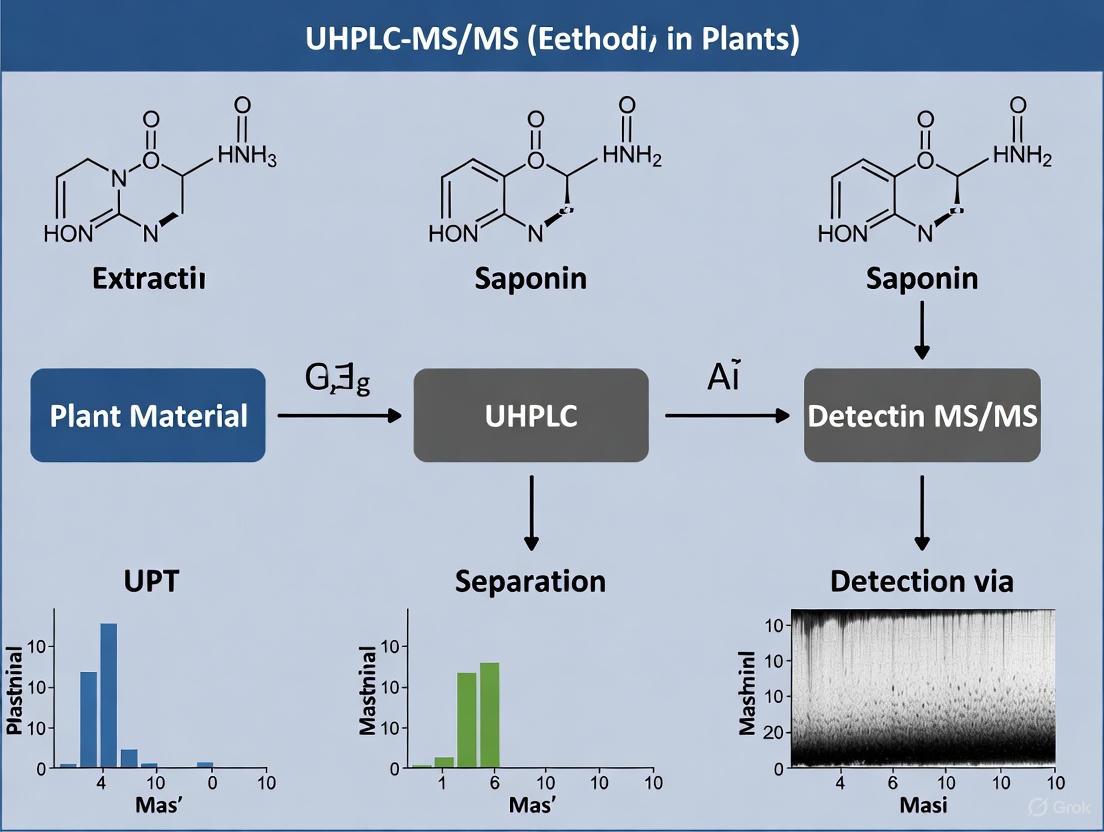
Development and Application of UHPLC-MS/MS for the Quantification of Bioactive Saponins in Plant Materials
This article provides a comprehensive overview of the UHPLC-MS/MS methodology for the analysis of plant saponins, a class of compounds with significant pharmacological importance.
Development and Application of UHPLC-MS/MS for the Quantification of Bioactive Saponins in Plant Materials
Abstract
This article provides a comprehensive overview of the UHPLC-MS/MS methodology for the analysis of plant saponins, a class of compounds with significant pharmacological importance. It covers the foundational principles of saponin chemistry and the advantages of UHPLC-MS/MS over traditional techniques. A detailed, step-by-step guide for method development is presented, including sample preparation, chromatographic separation, and mass spectrometric detection. The content further addresses critical troubleshooting and optimization strategies to enhance analytical performance and delves into rigorous method validation protocols. Finally, it explores the application of this technology in comparative phytochemical studies, such as assessing the impact of plant processing and geographical origin on saponin profiles, providing a vital resource for researchers and professionals in phytochemistry and drug development.
Saponin Chemistry and the Analytical Power of UHPLC-MS/MS
Structural Classification and Diversity
Saponins are a vast group of non-volatile, surface-active plant secondary metabolites, structurally defined as glycosides consisting of a hydrophobic aglycone (sapogenin) coupled with one or more hydrophilic monosaccharide moieties [1] [2]. This combination of polar and non-polar structural elements explains their soap-like behavior in aqueous solutions [2]. The primary classification of saponins is based on the chemical structure of the sapogenin, dividing them into two major classes: triterpenoid saponins and steroidal saponins [2].
Table 1: Fundamental Characteristics of Triterpenoid and Steroidal Saponins
| Characteristic | Triterpenoid Saponins | Steroidal Saponins |
|---|---|---|
| Sapogenin Carbon Skeleton | Pentacyclic C30 or tetracyclic C30 (Triterpene) [3] [2] | C27 Steroid (derived from cholesterol) [4] [5] |
| Biosynthetic Precursor | 2,3-oxidosqualene (30 carbon atoms) [6] [2] | 2,3-oxidosqualene (with three methyl groups removed) [2] |
| Common Aglycone Examples | Oleanolic acid, Hederagenin, Echinocystic acid [3] [7] | Spirostanol, Furostanol [4] [5] |
| Typical Sugar Attachment Points | Often at C-3 and/or C-28 positions [3] | Often at C-3 and/or C-26 positions [5] |
| Predominant Occurrence | Dicotyledonous plants [4] | Monocotyledonous plants [4] |
Triterpenoid Saponins
Triterpenoid saponins possess an aglycone composed of 30 carbon atoms arranged in a pentacyclic (less commonly tetracyclic) structure [3] [2]. The oleanane-type is one of the most common skeletons [3]. Their structural diversity arises from variations in the sapogenin structure and the composition of the sugar chains, which can be mono-, di-, or tri-saccharides attached at one or more positions, most commonly the C-3 and C-28 hydroxyl groups [3] [7]. Common monosaccharides include glucose, rhamnose, glucuronic acid, arabinose, galactose, and xylose [3] [7].
Steroidal Saponins
Steroidal saponins have an aglycone based on a C27 steroidal skeleton, biogenetically derived from cholesterol [5]. They are primarily categorized into two types:
- Spirostanol saponins: Characterized by a spiroacetal moiety (a tetrahydrofuran ring fused with a tetrahydropyran ring) at C-22 [4] [5].
- Furostanol saponins: Contain a hemiacetal moiety (a single tetrahydrofuran ring) and are often considered biosynthetic precursors to spirostanols [4] [5].
Modifications such as oxidation at various carbon atoms (e.g., C-1, C-12, C-23) and the introduction of double bonds further contribute to their structural complexity [5].
Biosynthesis and Key Enzymes
The biosynthesis of all saponins begins with the mevalonate pathway, leading to the common precursor 2,3-oxidosqualene [6] [2]. This compound is then cyclized by oxidosqualene cyclases (OSCs) into various triterpene or steroidal skeletons [6]. The subsequent structural diversification is primarily mediated by two key enzyme families:
- Cytochrome P450 monooxygenases (P450s): These enzymes catalyze a wide range of oxidation reactions on the sapogenin backbone, including hydroxylation, carboxylation, and epoxidation, thereby creating functional sites for glycosylation [6].
- UDP-dependent glycosyltransferases (UGTs): These enzymes are responsible for attaching sugar moieties to the oxidized aglycone, determining the final glycosylation pattern of the saponin [6].
The following diagram illustrates the core biosynthetic pathway shared by triterpenoid and steroidal saponins.
Detailed UHPLC-MS/MS Protocol for Saponin Quantification
The following section provides a detailed methodology for the simultaneous quantification of specific triterpenoid saponins in plant material, as exemplified by the analysis of Calenduloside E (CE) and Chikusetsusaponin IVa (ChIVa) in Amaranthaceae species [8] [9].
Sample Preparation and Extraction
- Plant Material Handling: Fresh plant material (roots, stems, leaves, fruits) should be lyophilized and ground to a homogeneous powder [9].
- Extraction Techniques:
- Weigh approximately 100 mg of dried powder accurately.
- Employ ultrasound-assisted extraction with 5 mL of 70-80% methanol in water (v/v) for 30-45 minutes at room temperature [9] [10].
- Centrifuge the extracts (e.g., 10,000 × g for 10 minutes) and collect the supernatant.
- Filter the supernatant through a 0.22 μm membrane filter prior to UHPLC-MS/MS analysis [9].
UHPLC-MS/MS Analytical Conditions
The developed method must be validated for specificity, linearity, precision, and accuracy according to ICH guidelines [9] [10].
Table 2: Exemplary UHPLC-MS/MS Conditions for Saponin Quantification
| Parameter | Specification |
|---|---|
| Chromatography System | Ultra-High Performance Liquid Chromatography (UHPLC) |
| Column | Reversed-Phase C18 (e.g., 100 mm x 2.1 mm, 1.7-1.8 μm particle size) |
| Mobile Phase | A: Water with 0.1% Formic AcidB: Acetonitrile |
| Gradient Elution | Linear gradient from 20% B to 95% B over 10-15 minutes [10] |
| Column Temperature | 35 °C [10] |
| Flow Rate | 0.26 - 0.4 mL/min [9] [10] |
| Injection Volume | 1-5 μL |
| Mass Spectrometer | Triple Quadrupole (QqQ) with Electrospray Ionization (ESI) |
| Ionization Mode | Negative Ion Mode (for oleanane-type saponins) [9] |
| Data Acquisition | Multiple Reaction Monitoring (MRM) |
Application: Quantitative Results from Amaranthaceae Species
The validated method was successfully applied to quantify CE and ChIVa in various plant parts of ten Amaranthaceae species [9]. The results demonstrate the variability of saponin content, highlighting rich botanical sources.
Table 3: Distribution of Calenduloside E and Chikusetsusaponin IVa in Selected Amaranthaceae Species (mg/g dry weight) [9]
| Plant Species | Plant Part | Calenduloside E (CE) | Chikusetsusaponin IVa (ChIVa) |
|---|---|---|---|
| Atriplex sagittata | Fruit | 7.84 | 13.15 |
| Chenopodium strictum | Fruit | 6.54 | 5.52 |
| Chenopodium strictum | Roots | Not Specified | 7.77 |
| Lipandra polysperma | Fruit | Not Specified | 12.20 |
| Chenopodium album | Fruit | Not Specified | 10.00 |
The Scientist's Toolkit: Essential Research Reagents and Materials
Table 4: Key Reagent Solutions for Saponin Analysis
| Reagent / Material | Function / Application | Exemplification |
|---|---|---|
| 70-80% Methanol | Extraction solvent for saponins from plant matrices [9]. | Optimal for simultaneous extraction of Calenduloside E and Chikusetsusaponin IVa [9]. |
| Reversed-Phase C18 UHPLC Column | High-efficiency chromatographic separation of saponin isomers [9] [10]. | Enables baseline separation of complex saponin mixtures from Achyranthes bidentata [10]. |
| Ammonium Acetate / Formic Acid | Mobile phase additives to modify pH and improve ionization efficiency in MS [9]. | 0.1% formic acid in water used for UPLC-ESI-MS/MS analysis of triterpenoid saponins [9]. |
| Saponin Reference Standards | Essential for method validation, calibration curves, and compound identification [10]. | Pure Calenduloside E and Chikusetsusaponin IVa are required for accurate quantification [9]. |
| Macroporous Resin (e.g., D101) | Purification and enrichment of saponins from crude plant extracts [3]. | Used for initial clean-up before detailed chromatographic analysis [3]. |
Saponins are a structurally diverse class of plant secondary metabolites with significant pharmaceutical potential, known for their amphiphilic nature due to the presence of hydrophobic aglycone (sapogenin) and hydrophilic sugar moieties [11]. This very combination of structural diversity and polarity presents formidable analytical challenges for researchers aiming to separate, identify, and quantify these compounds in complex plant matrices. Modern analytical techniques, particularly ultra-high-performance liquid chromatography coupled with tandem mass spectrometry (UHPLC-MS/MS), have emerged as powerful tools to address these challenges, enabling comprehensive metabolomic profiling and precise quantification even at trace concentrations [12] [13].
The analysis of saponins is crucial for advancing research on bioactive natural products, as these compounds demonstrate a broad spectrum of pharmacological activities, including anti-tumor, anti-inflammatory, immunomodulatory, and antiviral effects [12] [13]. This application note details the specific challenges associated with saponin analysis and provides structured protocols and workflows developed within the context of a broader thesis on UHPLC-MS/MS method development for saponin quantification in plant research.
The Analytical Challenge: Structural Complexity and Diversity
Saponins are broadly classified into two main groups based on their sapogenin structure: triterpenoid saponins (30 carbon atoms) and steroidal saponins (27 carbon atoms) [14]. This fundamental classification belies an immense structural complexity that directly impacts analytical method development.
The challenges in analysis stem from several factors that create a vast landscape of structurally similar compounds:
- Sapogenin Backbone Variations: The aglycone structure can occur as furostane, spirostane, cholestane, or pregnane skeletons, each with different functional group modifications including hydroxylations, ketones, and double bonds at various positions [15].
- Glycosylation Patterns: Saponins contain 1-6 sugar units, including hexoses (glucose, galactose), deoxyhexoses (rhamnose), pentoses (xylose, arabinose), and uronic acids (glucuronic acid). Isomeric forms arise from the type of sugar, glycosylation position, linkage between sugars, and anomeric configuration [16] [15].
- Acyl Modifications: Further complexity is added by ester-linked acyl groups (acetyl, angeloyl, cinnamyl, or tigloyl) at various positions on the sapogenin or sugar chains [16].
Impact of Polarity on Analysis
The polarity of saponins is primarily governed by the number and nature of the attached sugar units. The hydrophilic sugar moieties make saponins water-soluble, while the lipophilic sapogenin backbone contributes to amphiphilic character [11]. This results in:
- A wide range of hydrophilic-lipophilic balance across different saponins within a single plant extract.
- Challenges in developing a single chromatographic method that effectively separates all structural analogs.
- Difficulties in extraction optimization, as recovery rates are highly dependent on the solvent system's polarity [11].
Table 1: Major Saponin Classes and Their Structural Characteristics
| Saponin Class | Sapogenin Backbone | Carbon Atoms | Common Sugar Attachments | Example Compounds |
|---|---|---|---|---|
| Triterpenoid Saponins | Oleanane, Ursane, Lupane | 30 | GlcA, Glc, Gal, Xyl, Ara, Rha [16] | Soyasaponins [11], Tea Saponins [16], Chikusetsusaponin IVa [13] |
| Steroidal Saponins | Spirostane, Furostane | 27 | Glc, Gal, Rha, Xyl [15] | Polyphyllins [12], Dioscin [12] |
| Steroidal Alkaloids | Solanidane, Spirosolane | 27 | Glc, Gal, Rha, Xyl | - |
Advanced UHPLC-MS/MS Solutions for Saponin Analysis
The combination of UHPLC's high separation power with the selectivity and sensitivity of MS/MS detection is the most effective approach to overcome challenges posed by saponin diversity and polarity.
Chromatographic Optimization and Separation
Successful separation of complex saponin mixtures requires careful optimization of chromatographic conditions:
- Stationary Phase: The Waters ACQUITY UPLC HSS T3 column (1.8 μm, 150 mm × 2.1 mm i.d.) has demonstrated superior separation efficiency for Camellia sinensis seed saponins compared to other C18 columns [17]. For highly polar saponins, Hydrophilic Interaction Liquid Chromatography (HILIC) provides an excellent alternative to reversed-phase chromatography, as demonstrated for soyasaponins in yoghurt alternatives [11].
- Mobile Phase: Acetonitrile is generally preferred over methanol due to lower UV absorption at short wavelengths, improved separation efficiency, and reduced column backpressure [17]. The addition of 0.1% formic acid to both aqueous and organic mobile phases improves peak shape, sensitivity, and retention time reproducibility [17].
- Elution Program: A typical gradient for saponin separation starts with a low organic phase percentage (e.g., 10% acetonitrile) and increases to a high percentage (e.g., 76% acetonitrile) over 10-20 minutes, effectively eluting saponins based on their polarity [16].
Mass Spectrometric Detection and Identification
Mass spectrometry, particularly high-resolution systems like Q-TOF, is indispensable for saponin identification and characterization:
- Ionization Mode: Electrospray Ionization (ESI) in negative mode generally provides superior signal intensity for saponins compared to positive mode [17]. This is particularly effective for saponins containing glucuronic acid residues.
- Fragmentation Patterns: Tandem mass spectrometry (MS/MS) reveals characteristic fragmentation pathways that provide structural information. Key fragmentation patterns include:
- Advanced Identification Strategies: Feature-based Molecular Networking (FBMN) clusters structurally related saponins through systematic analysis of MS/MS spectral data, greatly facilitating the identification of analogs within complex mixtures [16].
Table 2: Key Mass Spectrometric Parameters for Saponin Analysis
| Parameter | Recommended Setting | Rationale | Application Example |
|---|---|---|---|
| Ionization Mode | ESI-Negative | Enhanced signal for most saponin types [17] | Analysis of tea saponins [16] |
| Scan Mode | Full Scan (MS1) + Data-Dependent MS/MS (ddMS2) | Comprehensive profiling and structural info | Untargeted saponin profiling [12] |
| Mass Resolution | High-Resolution (>20,000) | Accurate mass measurement for formula assignment | UPLC-IM-Q-TOF-MS/MS [10] |
| Collision Energy | Ramped (20-50 eV) | Optimized fragmentation across different saponins | Structural characterization [15] |
| Data Analysis | Molecular Networking | Groups structurally related compounds | Tea saponin profiling [16] |
Detailed Experimental Protocols
Sample Preparation and Extraction Protocol
Principle: Efficient extraction of saponins from plant matrices while minimizing degradation and maximizing recovery.
Reagents: Methanol (HPLC grade), acetonitrile (HPLC grade), formic acid, ultrapure water, ammonia solution (5%, v/v).
Procedure:
- Homogenization: Dry plant material (e.g., rhizomes, leaves, seeds) at 60°C and grind to a fine powder that passes through a 3-mesh sieve [12].
- Weighing: Accurately weigh approximately 0.1 g of dried powder into a 2.0 mL centrifuge tube.
- Extraction: Add 1 mL of methanol. Soak the mixture overnight, then perform ultrasonic extraction for 30 minutes [12].
- pH Adjustment (for certain matrices): For acidic food matrices like yoghurt alternatives, adjust the sample pH to 8 ± 0.25 using aqueous ammonia solution to optimize saponin solubility and recovery [11].
- Reconstitution: After cooling to room temperature, adjust the methanol volume to the initial level. Centrifuge at 12,000 × g for 10 minutes.
- Clean-up: Pass the supernatant through a 0.22 μm microporous membrane to obtain the test solution for UHPLC-MS/MS analysis [12].
- Quality Control: Prepare a pooled quality control (QC) sample by combining equal volumes of all test solutions to monitor instrument performance during the analysis sequence.
UHPLC-MS/MS Analytical Protocol
Principle: High-resolution separation and sensitive detection of saponins in plant extracts.
Equipment: UHPLC system coupled to Q-TOF mass spectrometer (e.g., Agilent 1290 Infinity II UHPLC with Agilent 6545 Q-TOF) [12].
Chromatographic Conditions:
- Column: Waters ACQUITY UPLC HSS T3 (1.8 μm, 150 mm × 2.1 mm i.d.) [17] or equivalent C18 column.
- Mobile Phase A: Ultrapure water containing 0.1% formic acid.
- Mobile Phase B: Acetonitrile containing 0.1% formic acid.
- Gradient Program:
- 0-2 min: 10% B
- 2-15 min: 10% B → 76% B (linear gradient)
- 15-17 min: 76% B → 95% B
- 17-19 min: 95% B (column cleaning)
- 19-22 min: 95% B → 10% B (re-equilibration) [16]
- Flow Rate: 0.26 mL/min [10]
- Column Temperature: 30°C
- Injection Volume: 2-5 μL
Mass Spectrometric Conditions:
- Ionization Mode: ESI-negative
- Drying Gas Temperature: 325°C
- Drying Gas Flow: 8 L/min
- Nebulizer Pressure: 40 psi
- Sheath Gas Temperature: 350°C
- Sheath Gas Flow: 11 L/min
- Capillary Voltage: 3500 V
- Nozzle Voltage: 500 V
- Fragmentor Voltage: 180 V
- Scan Range: m/z 100-1700
- Collision Energies: 10-40 eV for MS/MS
Data Processing and Analysis Workflow
Software: Use instrument vendor software and specialized platforms like MS-DIAL or GNPS for molecular networking.
Procedure:
- Peak Picking and Alignment: Perform automated peak detection, deconvolution, and alignment across all samples.
- Compound Identification:
- Match accurate mass and isotopic pattern against databases (e.g., PlantCyc, KNApSAcK).
- Interpret MS/MS spectra for characteristic saponin fragmentation patterns.
- Confirm identities using authentic standards when available.
- Molecular Networking: Upload MS/MS data to the GNPS platform to create molecular networks that visualize structurally related saponins and identify novel analogs [16].
- Multivariate Statistical Analysis: Apply Principal Component Analysis (PCA) and Hierarchical Clustering Analysis (HCA) to identify metabolic patterns and group samples based on their saponin profiles [12].
- Quantitation: Generate calibration curves using reference standards for absolute quantitation, or use peak areas for relative quantitation when standards are unavailable.
Visualizing the Analytical Workflow
The following diagram illustrates the comprehensive workflow for saponin analysis from sample preparation to data interpretation:
Diagram 1: Comprehensive workflow for saponin analysis in plant materials, covering sample preparation, instrumental analysis, and data interpretation stages.
The Scientist's Toolkit: Essential Research Reagents and Materials
Table 3: Key Research Reagent Solutions for Saponin Analysis
| Reagent/Material | Function/Purpose | Application Note |
|---|---|---|
| HSS T3 UPLC Column (1.8 μm, 150 mm × 2.1 mm) | Superior separation of complex saponin mixtures [17] | Provides optimal resolution for structural analogs |
| MS-grade Acetonitrile with 0.1% Formic Acid | Mobile phase for UHPLC separation | Enhances ionization and improves peak shape [17] |
| Saponin Reference Standards (e.g., Polyphyllin I, II, VII) | Method validation and absolute quantification | Essential for calibration curves and compound verification [12] |
| Solid Phase Extraction (SPE) Cartridges (e.g., Biotage Isolute PLD+) | Sample clean-up and pre-concentration | Removes interfering matrix components [11] |
| Quality Control (QC) Reference Material (e.g., digitoxin) | Internal standard for relative quantification | Monitors instrument performance and normalizes data [18] |
| Ultrasonic Extraction Bath | Efficient extraction of saponins from plant matrix | Maximizes recovery while minimizing degradation [12] |
The structural diversity and polarity of saponins present significant but surmountable challenges for analytical scientists. Through the implementation of optimized UHPLC-MS/MS methods—incorporating careful sample preparation, advanced chromatographic separation, high-resolution mass spectrometry, and comprehensive data analysis strategies—researchers can effectively navigate this complexity. The protocols and workflows detailed in this application note provide a robust framework for the accurate identification and quantification of saponins in plant materials, supporting ongoing research into their considerable pharmacological potential. Future methodological advances will likely focus on increasing throughput, enhancing sensitivity for trace-level saponins, and improving the structural annotation of novel saponins through integrated computational approaches.
The Evolution from Traditional Methods to Modern Hyphenated Techniques
The quantification of saponins in plant materials has undergone a significant methodological evolution, moving from traditional techniques like open-column chromatography and spectrophotometry to advanced hyphenated systems such as Ultra-High Performance Liquid Chromatography coupled with Tandem Mass Spectrometry (UHPLC-MS/MS). This transition has addressed critical limitations in selectivity, sensitivity, and analytical efficiency. Modern UHPLC-MS/MS methods now enable the simultaneous quantification of multiple bioactive saponins, such as calenduloside E and chikusetsusaponin IVa, even at trace levels in complex plant matrices. These integrated approaches provide researchers with powerful tools for comprehensive quality assessment, authentication of medicinal plants, and accelerated drug development from natural products.
The analysis of bioactive plant compounds, particularly triterpene saponins, presents substantial analytical challenges due to their complex structures, occurrence in intricate plant matrices, and frequently low concentrations despite significant biological activity. Traditional methods, including high-performance liquid chromatography (HPLC) with ultraviolet (UV) or diode-array detection (DAD) and evaporative light scattering detection (ELSD), often proved inadequate for reliable saponin quantification. These techniques were characterized by lengthy analysis times, significant solvent consumption, and poor selectivity, often providing insufficient resolution for structurally similar compounds and isomers [19].
The integration of separation techniques with mass spectrometry, creating "hyphenated systems," marks the most significant advancement in this field. UHPLC-MS/MS combines the superior resolving power of ultra-high-performance liquid chromatography with the exceptional sensitivity and specificity of tandem mass spectrometry. This combination is particularly crucial for saponin analysis, as many triterpene saponins show poor absorbance in the short-wavelength UV range (below 210 nm), preventing their reliable quantification in complex plant extracts using traditional spectroscopic techniques [9]. The evolution to UHPLC-MS/MS has thus become the cornerstone of modern phytochemical analysis, enabling precise quantification and robust quality control for herbal medicines and drug development.
Comparative Analysis of Traditional vs. Modern Techniques
The transition from traditional to modern hyphenated techniques represents a paradigm shift in analytical capabilities. The table below summarizes the key differences in performance characteristics.
Table 1: Comparison of Traditional and Modern Techniques for Saponin Quantification
| Analytical Characteristic | Traditional Methods (HPLC-UV/ELSD) | Modern Hyphenated Techniques (UHPLC-MS/MS) |
|---|---|---|
| Analysis Time | Long analysis times (often >30 minutes) [19] | Short analysis times (e.g., 10 minutes) [19] |
| Sensitivity | Poor sensitivity, especially for non-chromophoric saponins [9] | Exceptional sensitivity (LOD in ng/L range for pharmaceuticals) [20] |
| Selectivity & Specificity | Poor selectivity, relying only on retention time [19] | High specificity using Multiple Reaction Monitoring (MRM) [20] |
| Solvent Consumption | High solvent consumption, resulting in waste [19] | Reduced solvent consumption, aligning with Green Chemistry [20] |
| Data Richness | Limited to retention time and UV spectrum | Provides mass fragments, structural information, and confirmatory data |
| Handling of Isomers | Difficulty separating isomers with identical m/z [19] | Capable of rapid and complete separation of polar isomers [19] |
The advantages of UHPLC-MS/MS extend beyond speed and sensitivity. Its mass-based detection does not depend on the presence of a chromophore, making it ideal for triterpenoid saponins [9]. Furthermore, the technique provides unambiguous identification based on molecular mass and specific fragmentation patterns, minimizing matrix interferences that commonly plague traditional methods [20].
Application Note: UHPLC-MS/MS for Saponin Quantification in Amaranthaceae Species
Background and Objective
Triterpene saponins like calenduloside E (CE) and chikusetsusaponin IVa (ChIVa) exhibit multidirectional bioactivity, including anti-inflammatory, cardioprotective, and neuroprotective effects [9]. Ensuring the efficacy and safety of plant-derived formulations requires accurate quantification of these bioactive compounds. This application note details a validated UHPLC-ESI-MS/MS method for the simultaneous quantification of CE and ChIVa in various plant parts of ten Amaranthaceae species, demonstrating the power of modern hyphenated techniques in phytochemical analysis.
Experimental Protocol
Sample Preparation and Extraction
- Plant Material: Roots, stems, leaves, and fruits from ten wild-growing Amaranthaceae species were collected and authenticated [9].
- Extraction Optimization: Four extraction methods were compared: maceration, shaking-assisted maceration, ultrasound-assisted extraction (UAE), and heat reflux extraction. UAE was determined to be optimal for the studied saponins [9].
- Optimized UAE Procedure:
- Solvent: Ethanol-H₂O (1:1, v/v) [21].
- Solvent-to-Sample Ratio: 1:8 [21].
- Extraction Process: Perform extraction 3 times, each for 30 minutes [21].
- Post-Extraction: The extract is subjected to liquid-liquid partitioning with petroleum ether followed by saturated n-butanol. The n-butanol fraction is concentrated and further purified using AB-8 macroporous resin column chromatography with ethanol-H₂O (7:3, v/v) as the eluent to enrich the saponin fraction [21].
UHPLC-MS/MS Analysis
- Chromatography:
- System: Ultra-High Performance Liquid Chromatography (UHPLC).
- Mobile Phase: Acetonitrile/water system with 0.3% acetic acid added to the water phase to enhance ionization and improve peak shape of the analytes [19].
- Gradient: Optimized gradient program to achieve satisfactory separation within a short analysis time [19] [9].
- Mass Spectrometry:
- Technique: Tandem Mass Spectrometry (MS/MS).
- Ionization: Electrospray Ionization (ESI), typically in negative mode for saponins, yielding intense deprotonated molecular ions [M−H]⁻ [21].
- Mode: Multiple Reaction Monitoring (MRM) for high sensitivity and selectivity. The method was developed and validated to be specific for CE and ChIVa, guaranteeing well-shaped peaks and appropriate resolution [9].
Method Validation
The developed UHPLC-MS/MS method was validated according to standard guidelines to ensure reliability, with results satisfying acceptance criteria [9]:
- Precision: Intra-day and inter-day variations (RSD) were desirably low, e.g., 1.57–2.46% and 1.51–3.00%, respectively, in a similar study [19].
- Accuracy: Recovery rates were demonstrated to be high, for instance, ranging from 98.58–101.48% in a related method [19].
- Linearity: The method demonstrated a linear response with correlation coefficients (R²) ≥ 0.999 [20].
- Specificity: The method was found to be selective for the investigated compounds, free from interference [9].
Key Research Reagent Solutions
Table 2: Essential Reagents and Materials for UHPLC-MS/MS Saponin Analysis
| Reagent/Material | Function/Application | Example from Protocol |
|---|---|---|
| Acetonitrile (HPLC-MS Grade) | Mobile phase component for UHPLC separation | Used in acetonitrile/water mobile phase system [19] |
| Acetic Acid | Mobile phase modifier to enhance analyte ionization | Added at 0.3% to water phase to increase signal intensity [19] |
| Ethanol & Purified Water | Extraction solvent for ultrasound-assisted extraction | Used as ethanol-H₂O (1:1, v/v) for saponin extraction [21] |
| AB-8 Macroporous Resin | Stationary phase for pre-cleaning and enriching saponins | Used in column chromatography to enrich saponin fraction [21] |
| Analytical Standards | Calibration and quantification reference | Calenduloside E and Chikusetsusaponin IVa pure standards [9] |
Results and Data Presentation
The validated method was successfully applied to quantify CE and ChIVa in different plant parts. The results highlighted significant interspecies and intraspecies variation in saponin content.
Table 3: Quantification of Calenduloside E (CE) and Chikusetsusaponin IVa (ChIVa) in Selected Amaranthaceae Species (mg/g dry weight) [9]
| Species | Plant Part | Calenduloside E (CE) | Chikusetsusaponin IVa (ChIVa) |
|---|---|---|---|
| A. sagittata | Fruit | 7.84 | 13.15 |
| L. polysperma | Fruit | Not Specified | 12.20 |
| Ch. album | Fruit | Not Specified | 10.00 |
| Ch. strictum | Roots | Not Specified | 7.77 |
| Ch. strictum | Fruit | 6.54 | 5.52 |
The data obtained demonstrates the utility of the UHPLC-MS/MS method for identifying rich sources of bioactive saponins. For instance, the fruit of A. sagittata was identified as a particularly convenient source of both CE and ChIVa [9]. Furthermore, this was the first report of CE and ChIVa in several species, including L. polysperma and A. patula [9].
Workflow and Data Interpretation Visualizations
UHPLC-MS/MS Saponin Analysis Workflow
Saponin Identification via Tandem Mass Spectrometry
The evolution from traditional chromatographic methods to modern hyphenated techniques like UHPLC-MS/MS has fundamentally transformed the landscape of saponin research. This advanced methodology provides an unparalleled combination of speed, sensitivity, and specificity, enabling the precise quantification of complex plant metabolites that were previously difficult to analyze. The successful application of UHPLC-MS/MS for profiling calenduloside E and chikusetsusaponin IVa across multiple Amaranthaceae species underscores its critical role in modern phytochemistry, quality control of herbal medicines, and the discovery of new plant-derived pharmaceutical compounds. As this technology continues to evolve alongside green chemistry principles, it will undoubtedly remain the gold standard for analytical scientists in natural product research and drug development.
Saponins are a diverse class of secondary metabolites found extensively in medicinal plants, known for their complex chemical structures and broad pharmacological activities, including anti-inflammatory, anticancer, antiviral, and cardioprotective effects. The analysis of these compounds presents significant challenges due to their structural diversity, low UV absorption, isomeric complexity, and frequently low concentrations within complex plant matrices. Additionally, saponins often occur as a series of analogues with nearly identical structures, differing only in their glycosylation patterns or slight modifications to the aglycone moiety. Ultra-High-Performance Liquid Chromatography coupled with Tandem Mass Spectrometry (UHPLC-MS/MS) has emerged as a powerful analytical platform that effectively addresses these challenges by combining high-resolution separation with selective and sensitive detection.
Core Analytical Challenges and UHPLC-MS/MS Solutions
Challenge 1: Structural Complexity and Isomer Separation
Saponins exhibit extensive structural complexity with numerous isomers that have identical molecular formulas but differ in sugar linkage types, positions, and stereochemistry. Traditional HPLC methods often lack the resolution to separate these compounds adequately.
UHPLC-MS/MS Solution: The use of sub-2µm particle columns in UHPLC provides superior chromatographic resolution and peak capacity. The enhanced efficiency allows for the separation of structurally similar saponins, such as calenduloside E (CE) and chikusetsusaponin IVa (ChIVa), which share oleanolic acid as an aglycone but differ by an additional glucose moiety ester-linked at the C-17 position in ChIVa [13] [9]. The high peak capacity of UHPLC systems enables resolution of these challenging compounds within significantly reduced analysis times.
Table 1: Representative Chromatographic Conditions for Saponin Separation
| Parameter | Condition 1 [13] | Condition 2 [22] | Condition 3 [23] |
|---|---|---|---|
| Column | Not specified | Agilent Zorbax SB-18 MS (3.0 × 50 mm, 1.8 µm) | Supelco C18 (3.0 × 50 mm, 2.7 µm) |
| Mobile Phase | Acetonitrile/0.1% formic acid | 0.1% formic acid in water (A) and methanol (B) | Acetonitrile (A) and 0.1% formic acid in water (B) |
| Gradient | Optimized for specific separation | 10-65% B (0-3 min), 65% B (3-6 min), 65-90% B (6-9 min) | 15% A (2 min), to 30% A (13 min), to 95% A (10 min) |
| Flow Rate | Not specified | 0.5 mL/min | 0.5 mL/min |
| Analysis Time | Rapid analysis achieved | 13 min total run time | 35 min total run time |
Challenge 2: Detection Sensitivity and Selectivity
Triterpene saponins typically show poor absorbance in the short-wavelength UV range (below 210 nm), which often prevents their reliable detection and quantification in complex plant extracts using conventional UV or DAD detectors [13] [9].
UHPLC-MS/MS Solution: Mass spectrometric detection provides exceptional sensitivity and selectivity independent of chromophore presence. The multiple reaction monitoring (MRM) mode available in triple quadrupole instruments offers unparalleled specificity by monitoring specific precursor-to-product ion transitions for each analyte. This effectively filters out matrix interference, enabling precise quantification even at trace levels. The limits of detection (LOD) and quantification (LOQ) reported for saponin analysis demonstrate remarkable sensitivity, with LODs ranging from 0.20 to 0.61 ng/mL and LOQs from 0.61 to 1.85 ng/mL in validated methods [24].
Challenge 3: Comprehensive Profiling in Complex Matrices
Plant extracts represent highly complex mixtures containing thousands of compounds with varying polarities and concentrations, creating significant matrix effects that can suppress or enhance ionization and compromise accurate quantification.
UHPLC-MS/MS Solution: The combination of high-resolution chromatographic separation with advanced mass spectrometric detection enables comprehensive profiling of saponins in complex plant materials. The two-stage mass spectrometry approach utilizes high-resolution instruments like Q-TOF for qualitative identification of novel saponins, followed by highly sensitive QQQ systems for precise quantification [22]. This integrated strategy was successfully applied to profile 63 saponins in different morphological regions of American ginseng, identifying marker compounds specific to main roots, lateral roots, and rhizomes [23].
Figure 1: Experimental workflow for saponin analysis in plant materials
Detailed Experimental Protocols
Sample Preparation and Extraction
Protocol 1: Standardized Extraction Procedure [13] [23]
- Plant Material Preparation: Separate fresh plant material into different morphological regions (main root, lateral root, rhizome, stem, leaves, fruits). Air-dry at 25-30°C, grind to fine powder, and sieve for uniform particle size.
- Extraction: Accurately weigh 0.1 g of fine powder into a suitable container. Add 4 mL of 70% methanol-water (v/v) extraction solvent.
- Sonication: Sonicate the mixture for 45 minutes at room temperature with controlled power settings.
- Filtration: Pass the extraction through a 0.22 μm membrane filter prior to UHPLC-MS/MS analysis.
- Alternative Methods: For comparative extraction efficiency, maceration, shaking-assisted maceration, and heat reflux extraction can be evaluated alongside ultrasound-assisted extraction [13].
Protocol 2: Tissue Distribution Studies [25]
- Biological Sample Preparation: Homogenize tissue samples (liver, kidney, heart, spleen) with appropriate buffer.
- Protein Precipitation: Employ a one-step protein precipitation using methanol for sample clean-up.
- Internal Standard Addition: Add chloramphenicol or glycyrrhetinic acid as internal standards to monitor extraction efficiency and matrix effects.
- Centrifugation: Centrifuge at high speed (≥10,000 × g) for 10 minutes and collect supernatant for analysis.
UHPLC-MS/MS Instrumental Conditions
Chromatographic System: [13] [22] [23]
- Column: C18 reversed-phase columns (50-100 mm length × 2.1-3.0 mm i.d., 1.8-2.7 µm particle size)
- Mobile Phase: Binary gradient systems combining (A) 0.1% formic acid in water and (B) acetonitrile or methanol with 0.1% formic acid
- Gradient Program: Optimized linear gradients ranging from 5-15% B to 90-100% B over 5-15 minutes
- Flow Rate: 0.3-0.5 mL/min
- Column Temperature: 35-40°C
- Injection Volume: 5-10 µL
Mass Spectrometric Detection: [26] [13] [22]
- Ionization Source: Electrospray Ionization (ESI) operating in negative or positive mode, depending on analyte characteristics
- Ion Source Parameters: Capillary voltage: 3.0-3.5 kV; Drying gas temperature: 300-350°C; Sheath gas flow: 8-12 L/min
- Scan Modes:
- Qualitative Analysis: Full scan and data-dependent MS/MS using Q-TOF instruments for structural elucidation
- Quantitative Analysis: Multiple Reaction Monitoring (MRM) using triple quadrupole instruments for sensitive quantification
- Collision Energies: Optimized individually for each analyte (typically 20-45 eV)
Figure 2: MRM detection principle for selective saponin quantification
Method Validation Parameters
Comprehensive validation following ICH guidelines ensures method reliability, with key parameters including:
- Linearity: Correlation coefficients (r²) > 0.99 over relevant concentration ranges [24] [26]
- Precision: Intra-day and inter-day precision with RSD < 5% [24] [26]
- Accuracy: Recovery rates of 88.3-104.8% for various matrices [24] [26] [25]
- Sensitivity: LOD and LOQ values in ng/mL range [24]
- Matrix Effects: Evaluation of ion suppression/enhancement with acceptable RSD values [26]
Table 2: Validation Data from Representative Saponin Quantification Methods
| Validation Parameter | Chenopodium bonus-henricus Method [24] | Achyranthes bidentata Method [26] | Dipsacus asper Method [25] |
|---|---|---|---|
| Linearity (R²) | > 0.99 | > 0.9998 | > 0.9991 |
| LOD Range | 0.20-0.61 ng/mL | Not specified | Not specified |
| LOQ Range | 0.61-1.85 ng/mL | 20.4-8500 ng/mL | Not specified |
| Precision (RSD) | Intra-day: 0.64-4.25% | Intra-day: < 3.95% | Intra-day: -4.62 to 4.93% |
| Accuracy (% Recovery) | 95.38-103.47% | 95.2-104.8% | 88.3-100.1% |
| Analysis Time | Significantly reduced | Not specified | Not specified |
The Scientist's Toolkit: Essential Research Reagents and Materials
Table 3: Key Research Reagent Solutions for Saponin Analysis
| Item | Function/Purpose | Examples/Specifications |
|---|---|---|
| UHPLC Columns | High-resolution separation of saponins | C18 reversed-phase (50-100mm, 1.8-2.7µm); Waters BEH C18 [26] |
| Mass Spectrometry Instruments | Sensitive detection and quantification | Triple quadrupole (QQQ) for MRM quantification; Q-TOF for structural identification [22] [23] |
| Reference Standards | Method development, calibration, identification | β-ecdysterone, ginsenoside Ro, chikusetsusaponin IV, calenduloside E [26] [13] |
| Internal Standards | Correction for matrix effects, recovery calculation | Chloramphenicol, glycyrrhetinic acid [13] [25] |
| Extraction Solvents | Efficient extraction of saponins from plant material | 70% methanol-water, ethanol-water (4:1, v/v) [13] [23] |
| Mobile Phase Additives | Improve chromatography and ionization | 0.1% formic acid in water and organic phase [26] [22] [23] |
Application Examples in Plant Research
Phytochemical Distribution Studies
UHPLC-MS/MS enables precise mapping of saponin distribution across different plant parts. In studies of Amaranthaceae species, the highest chikusetsusaponin IVa content was found in the fruit of A. sagittata (13.15 mg/g dw), L. polysperma (12.20 mg/g dw), and C. album (10.0 mg/g dw), while the highest calenduloside E content was determined in the fruit of A. sagittata (7.84 mg/g dw) and C. strictum (6.54 mg/g dw) [13]. Such distribution studies help identify optimal plant parts and harvesting times for maximum saponin yield.
Pharmacokinetic and Bioavailability Studies
The high sensitivity of UHPLC-MS/MS enables pharmacokinetic studies of saponins in biological matrices. A comparative study of raw and salt-processed Achyranthes bidentata revealed that salt-processing significantly increased the bioavailability of β-ecdysterone, 25S-inokosterone, ginsenoside Ro, and chikusetsusaponin IVa, as evidenced by elevated Cmax and AUC0-t parameters [26]. Similarly, tissue distribution studies of Dipsacus asper constituents demonstrated that wine-processing altered the disposition of bioactive compounds, leading to increased accumulation in liver and kidney tissues, which correlates with enhanced therapeutic effects on these organs [25].
Chemotaxonomic and Metabolomic Studies
The technique supports chemotaxonomic classifications by revealing genus-specific and species-specific saponin profiles. Metabolomic studies of Banisteriopsis and Stigmaphyllon genera using UHPLC-QTOF-MS/MS revealed distinct chemical profiles influenced by environmental factors, humidity levels, and plant habit, with discriminant metabolites including coumaroyl hexoside, myricetin-3-galactoside, and quercetin derivatives [27]. Such comprehensive profiling provides valuable insights for phylogenetic studies and quality control of medicinal plants.
UHPLC-MS/MS has revolutionized saponin analysis by effectively addressing the core challenges of structural complexity, low detection sensitivity, and matrix interference. The integration of high-resolution chromatographic separation with advanced mass spectrometric detection provides researchers with a powerful tool for qualitative and quantitative analysis of these biologically significant compounds. As evidenced by the diverse applications in phytochemistry, pharmacognosy, and pharmacokinetics, this analytical platform continues to drive advancements in natural product research, enabling more comprehensive characterization of medicinal plants and facilitating quality control in phytopharmaceutical development.
The analysis of plant saponins represents a significant challenge in natural product research due to the structural complexity and diversity of these bioactive compounds. Ultra-High Performance Liquid Chromatography coupled with Tandem Mass Spectrometry (UHPLC-MS/MS) has emerged as a powerful analytical platform that enables researchers to address key challenges in saponin quantification, identification, and dereplication within complex plant matrices. This technology provides the sensitivity, resolution, and speed required to analyze these often closely-related compounds in plant extracts, supporting research from basic phytochemical characterization to drug discovery applications.
The fundamental advantage of UHPLC-MS/MS lies in its ability to separate complex mixtures with high efficiency while providing structural information through mass spectrometry. For saponins, which frequently lack strong chromophores and exhibit poor UV absorption, the MS/MS detection offers superior sensitivity and specificity compared to conventional UV-based detection methods [9]. This technical capability has become increasingly valuable as research continues to reveal the multifaceted bioactivities of saponins, including their anti-inflammatory, anti-tumor, neuroprotective, and cardioprotective effects [9] [28].
Key Application Areas
Quantitative Analysis of Bioactive Saponins
Targeted quantification of specific, biologically relevant saponins represents a cornerstone application of UHPLC-MS/MS in phytochemical research. The technology enables precise measurement of individual saponins across different plant species, tissues, and cultivation conditions, providing critical data for assessing plant material quality and selecting optimal sources for further development.
A recent investigation into ten wild-growing species of Amaranthaceae exemplifies this approach, focusing on the simultaneous quantification of two oleanolic acid-type saponins: calenduloside E (CE) and chikusetsusaponin IVa (ChIVa) [9]. The study developed and validated a specific UPLC-ESI-MS/MS method that achieved appropriate peak resolution, repeatability, and shortened analysis time. This methodological rigor enabled the discovery that these saponins coexist in most species analyzed, with particularly high concentrations found in specific plant organs, thereby identifying promising sources for these bioactive compounds [9].
Table 1: Distribution of Calenduloside E and Chikusetsusaponin IVa in Selected Amaranthaceae Species
| Species | Plant Part | Calenduloside E (mg/g dw) | Chikusetsusaponin IVa (mg/g dw) |
|---|---|---|---|
| A. sagittata | Fruit | 7.84 | 13.15 |
| L. polysperma | Fruit | Not specified | 12.20 |
| Ch. album | Fruit | Not specified | 10.00 |
| Ch. strictum | Roots | Not specified | 7.77 |
| Ch. strictum | Fruit | 6.54 | 5.52 |
The quantitative data revealed significant variation in saponin content between species and plant parts, highlighting the importance of selective harvesting and the value of UHPLC-MS/MS in guiding these decisions. For instance, fruits of A. sagittata accumulated the highest levels of both CE (7.84 mg/g dw) and ChIVa (13.15 mg/g dw), identifying this species and tissue as particularly valuable for obtaining these saponins [9].
Comprehensive Profiling and Dereplication of Saponins
Beyond targeted quantification, UHPLC-MS/MS serves as an essential tool for untargeted profiling and dereplication of complex saponin mixtures in plant extracts. Dereplication—the rapid identification of known compounds in complex mixtures—is crucial for avoiding redundant research and prioritizing novel bioactive constituents.
The integration of molecular networking with UHPLC-HRMS/MS has significantly advanced dereplication strategies for natural products [29]. This approach visualizes complex MS/MS data as molecular families, grouping structurally related compounds based on similar fragmentation patterns. In a study on Hypericum species, molecular networking efficiently clustered polycyclic polyprenylated acylphloroglucinols (PPAPs), enabling rapid comparison of chemical compositions between species and annotation of both known and potentially novel compounds [29].
Advanced LC configurations further enhance separation power for complex saponin mixtures. An offline HILIC × RP LC/QTOF-MS system demonstrated exceptional utility in characterizing triterpene saponins from Rhizoma Panacis Japonici (RPJ) [30]. This two-dimensional separation approach significantly increased peak capacity to 1,249, with an orthogonality of 0.61, enabling the characterization of 307 saponins—76 of which were identified for the first time in Panax japonicus [30]. The integration of an in-house database containing 612 known saponins from the Panax genus and 228 predicted metabolites facilitated efficient annotation of the detected compounds.
Table 2: Saponin Characterization in Herbal Medicines Using Advanced UHPLC-MS/MS Approaches
| Herbal Medicine | Analytical Approach | Number of Saponins Characterized | Novel Identifications | Reference |
|---|---|---|---|---|
| Rhizoma Panacis Japonici | Offline HILIC × RP LC/QTOF-MS | 307 | 76 | [30] |
| Achyranthes bidentata | UHPLC-ELSD / UPLC-IM-Q-TOF-MS/MS | 9 (quantified) | 2 new triterpenoid saponins | [10] |
| Lilium lancifolium | NADES-UHPLC-MS/MS | 9 target steroidal saponins | 31 total compounds identified | [31] |
Bioanalytical Applications
UHPLC-MS/MS also finds critical application in pharmacokinetic and bioavailability studies of saponins, which often exhibit poor absorption and complex metabolism. A validated LC-MS/MS method for quantifying 20(S)-protopanaxadiol (PPD)—a saponin derivative with potent biological activities—demonstrated the application of this technology across multiple biological matrices, including plasma, tissues, bile, urine, and fecal samples [32]. The method achieved a low quantification limit of 2.5 ng/mL and was successfully applied to kinetic studies in both rats and dogs, providing comprehensive absorption, distribution, metabolism, and excretion (ADME) profiles [32].
Experimental Protocols
Protocol 1: UPLC-ESI-MS/MS Method for Simultaneous Saponin Quantification
This protocol describes the simultaneous quantification of calenduloside E and chikusetsusaponin IVa in plant material, adapted from the validated method reported for Amaranthaceae species [9].
Sample Preparation:
- Extraction Optimization: Compare extraction techniques (maceration, shaking-assisted maceration, ultrasound-assisted extraction, and heat reflux extraction) to determine optimal conditions for target saponins.
- Plant Material Processing: Lyophilize and pulverize plant material to a fine powder. Precisely weigh 100 mg of powder for extraction.
- Extraction Procedure: Extract with 5 mL of suitable solvent (e.g., methanol-water mixture) using the optimized extraction method.
- Sample Cleanup: Centrifuge at 10,000 × g for 10 minutes and filter through a 0.22 μm membrane prior to UHPLC-MS/MS analysis.
UHPLC-MS/MS Analysis:
- Chromatographic Conditions:
- Column: Acquity UPLC BEH C18 (100 mm × 2.1 mm, 1.7 μm)
- Mobile Phase: A) 0.1% formic acid in water; B) 0.1% formic acid in acetonitrile
- Gradient: 5-95% B over 10 minutes
- Flow Rate: 0.4 mL/min
- Column Temperature: 40°C
- Injection Volume: 2 μL
Mass Spectrometry Parameters:
- Ionization Mode: Electrospray ionization (ESI) in negative mode
- Ion Source Temperature: 150°C
- Desolvation Temperature: 500°C
- Cone Gas Flow: 50 L/h
- Desolvation Gas Flow: 1000 L/h
- Multiple Reaction Monitoring (MRM) transitions optimized for each saponin
Method Validation:
- Establish linearity, precision, accuracy, limit of detection (LOD), and limit of quantification (LOQ) according to ICH guidelines.
- Determine specificity by analyzing blank samples and assessing potential interference.
- Evaluate extraction recovery and matrix effects.
Protocol 2: Molecular Networking for Saponin Dereplication
This protocol outlines the creation and analysis of molecular networks for saponin dereplication, adapted from studies on Hypericum and Gliricidia sepium [29] [33].
Sample Preparation and Data Acquisition:
- Extract Preparation: Prepare comprehensive plant extracts using 70% ethanol or other appropriate solvents.
- UHPLC-QTOF-MS/MS Analysis:
- Column: Kinetex phenyl-hexyl (1.7 μm, 2.1 × 50 mm) or similar
- Mobile Phase: A) 0.1% formic acid in water; B) 0.1% formic acid in acetonitrile
- Gradient: 5-100% B over 15 minutes
- Data Acquisition: Use data-dependent acquisition (DDA) or data-independent acquisition (DIA) modes
- Mass Range: 100-1500 m/z
Molecular Networking Workflow:
- Data Conversion: Convert raw data to open formats (e.g., mzML) using MSConvert.
- Feature Detection: Use MZmine or XCMS for feature detection and alignment.
- GNPS Analysis:
- Upload MS/MS data to the GNPS platform (http://gnps.ucsd.edu)
- Set parameters: minimum pairs cosine score 0.7, fragment ion mass tolerance 0.02 Da
- Create molecular network using molecular networking workflow
- Network Visualization and Analysis:
- Import network data into Cytoscape for visualization
- Identify clusters of structurally related saponins
- Annotate nodes using spectral library matches and in-house databases
Advanced Dereplication:
- In-House Database Creation: Compile known saponins from literature and predicted metabolites based on common substitutions.
- Fragmentation Pattern Analysis: Study characteristic fragmentation pathways of saponin classes.
- Targeted Isolation: Prioritize unknown clusters for further isolation and structural elucidation.
The Scientist's Toolkit: Essential Research Reagents and Materials
Table 3: Essential Research Reagents and Materials for Saponin Analysis by UHPLC-MS/MS
| Category | Item | Specification/Function | Application Examples |
|---|---|---|---|
| Chromatography | UHPLC System | High-pressure capable (≥15,000 psi) | All separation applications |
| C18 Column | 1.7-1.8 μm particle size, 100 × 2.1 mm | Primary reversed-phase separation [9] | |
| HILIC/Amide Column | For orthogonal separation | 2D-LC applications [30] | |
| Mass Spectrometry | QTOF Mass Spectrometer | High resolution (>30,000) and mass accuracy (<5 ppm) | Untargeted profiling, dereplication [29] [33] |
| Tandem Quadrupole | Multiple reaction monitoring (MRM) capability | Targeted quantification [9] [32] | |
| ESI Ion Source | Electrospray ionization in negative/positive mode | Optimal saponin ionization [9] | |
| Reference Standards | Saponin Standards | High-purity (>95%) reference compounds | Method validation, quantification [9] [10] |
| Internal Standards | Stable isotope-labeled or structural analogs | Quantification normalization [32] | |
| Extraction Materials | Natural Deep Eutectic Solvents | Green extraction alternative | Enhanced saponin extraction [31] |
| Solid Phase Extraction | Cartridges for sample cleanup | Matrix complexity reduction |
Visualization of Key Workflows
Two-Dimensional LC-MS Workflow for Comprehensive Saponin Characterization
Integrated Dereplication Strategy for Saponin Discovery
UHPLC-MS/MS technologies have revolutionized saponin research by providing powerful tools for quantification, profiling, and dereplication. The applications outlined in this document—from targeted quantification of specific bioactive saponins to comprehensive characterization of complex saponin mixtures—demonstrate the versatility and power of these analytical platforms. As research continues to uncover the pharmacological potential of plant saponins, these methodologies will play an increasingly critical role in accelerating natural product discovery and development, ultimately supporting the creation of new therapeutic agents from plant sources.
A Step-by-Step Guide to UHPLC-MS/MS Method Development for Saponins
The precision of any Ultra-High Performance Liquid Chromatography-Tandem Mass Spectrometry (UHPLC-MS/MS) method for saponin quantification is fundamentally dependent on the initial sample preparation stages. Extraction efficiency and selectivity are paramount, as they directly influence the sensitivity, accuracy, and reproducibility of the final analytical results. Saponins, being amphipathic glycosides with diverse chemical structures and polarities, present a significant challenge for efficient extraction from complex plant matrices [28] [34]. This application note, framed within a broader thesis on UHPLC-MS/MS method development, provides a detailed protocol for optimizing extraction solvents and techniques for saponin analysis, leveraging the most recent advancements in green chemistry and analytical science.
State of the Art in Saponin Extraction
The field of saponin extraction has progressively evolved from traditional methods like maceration and reflux extraction towards more efficient, environmentally sustainable techniques [28]. The current landscape is characterized by a shift to methods that enhance mass transfer, reduce solvent consumption, and improve selectivity.
Table 1: Comparison of Modern Saponin Extraction Techniques
| Extraction Technique | Key Principle | Optimal Conditions for Saponins | Key Advantages | Reported Performance |
|---|---|---|---|---|
| Ultrasound-Assisted Extraction (UAE) | Uses ultrasonic waves to cavitate cells, enhancing solvent penetration. | Ethanol concentration: 40-60%; Time: 89 min [35]. | Reduced extraction time and solvent volume; improved efficiency over maceration. | Higher extraction rate compared to traditional maceration [34]. |
| Microwave-Assisted Extraction (MAE) | Microwave energy heats solvents and plant tissues internally, causing cell rupture. | Not specified in results. | Rapid heating; reduced solvent volume; high sample throughput. | Alternative to conventional methods with increased throughput [34]. |
| Natural Deep Eutectic Solvents (NADES) | Uses mixtures of HBDs and HBAs to form a solvent with high solubility for target compounds. | Choline Chloride: 1,2-Propylene glycol (1:1); Water content: 40% [35]. | Green, tunable solvent; high extraction yield and selectivity; biodegradability. | 46.6 mg/g from Lilium lancifolium vs. lower yield for ethanol [31]. |
| Ionic Liquid-Based UAE (IL-UAE) | Combines ionic liquids with ultrasonication. | Specific ILs tailored to target saponins. | High extraction efficiency; significantly shortened extraction time. | Higher efficiency and shorter time vs. traditional UAE [34]. |
The selection of the extraction solvent is equally critical. While conventional solvents like methanol and ethanol are widely used, Natural Deep Eutectic Solvents (NADES) have emerged as a superior green alternative. NADES are typically composed of natural primary metabolites, such as choline chloride (a hydrogen bond acceptor, HBA) and organic acids, sugars, or alcohols (hydrogen bond donors, HBD), which form a low-temperature eutectic mixture through hydrogen bonding [31]. Their advantages include low toxicity, biodegradability, low cost, and tunable physicochemical properties, allowing for customization to extract a wide range of saponin polarities [31] [35]. For instance, a study on Lilium lancifolium demonstrated that a NADES composed of choline chloride and anhydrous citric acid (2:1) yielded a total saponin content of 46.6 mg/g, significantly surpassing the yield obtained with conventional ethanol extraction [31].
Detailed Experimental Protocols
Protocol 1: Optimization of Saponin Extraction fromPanaxLeaves Using Response Surface Methodology
This protocol, adapted from recent research, details a systematic approach to optimizing the extraction of triterpenoid saponins from Panax notoginseng leaves (PNL) and Panax quinquefolium leaves (PQL) [36].
3.1.1 Materials and Reagents
- Dried, powdered leaves of Panax notoginseng (PNL) or Panax quinquefolium (PQL).
- Ethanol solutions (0% to 99.70%, v/v).
- Vanillin, perchloric acid, sulfuric acid, and glacial acetic acid (for colorimetric analysis).
- Distilled or deionized water.
3.1.2 Equipment
- Analytical balance.
- UV-Vis spectrophotometer.
- Centrifuge.
- Ultrasonic bath or shaker.
- Water bath.
3.1.3 Single-Factor Experimentation The first step involves a single-factor experiment to identify the preliminary range of key variables. For each factor listed below, keep the others constant.
- Ethanol Concentration: Extract 1 g of sample with 20 mL of ethanol at concentrations of 0%, 20%, 40%, 60%, 80%, and 99.70% (v/v) for 36 hours.
- Solid-Liquid Ratio: Extract 1 g of sample with ethanol at the optimal concentration from step 1, using ratios of 1:5, 1:10, 1:15, 1:20, 1:25, and 1:30 (g:mL) for 36 hours.
- Extraction Time: Extract 1 g of sample with the optimal ethanol concentration and solid-liquid ratio for 12, 24, 36, 48, 60, and 72 hours.
After each extraction, centrifuges the samples, collect the supernatant, and determine the total saponin content using the vanillin-sulfuric acid colorimetric method. Measure the absorbance at 548 nm and calculate the content using a pre-established standard curve (e.g., C = 349.93x - 9.9272, R² = 0.9993) [36].
3.1.4 Response Surface Methodology (RSM) Optimization Based on the single-factor results, a three-factor, three-level Box-Behnken Design (BBD) is implemented using statistical software (e.g., Design Expert).
- For PNL, the factors and levels could be: Ethanol concentration (20%, 40%, 60%), Solid-liquid ratio (1:20, 1:25, 1:30), and Extraction time (40 h, 56 h, 72 h) [36].
- The model proposes 17 randomized experimental runs. The saponin extraction rate is the response value.
- Analyze the results to generate a predictive model and 3D response surface plots to identify the optimal interaction of factors and establish the final extraction process.
Protocol 2: Green Extraction Using Natural Deep Eutectic Solvents (NADES)
This protocol describes the synthesis of an optimized NADES and its application for the efficient extraction of steroidal saponins from Lilium lancifolium bulbs [31] [35].
3.2.1 Synthesis and Screening of NADES
- Preparation: Synthesize the NADES by combining Choline Chloride (HBA) and 1,2-Propylene Glycol (HBD) in a 1:1 molar ratio in a sealed flask [35]. Heat the mixture in a water bath at 80°C with continuous stirring until a clear, homogeneous liquid forms.
- Hydration: Add 40% (w/w) water to the synthesized NADES to reduce its viscosity and improve extraction efficiency [35].
- Stability Test: Store the prepared NADES at 4°C, protected from light, for an extended period (e.g., 100 days) to confirm stability before use [31].
3.2.2 NADES-Based Extraction
- Weigh 1 g of finely powdered Lilium lancifolium bulb.
- Use a solid-to-solvent ratio of 0.05 g/mL (e.g., 1 g sample with 20 mL of hydrated NADES) [35].
- Perform extraction using a suitable technique such as:
- Shaking-Assisted Extraction: Place the mixture on a shaker and extract for 89 minutes at 75°C [35].
- Ultrasound-Assisted Extraction (UAE): Subject the mixture to ultrasonication at a controlled temperature.
- Centrifuge the extract and collect the supernatant for analysis.
3.2.3 Quantitative and Qualitative Analysis
- Total Saponin Content: Use the vanillin glacial acetic acid-perchloric acid method. Measure absorbance at 560 nm and calculate the content against a diosgenin standard curve (e.g., y = 6.9174x + 0.0193, R² = 0.9992) [31].
- Component Identification: Analyze the NADES extract via UHPLC-MS/MS. The superior efficiency of NADES-15 was demonstrated by the tentative identification of 31 compounds, including all nine target steroidal saponins, compared to only 17 compounds and six saponins in the ethanol extract [31].
The Scientist's Toolkit: Research Reagent Solutions
Table 2: Essential Reagents and Materials for Saponin Extraction and Analysis
| Item | Function/Application | Example from Literature |
|---|---|---|
| Choline Chloride | A common Hydrogen Bond Acceptor (HBA) for synthesizing NADES. | Used in NADES-15 with citric acid [31] and with 1,2-propylene glycol for quinoa saponin extraction [35]. |
| 1,2-Propylene Glycol / Citric Acid | Hydrogen Bond Donors (HBD) for NADES formation, determining solvent properties. | 1,2-propylene glycol (HBD) with choline chloride [35]; Citric acid (HBD) with choline chloride [31]. |
| Ethanol (Aqueous Solutions) | Conventional extraction solvent; concentration optimization is critical. | Optimized at 40-60% for Panax leaf saponins [36]; used as a benchmark against NADES [31] [35]. |
| Authentic Saponin Standards | Essential for constructing calibration curves and definitive metabolite identification in UHPLC-MS/MS. | Calenduloside E (CE) and chikusetsusaponin IVa (ChIVa) for quantitative UPLC-MS/MS [13]. |
| Isotopically-Labelled Internal Standards | Added to samples to correct for matrix effects and losses during preparation in targeted quantification. | Ginsenoside Rh2 used as an internal standard for quantifying 20(S)-protopanaxadiol [32]. |
| Vanillin & Perchloric Acid | Key reagents for the colorimetric determination of total saponin content. | Used in vanillin-perchloric acid-sulfuric acid assay for Panax leaves [36] and for quinoa saponins [35]. |
Concluding Remarks for UHPLC-MS/MS Research
The integration of optimized sample preparation is the foundation of a robust UHPLC-MS/MS method for saponin quantification. The move towards green solvents like NADES, especially when coupled with assisted extraction techniques, provides a pathway to achieving higher yields, a more comprehensive metabolite profile, and reduced environmental impact. The protocols outlined here offer a validated, systematic approach to sample preparation, enabling researchers to generate highly reproducible and reliable quantitative data for their thesis research and beyond. By carefully selecting and optimizing the extraction process as detailed, scientists can ensure that the full potential of their UHPLC-MS/MS analysis is realized.
Chromatographic Column Selection and Mobile Phase Optimization
The development of robust, sensitive, and selective analytical methods for saponin quantification in plant materials remains a significant challenge in natural product research and drug development. This application note provides detailed protocols for chromatographic column selection and mobile phase optimization specifically tailored for UHPLC-MS/MS analysis of saponins. These compounds exhibit considerable structural diversity and often lack strong chromophores, making their separation and detection particularly demanding [37]. The methodologies presented herein support a broader thesis research endeavor focused on establishing a validated UHPLC-MS/MS method for precise saponin quantification in complex plant matrices.
Experimental Protocols
Column Selection Criteria and Stationary Phases
The selection of an appropriate chromatographic column is paramount for achieving optimal resolution of saponin compounds, which typically exist in plant extracts as complex mixtures of structurally similar analogs.
- Column Dimensions: For UHPLC-MS/MS applications, columns with dimensions of 100-150 mm in length × 2.1 mm internal diameter are widely recommended, packed with sub-2µm particles to achieve high efficiency separations with improved peak capacity [38].
- Stationary Phase Chemistry: The reversed-phase C18 chemistry remains the most extensively employed stationary phase for saponin separations. The Waters ACQUITY UPLC BEH C18 column (100 mm × 2.1 mm, 1.7 µm) has been successfully implemented for separating 31 saponins from Shizhu ginseng, demonstrating excellent performance under gradient elution conditions [38].
- Temperature Control: Maintaining consistent column temperature (typically 40°C) enhances chromatographic reproducibility by improving mobile phase viscosity and mass transfer properties [38].
Mobile Phase Composition and Optimization
Mobile phase optimization focuses on achieving baseline separation of target saponins while maintaining compatibility with MS detection systems.
- Organic Modifier Selection: Acetonitrile is generally preferred over methanol due to its lower viscosity and background signal in MS detection. Gradient elution programs typically initiate with 20% acetonitrile, gradually increasing to 30-35% over 10-30 minutes, effectively separating saponins of varying hydrophobicity [38].
- Acidic Additives: The incorporation of 0.1% formic acid in the aqueous mobile phase component enhances ionization efficiency in positive ESI mode by promoting protonation of saponin molecules [38]. This additive also improves peak symmetry by suppressing silanol interactions.
- Alternative Buffers: For negative ionization mode, 8 mM aqueous ammonium acetate serves as an effective mobile phase modifier, facilitating the formation of deprotonated molecular ions [M-H]- and chloride adducts [M+Cl]- which provide valuable structural information during MS/MS fragmentation studies [39].
Table 1: Optimized Mobile Phase Compositions for Saponin Analysis
| Application Focus | Organic Phase | Aqueous Phase | Gradient Program | Flow Rate | Citation |
|---|---|---|---|---|---|
| Ginseng Saponins (31 compounds) | Acetonitrile | 0.1% Formic Acid in Water | 20-30% ACN in 10 min, 30-35% in 30 min | 0.25 mL/min | [38] |
| Panax notoginseng Saponins | Acetonitrile | 8 mM Ammonium Acetate | Gradient elution over 50 min | Not specified | [39] |
| General Saponin Analysis | Acetonitrile | Water with acid or buffer additives | Optimized for specific saponin classes | 0.2-0.5 mL/min | [37] [34] |
Sample Preparation and Extraction Methodology
Proper sample preparation is critical for accurate saponin quantification and maintaining instrument performance.
- Extraction Solvent Optimization: 70% ethanol demonstrates superior extraction efficiency for most saponins when applied to powdered plant material (3g) using ultrasonic assistance at room temperature for 30 minutes [38]. This concentration effectively balances solubility with minimal co-extraction of non-target compounds.
- Ultrasonic vs. Reflux Extraction: Ultrasonic extraction at ambient temperature is preferred over reflux methods for preserving structural integrity of thermally labile saponins, particularly malonyl-ginsenosides which can degrade under elevated temperatures [38].
- Extract Processing: Following extraction, filtration and concentration under reduced pressure precedes reconstitution in 20% acetonitrile (10mL final volume) with subsequent filtration through 0.22µm membranes to eliminate particulate matter that could compromise UHPLC system performance [38].
MS/MS Detection Parameters
Mass spectrometric detection provides the specificity and sensitivity required for accurate saponin quantification in complex plant matrices.
- Ionization Mode Selection: Both positive and negative electrospray ionization (ESI) modes are employed, with negative mode often providing superior sensitivity and clearer structural information through characteristic fragmentation patterns [39].
- Source Parameters: Optimal MS parameters typically include capillary voltage of 2.9-3.0 kV, cone voltage of 50 V, source temperature of 105°C, and desolvation temperature of 350°C with nitrogen desolvation gas flow rates of 600 L/h [38] [39].
- Data Acquisition: Full scan MS data collection across 100-1200 m/z range in both ionization modes enables comprehensive saponin profiling, while MS/MS fragmentation through collision-induced dissociation (CID) provides structural elucidation capabilities [38].
Table 2: MS/MS Instrument Parameters for Saponin Analysis
| Parameter | Optimal Setting | Impact on Analysis | Application Note |
|---|---|---|---|
| Capillary Voltage | 2.9-3.0 kV | Influences ionization efficiency and signal intensity | Critical for negative ion mode [38] [39] |
| Cone Voltage | 50 V | Affects precursor ion transmission and fragmentation | Optimized for ginsenoside standards [38] |
| Ion Source Temperature | 105°C | Impacts desolvation process | Higher temperatures improve signal stability [38] |
| Desolvation Temperature | 350°C | Enhances solvent removal | Requires higher flow rates for saponins [38] |
| Desolvation Gas Flow | 600 L/h | Facilitates droplet disintegration and ion formation | Nitrogen typically used [38] |
| Collision Gas Pressure | 2.50 × 10⁻³ mbar (He) | Controls CID fragmentation patterns | Structural elucidation [38] [39] |
Visualized Workflows and Relationships
The Scientist's Toolkit: Research Reagent Solutions
Table 3: Essential Research Reagents and Materials for Saponin Analysis
| Reagent/Material | Specification | Function in Analysis | Application Example |
|---|---|---|---|
| UHPLC Column | C18, 100-150 × 2.1mm, 1.7-1.8µm | Stationary phase for compound separation | Waters ACQUITY UPLC BEH C18 [38] |
| Acetonitrile | HPLC-MS Grade | Organic mobile phase component | Gradient elution with 0.1% formic acid [38] |
| Formic Acid | LC-MS Grade (0.1%) | Mobile phase additive for positive ionization | Enhances [M+H]+ ion formation in ESI+ [38] |
| Ammonium Acetate | Analytical Grade (8mM) | Mobile phase buffer for negative ionization | Promotes [M-H]- and [M+Cl]- formation [39] |
| Ethanol | Analytical Grade (70%) | Extraction solvent for saponins | Ultrasonic extraction of plant material [38] |
| Reference Standards | Ginsenosides Rg1, Re, Rb1, etc. | Method calibration and compound identification | National Institute for Food and Drug Control [38] |
| Solid Phase Extraction | C18 or C8 cartridges | Extract clean-up and concentration | Removal of interfering compounds [37] |
This application note provides comprehensive protocols for chromatographic column selection and mobile phase optimization specifically designed for UHPLC-MS/MS analysis of plant saponins. The detailed methodologies cover the critical aspects of stationary phase chemistry, mobile phase composition, sample preparation, and MS detection parameters that collectively enable robust separation and accurate quantification of these challenging compounds. Implementation of these optimized conditions supports research objectives in natural product analysis, phytochemical characterization, and quality control of saponin-containing botanicals and herbal medicines.
The quantification of saponins in plant matrices using Ultra-High Performance Liquid Chromatography coupled with Tandem Mass Spectrometry (UHPLC-MS/MS) presents significant analytical challenges due to the structural diversity, wide polarity range, and often low abundance of these compounds within complex biological samples. The sensitivity, accuracy, and reproducibility of the analysis are profoundly influenced by the mass spectrometric parameters, particularly the ionization mode and source conditions. This document provides detailed application notes and protocols for optimizing these critical parameters, framed within the context of developing a robust UHPLC-MS/MS method for saponin quantification in plant research, supporting drug development endeavors.
Ionization Mode Selection for Saponin Analysis
The first critical step in method development is selecting the appropriate ionization mode. Saponins, being glycosides, can be effectively ionized in both positive and negative modes, but the choice significantly impacts the spectrum and quality of the data.
Positive Ionization Mode (+ESI): In this mode, saponins typically form adducts with alkali metal ions such as sodium or potassium, resulting in prominent [M+Na]⁺ or [M+K]⁺ ions [38] [40]. This is a highly reliable and sensitive mode for many ginsenosides and triterpenoid saponins. The formation of these stable adducts provides a consistent target for Selected Reaction Monitoring (SRM) transitions, which is crucial for quantitative work.
Negative Ionization Mode (-ESI): Saponins can also be efficiently analyzed by detecting deprotonated [M-H]⁻ ions [41] [40]. This mode is often preferred for saponins with glucuronic acid moieties or other acidic functional groups, as it facilitates clean and sensitive detection. The affinity for negative mode can vary with the saponin's specific structure.
For comprehensive screening and quantification of diverse saponin profiles, it is highly recommended to acquire data in both positive and negative modes, as demonstrated in the separation of 31 saponins from Shizhu ginseng [38]. The table below summarizes the characteristics of each ionization mode.
Table 1: Comparison of Ionization Modes for Saponin Analysis
| Ionization Mode | Typical Ion Formed | Advantages | Common Saponin Applications |
|---|---|---|---|
| Electrospray Positive (+ESI) | [M+Na]⁺, [M+K]⁺ | Stable, reproducible adducts; High sensitivity for many ginsenosides | Ginsenosides (Rg1, Re, Rb1, etc.) [38] |
| Electrospray Negative (-ESI) | [M-H]⁻ | Ideal for acidic saponins; Clean spectra | Saponins with glucuronic acid; Steroidal saponins [41] |
Optimization of ESI Source Conditions
The electrospray ionization source conditions are pivotal for achieving stable and efficient ion generation, which directly affects signal intensity and method robustness. The following parameters must be systematically optimized for a specific saponin-plant matrix combination.
- Capillary Voltage: This parameter (also referred to as spray voltage) applies a high potential to the LC effluent to generate charged droplets. Optimal values typically range from 2.5 to 3.5 kV for both polarities [38]. Insufficient voltage leads to poor ionization, while excessive voltage can cause electrical discharge and increased background noise.
- Ion Source Temperature: This controls the temperature at which the ESI probe is heated to assist in solvent evaporation from the charged droplets. A common setting is 105°C [38]. Too low a temperature can result in inefficient desolvation, while excessively high temperatures may promote thermal degradation of labile saponins.
- Desolvation Gas Temperature and Flow: These parameters are critical for complete evaporation of the mobile phase. A higher temperature (350°C is typical) and flow rate (600 L/h, often of nitrogen) help ensure efficient droplet desolvation and ion release [38].
- Cone Voltage: This voltage guides ions from the atmospheric pressure source region into the high-vacuum mass analyzer. It can be tuned to induce mild in-source fragmentation, which is useful for generating characteristic precursor ions. A value of 50 V has been successfully applied for ginsenoside analysis [38].
Table 2: Optimized ESI Source Parameters for Saponin Analysis Based on Literature
| MS Parameter | Typical Value / Range | Function & Optimization Impact |
|---|---|---|
| Capillary Voltage | 2.9 kV [38] | Applies high potential to generate charged spray; critical for signal stability. |
| Cone Voltage | 50 V [38] | Guides ions into vacuum; can be optimized for precursor ion intensity. |
| Ion Source Temp | 105°C [38] | Heats the ESI probe to assist droplet desolvation. |
| Desolvation Temp | 350°C [38] | Evaporates solvent from charged droplets; crucial for signal intensity. |
| Desolvation Gas Flow | 600 L/h [38] | (Nitrogen) flow rate to assist in desolvation. |
| Cone Gas Flow | 300 L/h [38] | (Nitrogen) flow to help focus the ion beam into the sampling cone. |
The following workflow outlines a systematic procedure for tuning these parameters to achieve optimal response for target saponins.
Detailed Experimental Protocol: Tuning MS Parameters for Saponin Quantification
This protocol provides a step-by-step guide for optimizing mass spectrometry parameters for the quantification of saponins in plant extracts, based on established methodologies [38] [9] [42].
Materials and Reagents
- Saponin Standards: High-purity reference compounds for target saponins (e.g., Ginsenosides Rg1, Re, Rb1, Calenduloside E, Chikusetsusaponin IVa) [38] [9].
- Solvents: LC-MS grade acetonitrile, methanol, and water. Formic acid (≥98% purity) for mobile phase modification.
- UHPLC System: Equipped with a binary or quaternary pump, autosampler, and column oven.
- Mass Spectrometer: Tandem quadrupole mass spectrometer with an ESI source and capability for direct infusion.
- Data Acquisition Software: Instrument control and data processing software (e.g., MassLynx).
Step-by-Step Procedure
Preparation of Standard Solutions and Instrument Setup
- Stock Solution Preparation: Accurately weigh individual saponin standards. Dissolve them in a suitable solvent, typically methanol or 30-70% aqueous acetonitrile, to prepare concentrated stock solutions (e.g., 1 mg/mL) [38] [9].
- Tuning Solution: Prepare a working tuning solution by combining and diluting the stock solutions to a concentration of 100-500 ng/mL in the initial mobile phase composition of your UHPLC method. This ensures the tuning matrix is representative of the analytical conditions.
- Initial UHPLC Method: Set up a preliminary chromatographic method. A common starting point is a C18 column (e.g., 100 mm x 2.1 mm, 1.7 μm) with a mobile phase of acetonitrile and 0.1% formic acid in water under gradient elution at 40°C [38].
- Initial MS Setup: Install the MS instrument and allow it to stabilize according to the manufacturer's guidelines. Set the MS to full-scan mode (e.g., m/z 100-1200) for initial discovery.
Direct Infusion and Ionization Mode Assessment
- Direct Infusion: Introduce the tuning solution directly into the MS via a syringe pump at a flow rate of 5-10 μL/min, bypassing the UHPLC system.
- Polarity Survey: Acquire spectra in both positive and negative ionization modes. Observe the base peak intensity and signal-to-noise ratio for the [M+Na]⁺ and [M-H]⁻ ions of your target saponins.
- Ionization Mode Selection: Based on the survey, select the ionization mode that provides the most intense and stable precursor ions for the majority of your target analytes. Plan for data acquisition in both modes if necessary for comprehensive coverage.
Systematic Optimization of Source Parameters
Using the direct infusion setup and the selected ionization mode(s), systematically vary the following parameters while monitoring the total ion current (TIC) and extracted ion chromatogram (XIC) intensity for the precursor ions.
- Capillary/Spray Voltage: Adjust in 0.1-0.2 kV increments over a range of 2.0-4.0 kV. Identify the voltage that yields the maximum stable signal.
- Desolvation Gas Flow and Temperature: Increase the desolvation gas flow (from 0 to 800 L/h) and temperature (from 150°C to 450°C) to find the point where the signal intensity plateaus, indicating efficient desolvation.
- Cone Voltage: Ramp the cone voltage (e.g., from 10 V to 80 V). Monitor the precursor ion intensity. The optimal value is typically where the precursor ion is strongest before significant in-source fragmentation occurs.
- Source Temperature: Optimize the ion source temperature (e.g., 80°C to 150°C) for robust operation and stable signal.
MS/MS Parameter Optimization and Final Method Integration
- Collision Energy (CE): For each optimized precursor ion, introduce collision gas (argon or nitrogen) and ramp the collision energy. Determine the optimal CE that produces the most intense product ion for each SRM transition.
- Chromatographic Integration: Couple the MS back to the UHPLC system. Inject the tuning solution and run the preliminary chromatographic method.
- Final Parameter Verification: Fine-tune all MS parameters using the UHPLC-MS/MS setup with a matrix-matched sample (a blank plant extract spiked with saponin standards) to account for potential matrix effects. Confirm peak shape, sensitivity, and reproducibility.
The Scientist's Toolkit: Research Reagent Solutions
The following table details essential materials and reagents required for the successful development and application of a UHPLC-MS/MS method for saponin quantification.
Table 3: Essential Research Reagents and Materials for Saponin Analysis by UHPLC-MS/MS
| Item | Function / Application | Example from Literature |
|---|---|---|
| Reference Saponin Standards | Method development, calibration, and quantification of target analytes. | Ginsenosides Rg1, Re, Rb1, Calenduloside E, Chikusetsusaponin IVa [38] [9]. |
| LC-MS Grade Solvents | Mobile phase preparation and sample reconstitution; minimizes background noise and ion suppression. | Acetonitrile, Methanol, Water [38] [41]. |
| Volatile Acid/Additive | Mobile phase modifier to improve chromatographic peak shape and enhance ionization efficiency in +ESI or -ESI. | Formic Acid (0.1%) [38]. |
| UHPLC C18 Column | Core separation component for resolving complex saponin mixtures. | ACQUITY UPLC BEH C18 (100 mm x 2.1 mm, 1.7 μm) [38]. |
| Solid Phase Extraction (SPE) | Sample clean-up and pre-concentration of saponins from crude plant extracts. | C18 or mixed-mode SPE cartridges [34]. |
| Syringe Filters | Filtration of final sample solutions prior to UHPLC-MS/MS injection to prevent column and system blockage. | 0.22 μm pore size, PVDF or Nylon [41]. |
Ultra-High-Performance Liquid Chromatography coupled with Tandem Mass Spectrometry (UHPLC-MS/MS) has become an indispensable analytical platform in plant metabolomics, particularly for the quantification of bioactive compounds like saponins [43] [44]. The choice of data acquisition strategy—Multiple Reaction Monitoring (MRM), Quadrupole Time-of-Flight (Q-TOF), or Full-Scan analysis—profoundly influences the sensitivity, selectivity, and scope of metabolites that can be detected and quantified [44]. Within the context of a broader thesis on UHPLC-MS/MS method development for saponin quantification in plants, this application note provides a detailed comparison of these core techniques. It further offers standardized protocols for their application in saponin analysis, supporting researchers and drug development professionals in selecting the optimal approach for their specific research objectives, whether for targeted quantification, untargeted biomarker discovery, or a hybrid pseudotargeted strategy.
The table below summarizes the key characteristics, advantages, and limitations of MRM, Q-TOF, and Full-Scan data acquisition methods.
Table 1: Comparison of Data Acquisition Strategies for Saponin Analysis
| Feature | MRM (on QQQ-MS) | Full-Scan (on Q-TOF/MS) | Q-TOF (Targeted/Untargeted) |
|---|---|---|---|
| Primary Purpose | Highly sensitive, precise quantification of known target analytes [44] | Untargeted detection and identification of known and unknown metabolites [44] | High-resolution accurate mass (HRAM) measurement for quantification and identification [43] |
| Selectivity & Specificity | High (via precursor/product ion pairs) [44] | Low to Moderate (mass accuracy only) | High (HRAM and MS/MS fragmentation) [43] |
| Sensitivity | Excellent (low picogram-femtogram) [44] | Moderate | Good to Very Good |
| Linear Dynamic Range | Wide (4-5 orders of magnitude) [44] | Narrow due to matrix effects/detector saturation [44] | Wider than Full-Scan, narrower than MRM |
| Ideal Use Case | Validated quantification of a defined panel of saponins (e.g., 18 saponins in P. notoginseng) [43] | Discovery-phase profiling, biomarker identification, and non-targeted metabolomics [43] [44] | Pseudotargeted metabolomics, confirmation of analyte identity, structural elucidation [44] |
| Key Limitation | Requires prior knowledge of analyte transitions; limited to pre-defined compounds [44] | Limited quantitative performance due to narrow linear range and matrix effects [44] | Higher instrument cost; data processing can be complex |
Detailed Experimental Protocols
Protocol 1: Targeted Quantification of Saponins using MRM on UHPLC-QQQ-MS
This protocol is optimized for the simultaneous quantification of multiple saponins, as demonstrated for 18 saponins in different parts of Panax notoginseng [43].
3.1.1 Research Reagent Solutions
Table 2: Essential Materials and Reagents for MRM-based Saponin Quantification
| Item | Function / Specification | Example / Source |
|---|---|---|
| Saponin Standards | Reference compounds for calibration curves; purity >98% [43] | Ginsenosides (Rg1, Rb1, etc.), Notoginsenosides (R1, etc.) [43] |
| Chromatographic Solvents | Mobile phase components; LC-MS grade [43] | Acetonitrile (Solvent B), Water with 0.1% Formic Acid (Solvent A) [43] |
| Extraction Solvent | Efficient extraction of saponins from plant matrix [43] | Methanol, analytical or HPLC grade [43] |
| UHPLC Column | Chromatographic separation of saponins; high efficiency for small molecules | ACQUITY UPLC BEH C18 Column (2.1 × 100 mm, 1.7 μm) [43] |
| Sample Filters | Clarification of sample extracts prior to injection | Nylon membrane filters, 0.22 μm pore size [43] |
3.1.2 Sample Preparation
- Homogenization: Weigh 20 mg of dried, powdered plant material (e.g., root, stem, leaf) [43].
- Extraction: Add 20 mL of methanol to the powder. Sonicate the mixture for 40 minutes [43].
- Clarification: Centrifuge the extract and filter the supernatant through a 0.22 μm nylon membrane [43].
- Storage: Store the filtered solutions at 4°C until UHPLC-MS/MS analysis [43].
3.1.3 Instrumental Analysis: UHPLC-MS/MS Parameters
- UHPLC System: Agilent 1290 UHPLC [43].
- Column: ACQUITY UPLC BEH C18 (2.1 × 100 mm, 1.7 μm), maintained at 25°C [43].
- Mobile Phase:
- A: 0.1% Formic acid in water
- B: Acetonitrile
- Gradient Program:
- 0 – 1 min: 25% B to 33% B
- 1 – 5 min: 33% B (hold)
- 5 – 7 min: 33% B to 41% B
- 7 – 9 min: 41% B (hold)
- 9 – 10 min: 41% B to 59% B
- 10 – 15 min: 59% B (hold) [43]
- Flow Rate: 0.3 mL/min [43].
- Injection Volume: 5 μL [43].
- Mass Spectrometer: Agilent 6470 triple quadrupole MS with ESI source [43].
- Ionization Mode: Negative ESI [43].
- Source Parameters:
- Gas Temperature: 300°C
- Gas Flow: 7 L/min
- Nebulizer: 35 psi
- Sheath Gas Temperature: 250°C
- Sheath Gas Flow: 12 L/min
- Capillary Voltage: 4000 V [43]
- Data Acquisition: MRM mode. Specific MRM transitions and optimized parameters (collision energy, fragmentor voltage) must be established for each saponin standard [43].
Figure 1: MRM-based saponin quantification workflow.
Protocol 2: Untargeted Profiling and Pseudotargeted Analysis using UHPLC-Q-TOF/MS
This protocol leverages the high-resolution and accurate mass capabilities of Q-TOF instruments for comprehensive saponin profiling and can be adapted for an improved pseudotargeted approach [44].
3.2.1 Sample Preparation
- Follow the sample preparation steps outlined in Protocol 3.1.2 [43].
3.2.2 Instrumental Analysis: UHPLC-Q-TOF/MS Parameters for Full-Scan and MIM
- UHPLC System: Agilent 1290 Infinity UHPLC [43].
- Column: Waters UPLC BEH C18 (2.1 × 100 mm, 1.7 μm), temperature at 40°C [43].
- Mobile Phase:
- A: 0.1% Formic acid in water
- B: Acetonitrile
- Gradient Program:
- 0 – 5 min: 5% B to 15% B
- 5 – 11 min: 15% B to 30% B
- 11 – 25 min: 30% B to 38% B
- 25 – 30 min: 38% B to 90% B
- 30 – 38 min: 90% B (hold) [43]
- Flow Rate: 0.3 mL/min [43].
- Mass Spectrometer: Agilent 6520 Q-TOF or equivalent [43].
- Ionization Mode: Negative or positive ESI, depending on the saponins of interest.
- Data Acquisition Modes:
- Full-Scan MS: Data-Dependent Acquisition (DDA) with a mass range of, for example, 50–1200 m/z to collect high-resolution MS and MS/MS spectra for metabolite identification [44].
- Multiple Ion Monitoring (MIM): An improved pseudotargeted method where a time-staggered ion list (tsMIM) is created from Full-Scan data. This list monitors precursor ions of interest, ensuring adequate data points across chromatographic peaks for reliable quantification without the need for instrument transfer [44].
Figure 2: Q-TOF pseudotargeted analysis workflow.
Strategic Application in Saponin Research
The choice of data acquisition strategy should align with the research goal.
Targeted Quantification for Quality Control: For routine analysis of specific saponins (e.g., ginsenosides in Panax species), the MRM-based protocol (3.1) is superior due to its exceptional sensitivity, wide linear dynamic range, and robust quantitative performance [43] [44]. It is ideal for comparing saponin content across different plant parts, geographical origins, or cultivation conditions [43].
Biomarker Discovery and Chemical Profiling: For comprehensive characterization of saponins in a plant extract or to discover novel compounds, Full-Scan DDA on a Q-TOF platform (3.2) is the recommended starting point. It provides the full chemical context and enables structural elucidation [43].
High-Throughput Pseudotargeted Analysis: The improved pseudotargeted approach using tsMIM on a single Q-TOF instrument effectively bridges the gap between untargeted and targeted methods. It offers better repeatability and a wider linear range than Full-Scan analysis, making it suitable for large-scale metabolomic studies where reliable quantification of hundreds of metabolites, including saponins, is required [44].
By implementing these standardized protocols, researchers can generate high-quality, reproducible data on saponin composition in plants, facilitating advances in phytochemistry, quality control of herbal medicines, and drug discovery.
The comprehensive analysis of saponins in plant materials presents significant challenges due to the structural similarity of these compounds and their frequent occurrence in complex matrices. This application note details a validated ultra-high performance liquid chromatography coupled with tandem mass spectrometry (UHPLC-MS/MS) method for the simultaneous quantification of specific bioactive triterpene saponins. The protocol was developed within the context of broader thesis research focusing on the advancement of UHPLC-MS/MS techniques for saponin quantification in plant research, addressing the critical need for specific, sensitive, and rapid analytical methods in phytochemical analysis and drug development [9].
The method exemplified here for calenduloside E (CE) and chikusetsusaponin IVa (ChIVa) demonstrates the application's versatility for quantifying low-abundance saponins in various plant parts across multiple species [9]. These saponins exhibit multidirectional bioactivity, including anti-inflammatory, cardioprotective, neuroprotective, and anti-tumor properties, making their accurate quantification essential for pharmacological research and quality control of herbal medicines [9].
Experimental Protocols
Sample Preparation and Extraction
Plant Material Selection and Preparation:
- Select plant material from known saponin-containing species such as Aralia elata or various Amaranthaceae species (Chenopodium album, Amaranthus retroflexus, Atriplex sagittata, etc.) [45] [9].
- Dry plant material (roots, stems, leaves, or fruits) under controlled conditions to preserve compound integrity.
- Pulverize dried material to a homogeneous fine powder using a laboratory-grade mill.
- Weigh approximately 3 g of powdered material accurately for extraction [38].
Extraction Optimization:
- Solvent Selection: Based on comparative studies, 70% ethanol demonstrates optimal extraction efficiency for most saponins, balancing polarity for effective compound recovery [38]. Natural Deep Eutectic Solvents (NADES) present a promising alternative; specifically, a 1:1 mixture of choline chloride and malic acid or a 1:3 mixture of choline chloride and lactic acid has shown superior extraction efficiency for triterpene saponins from Aralia elata compared to conventional solvents [45].
- Extraction Technique: Ultrasonic-assisted extraction at room temperature is preferred for thermolabile saponins [38]. Extract with 25-50 mL of chosen solvent using ultrasonic treatment for 30 minutes [38]. Alternatively, shaking-assisted maceration or heat reflux extraction can be employed based on equipment availability and saponin stability [9].
- Post-Extraction Processing: Filter extracts through qualitative filter paper (Whatman No. 1 or equivalent). Concentrate filtrate under reduced pressure using a rotary evaporator at 40°C. Reconstitute dried extract in 10 mL of 20% acetonitrile. Filter through a 0.22-μm membrane filter prior to UHPLC-MS/MS analysis to remove particulate matter [38].
UHPLC-MS/MS Analysis
Chromatographic Conditions:
- Column: Waters ACQUITY UPLC BEH C18 column (100 mm × 2.1 mm, 1.7 μm) or equivalent reverse-phase column [38].
- Column Temperature: Maintain at 40°C for consistent retention times and separation efficiency [38].
- Mobile Phase: Utilize a binary gradient system consisting of (A) acetonitrile and (B) 0.1% formic acid in water [38]. The addition of formic acid improves peak shape and enhances ionization efficiency in MS detection.
- Gradient Elution Program:
- 0–5.0 min: 20% A (isocratic)
- 5–10 min: 20% to 30% A (linear gradient)
- 10–30 min: 30% to 35% A (linear gradient) [38]
- Flow Rate: 0.25 mL/min [38]
- Injection Volume: 10 μL [38]
Mass Spectrometric Parameters:
- Instrumentation: Micromass Quattro Micro API mass spectrometer or equivalent triple quadrupole mass spectrometer [38].
- Ionization Mode: Electrospray Ionization (ESI) in both positive and negative modes for comprehensive compound coverage [38]. Negative mode often provides better sensitivity for saponins [46].
- Operation Parameters:
- Data Acquisition: Operate in Multiple Reaction Monitoring (MRM) mode for optimal sensitivity and selectivity. Collect full-scan MS data from 100-1200 m/z for compound identification [38].
Method Validation
Specificity: Confirm no interference from matrix components at retention times of target analytes [9].
Linearity and Calibration: Prepare calibration curves using authentic standards across concentration ranges relevant to expected sample levels (e.g., 0.1-100 ng/mL). Acceptable linearity requires correlation coefficient (R²) ≥0.995 [9].
Precision: Evaluate intra-day and inter-day precision using quality control samples at low, medium, and high concentrations. Relative Standard Deviation (RSD) should not exceed 15% [9].
Accuracy: Determine recovery rates using spiked samples. Acceptable recovery ranges from 85-115% [9].
Limit of Detection (LOD) and Quantification (LOQ): Establish based on signal-to-noise ratios of 3:1 and 10:1, respectively [9].
Robustness: Test method resilience to minor variations in flow rate (±0.02 mL/min), mobile phase composition (±2%), and column temperature (±2°C) [9].
Data Presentation and Results
Quantitative Analysis of Saponins in Plant Materials
Table 1: Content of Calenduloside E and Chikusetsusaponin IVa in Different Plant Parts of Amaranthaceae Species (mg/g dry weight) [9]
| Species | Plant Part | Calenduloside E Content (mg/g) | Chikusetsusaponin IVa Content (mg/g) |
|---|---|---|---|
| A. sagittata | Fruit | 7.84 | 13.15 |
| C. strictum | Fruit | 6.54 | 5.52 |
| C. strictum | Roots | - | 7.77 |
| L. polysperma | Fruit | - | 12.20 |
| C. album | Fruit | - | 10.00 |
Table 2: Optimal NADES Formulations for Triterpene Saponin Extraction from Aralia elata Roots [45]
| NADES Components | Molar Ratio | Extraction Efficiency | Key Advantages |
|---|---|---|---|
| Choline Chloride : Malic Acid | 1:1 | Highest efficiency for number and recovery of analytes | Low toxicity, biodegradable |
| Choline Chloride : Lactic Acid | 1:3 | High efficiency comparable to malic acid system | Renewable sourcing, low cost |
Saponin Analysis Workflow
The following diagram illustrates the complete experimental workflow for saponin analysis from sample preparation to data interpretation:
Key Saponin Fragmentation Pathways
The structural characterization of saponins relies on understanding their fragmentation patterns in mass spectrometry:
The Scientist's Toolkit: Research Reagent Solutions
Table 3: Essential Materials and Reagents for Saponin Analysis via UHPLC-MS/MS
| Reagent/ Material | Specification | Function/Application | Recommendation |
|---|---|---|---|
| UHPLC Column | Waters ACQUITY UPLC BEH C18 (100 mm × 2.1 mm, 1.7 μm) | Stationary phase for compound separation | Maintain at 40°C for consistent retention times [38] |
| Mobile Phase A | Acetonitrile (HPLC grade) | Organic modifier for gradient elution | Use with 0.1% formic acid to enhance ionization [38] |
| Mobile Phase B | 0.1% Formic acid in water | Aqueous component for gradient elution | Prepare daily with LC-MS grade water [38] |
| Extraction Solvent | 70% Ethanol or NADES | Extraction of saponins from plant matrix | NADES (ChCl:Malic Acid 1:1) shows superior recovery for some saponins [45] |
| Saponin Standards | Calenduloside E, Chikusetsusaponin IVa | Method calibration and quantification | Source from certified reference material suppliers [9] |
| Filter Membrane | 0.22-μm PTFE or nylon | Sample filtration prior to injection | Prevents column clogging and system damage [38] |
Discussion
The developed UHPLC-MS/MS method demonstrates exceptional performance for simultaneous quantification of multiple saponins in complex plant extracts. The application of this methodology to various Amaranthaceae species revealed significant interspecies and intra-plant variation in saponin content, with fruits generally containing higher concentrations of both CE and ChIVa compared to other plant parts [9]. These findings highlight the importance of selective plant part harvesting for maximizing saponin yield.
The extraction solvent plays a critical role in saponin recovery. While 70% ethanol provides robust extraction efficiency for most applications [38], NADES formulations present a promising green alternative with potentially superior recovery for specific triterpene saponins [45]. The 1:1 choline chloride-malic acid NADES achieved higher recovery for 13 metabolites compared to conventional solvents in Aralia elata extraction [45].
The method's success in identifying nine previously unreported triterpene saponins in Aralia elata roots [45] and documenting CE and ChIVa in five Amaranthaceae species for the first time [9] underscores the sensitivity and applicability of this approach for phytochemical discovery. The validated protocol provides researchers with a reliable framework for saponin quantification that can be adapted to various plant matrices and research objectives.
This methodology supports quality control in herbal medicine preparation, enables comparative phytochemical studies across plant species and tissues, and facilitates research into the relationship between saponin composition and biological activity. The continued refinement of UHPLC-MS/MS approaches for saponin analysis will accelerate drug discovery efforts leveraging these versatile bioactive compounds.
Solving Common Problems and Enhancing Method Performance
Overcoming Matrix Effects and Ion Suppression
The accurate quantification of saponins in plant materials using Ultra-High-Performance Liquid Chromatography-Tandem Mass Spectrometry (UHPLC-MS/MS) is paramount for ensuring the quality, efficacy, and safety of herbal medicines and natural product-based pharmaceuticals [47]. However, this analytical process is significantly hampered by two major technical challenges: matrix effects and ion suppression [48] [49]. Matrix effects occur when co-eluting compounds from the complex plant matrix alter the ionization efficiency of the target saponins, leading to inaccurate quantification [48]. A prevalent form of this interference is ion suppression, where the signal of the target analyte is reduced due to competition during the ionization process [49]. These issues are particularly acute in saponin analysis due to the structural diversity of these compounds, their often-low abundance in complex plant extracts, and the absence of blank matrices for calibration [48] [34]. This application note provides detailed protocols and strategies to overcome these challenges, framed within the context of developing a robust UHPLC-MS/MS method for saponin quantification in plant research.
Background and Challenges
The Nature of Matrix Effects and Ion Suppression in Saponin Analysis
Matrix effects and ion suppression originate from the complex chemical composition of plant extracts. When a crude sample is injected into the UHPLC-MS/MS system, thousands of compounds co-elute with the target saponins. During ionization, these compounds can compete for available charge, thereby suppressing or, less commonly, enhancing the ionization of the analytes of interest [49]. Phospholipids have been identified as a major class of compounds causing ion suppression in LC-MS analyses [49]. Furthermore, saponins with multiple sugar moieties can undergo in-source dissociation, where they fragment before the first mass analyzer, generating signals for lower glycosylated species and leading to misidentification and inaccurate quantification [50].
Consequences for Saponin Quantification
The repercussions of unaddressed matrix effects are severe. They can compromise the reliability, selectivity, precision, and trueness of an assay, ultimately leading to erroneous data that undermines quality control and pharmacological research [48]. For instance, in the analysis of Paris species, the accurate measurement of steroidal saponins like Polyphyllin I, II, and VII is critical for evaluating medicinal potential [12]. Similarly, the quantification of calenduloside E and chikusetsusaponin IVa in Amaranthaceae species requires methods resilient to matrix interferences to correctly identify rich natural sources of these bioactive saponins [13].
Strategies and Protocols for Mitigation
Overcoming these challenges requires a holistic approach, integrating advanced sample preparation, optimized chromatographic separation, and vigilant method validation. The following sections outline detailed protocols for each critical stage.
Advanced Sample Preparation Techniques
The primary goal of sample preparation is to remove interfering compounds while efficiently extracting the target saponins.
Protocol 3.1.1: Selective Phospholipid Removal
This protocol is adapted from methods designed to eliminate phospholipid-induced interferences in bioanalytical samples [49].
- Principle: Phospholipids are a major source of ion suppression. This method uses a specialized phospholipid-removal plate to selectively bind these interferents while allowing saponins to pass through.
- Materials:
- Procedure:
- Extraction: Accurately weigh 0.1 g of dried plant powder. Extract with 1 mL of 80% ethanol via ultrasonication for 30 minutes. Cool and adjust the volume back to the initial level with solvent [12].
- Centrifugation: Centrifuge the extract at 12,000 × g for 10 minutes [12].
- Phospholipid Removal: Load the supernatant onto the phospholipid removal plate.
- Elution: Apply a vacuum to collect the purified eluent into a collection plate.
- Filtration: Pass the eluent through a 0.22 μm microporous membrane prior to UHPLC-MS/MS analysis [12].
- Comparison: Studies show that this method yields a dramatic reduction in phospholipid signal compared to protein precipitation and provides a 2.5-fold increase in sensitivity and a significantly extended column lifetime [49].
Protocol 3.1.2: Standard Superposition Method (SSM) for Quantification
This calibration strategy accounts for matrix effects when a blank matrix is unavailable [48].
- Principle: The calibration curve is prepared by spiking standard solutions into the sample extract itself, ensuring that the standards experience the same matrix environment as the native analytes.
- Materials:
- Purified sample extract (from Protocol 3.1.1)
- Standard stock solutions of target saponins (e.g., Polyphyllin I, II, VII)
- Procedure:
- Prepare a concentrated stock solution of the target saponin standards.
- Spike a series of known concentrations of the standard mixture into several aliquots of the purified sample extract.
- Analyze these spiked samples via UHPLC-MS/MS.
- Construct the calibration curve using the peak areas of the spiked standards against their concentrations. The slope of this curve inherently corrects for the matrix effect.
- Advantages: SSM is a simple and practical technique that provides more accurate results than traditional neat solvent-based calibration curves by directly compensating for ionization interference from the matrix [48].
Optimized Chromatographic Separation
Enhancing the separation of saponins from each other and from matrix components is a powerful way to reduce matrix effects at the source.
Protocol 3.2.1: Minimizing In-Source Dissociation
This protocol is based on strategies for analyzing crocins, which face similar in-source fragmentation challenges as saponins [50].
- Principle: Improve chromatographic resolution to prevent saponin isomers and congeners from co-eluting, which can cause in-source dissociation and mask the signals of less abundant compounds.
- UHPLC Conditions:
- Column: Supelco C18 (3.0 × 50 mm, 2.7 μm) or equivalent [23].
- Mobile Phase: (A) Acetonitrile, (B) 0.1% Formic acid in water [23].
- Gradient: Optimize for peak shape and resolution. Example: 15% A to 30% A in 13 min, then to 95% A in 10 min, followed by re-equilibration [23].
- Flow Rate: 0.5 mL/min [23].
- Column Temperature: 35 °C [23].
- Injection Volume: 10 µL [23].
- Outcome: Proper optimization, as demonstrated in the analysis of Buddlejae flos, allows for the clear identification of compounds that are typically masked by in-source dissociation species, leading to a more comprehensive and accurate metabolic profile [50].
Comprehensive Method Validation
Any developed UHPLC-MS/MS method must be rigorously validated to confirm its reliability despite the complex matrix. The following table summarizes key validation parameters and typical acceptance criteria, based on validated methods for saponin analysis [52] [13].
Table 1: Key Validation Parameters for a UHPLC-MS/MS Saponin Method
| Parameter | Description | Acceptance Criteria | Reference |
|---|---|---|---|
| Specificity | Ability to unequivocally assess the analyte in the presence of matrix. | No interference ≥ 5% of LOD level. | [13] |
| Linearity | The ability to obtain test results proportional to analyte concentration. | Correlation coefficient (R²) ≥ 0.999. | [52] |
| Precision | Degree of scatter between a series of measurements. | Relative Standard Deviation (RSD) < 5.0%. | [52] |
| Accuracy | Closeness of agreement between measured and accepted true value. | Recovery rates within 77-160% (depending on context). | [52] |
| LOD/LOQ | Limit of Detection/Degree of Detection. | Signal-to-noise ratio of 3:1 for LOD and 10:1 for LOQ. | [52] [13] |
The Scientist's Toolkit
Table 2: Essential Research Reagents and Materials for Saponin Analysis by UHPLC-MS/MS
| Item | Function / Application | Example from Literature |
|---|---|---|
| Phospholipid Removal Plate | Selectively removes phospholipids from sample extracts to mitigate a primary cause of ion suppression. | Phree (Phenomenex) [49] |
| Core-Shell C18 UHPLC Column | Provides high-efficiency chromatographic separation to resolve saponin isomers and separate analytes from matrix. | Kinetex C18; Supelco C18 [23] [49] |
| Saponin Reference Standards | Essential for method development, calibration, and identification based on retention time and fragmentation. | Polyphyllin I, II, VII; Calenduloside E; Chikusetsusaponin IVa [12] [13] |
| High-Purity Solvents & Additives | Ensure low background noise and consistent ionization; acids like formic acid improve protonation and peak shape. | HPLC-grade MeOH, ACN; 0.1% Formic acid [12] [23] |
| Internal Standard | Corrects for variability in sample preparation and ionization efficiency. | Chloramphenicol (for triterpene saponins) [13] |
Experimental Workflow and Decision Pathway
The following diagram illustrates the integrated experimental workflow and key decision points for developing a robust UHPLC-MS/MS method for saponin analysis, incorporating the strategies and protocols detailed in this document.
Saponin Analysis Workflow
Matrix effects and ion suppression are inevitable challenges in the UHPLC-MS/MS analysis of saponins in complex plant matrices. However, they can be effectively managed through a systematic strategy that combines selective sample cleanup to remove interferents like phospholipids, advanced calibration techniques like the Standard Superposition Method to account for residual matrix effects, and high-resolution chromatographic separation to minimize in-source dissociation. Rigorous method validation is non-negotiable to confirm the reliability of the final analytical method. By adhering to the detailed protocols and workflows outlined in this application note, researchers can generate accurate, reproducible, and reliable quantitative data on saponin content, thereby strengthening the scientific foundation for the development of herbal medicines and plant-derived pharmaceutical products.
Strategies for Separating Saponin Isomers and Complex Glycoforms
The structural characterization of natural products, particularly saponin isomers and protein glycoforms, presents significant analytical challenges due to their complex isomeric nature and subtle structural differences. These challenges are especially pertinent in pharmaceutical and botanical research, where precise structural determination directly impacts bioactivity, safety, and efficacy. Saponin isomers, often differing only in sugar type or linkage position, and complex glycoforms, varying in glycan structure and distribution, require sophisticated separation strategies beyond standard chromatographic approaches.
The integration of advanced chromatographic techniques with mass spectrometry has revolutionized this field, enabling researchers to achieve the necessary resolution for accurate identification and quantification. This document outlines practical protocols and application notes for separating these challenging compounds, with specific focus on their application within UHPLC-MS/MS frameworks for saponin quantification in plant research.
Analytical Strategies and Separation Fundamentals
Core Separation Mechanisms
Hydrophilic Interaction Liquid Chromatography (HILIC) has emerged as a powerful technique for separating hydrophilic compounds like saponins and glycoproteins. Unlike reversed-phase chromatography that separates primarily by hydrophobicity, HILIC leverages compound hydrophilicity, providing complementary selectivity that is particularly effective for resolving isomers with subtle differences in sugar moieties [53] [54] [55].
For saponin isomers, the sugar type and linkage position significantly impact HILIC retention. Research on Quillaja saponins demonstrates that HILIC effectively separates QS-21Xyl and QS-21Api isomers, which differ only in their terminal sugar (xylose versus apiose) [54] [55]. Similarly, for intact antibody analysis, HILIC provides superior resolution for glycoforms that differ by minute structural variations such as fucosylation or galactosylation, which are often poorly resolved by reversed-phase methods [53].
Optimized HILIC conditions for these separations typically involve:
- Stationary phases: Polyacrylamide monolithic columns or amide-based chemistries
- Mobile phases: Acetonitrile/water gradients with acidic modifiers (formic acid, TFA)
- Key parameters: Careful control of column temperature, buffer concentration, and gradient profile
Orthogonal Separation Approaches
Implementing multidimensional chromatography that combines orthogonal separation mechanisms significantly enhances resolution for complex samples. The combination of reversed-phase and HILIC in sequential or comprehensive 2D-LC setups provides greater peak capacity, effectively addressing challenging separations where single-dimension chromatography proves insufficient [55].
Ultra-high-performance liquid chromatography (UHPLC) coupled with advanced mass spectrometry offers improved efficiency, resolution, and sensitivity compared to conventional HPLC. The use of sub-2μm particles and higher operating pressures decreases analysis time while maintaining separation quality, making UHPLC-MS/MS particularly valuable for complex plant extracts containing multiple saponin isomers [9] [10] [33].
Application Note: Saponin Isomer Separation in Plant Extracts
UHPLC-MS/MS Protocol for Triterpene Saponin Quantification
Objective: Simultaneous quantification of bioactive triterpene saponins Calenduloside E and Chikusetsusaponin IVa in Amaranthaceae plant parts using UHPLC-ESI-MS/MS [9].
Table 1: UHPLC-MS/MS Instrumentation Parameters for Saponin Analysis
| Component | Parameter | Specification |
|---|---|---|
| Chromatography | Column | Kinetex phenyl-hexyl (1.7 μm, 2.1 × 50 mm) or equivalent |
| Mobile Phase A | Water + 0.1% formic acid | |
| Mobile Phase B | Acetonitrile/Water (95:5) + 0.1% formic acid | |
| Gradient | 1 min isocratic 90% A, then to 100% B over 6 min | |
| Flow Rate | 0.4 mL/min | |
| Column Temperature | 25-35°C | |
| Mass Spectrometry | Ionization | Electrospray Ionization (ESI) negative mode |
| Capillary Temperature | 320°C | |
| Source Voltage | 3.5 kV | |
| Sheath Gas Flow Rate | 11 L/min | |
| Scan Mode | Multiple Reaction Monitoring (MRM) |
Sample Preparation Protocol:
- Plant Material Processing: Homogenize dried plant material (roots, stems, leaves, fruits) to fine powder using a laboratory mill.
- Extraction Optimization: Test multiple extraction techniques (maceration, shaking-assisted, ultrasound-assisted, heat reflux) to determine optimal recovery for target saponins.
- Extraction Procedure: Weigh 100 mg of plant powder and extract with 10 mL of 70% ethanol using ultrasound assistance (30 min, 50°C).
- Sample Concentration: Evaporate extracts under vacuum at <50°C and reconstitute in 1 mL methanol for UHPLC-MS/MS analysis.
- Centrifugation and Filtration: Centrifuge at 14,000 × g for 10 min and filter through 0.22 μm membrane before injection.
Method Validation Parameters:
- Specificity: Verify no interference at retention times of target analytes
- Linearity: Establish calibration curves (r² > 0.99) over appropriate concentration range
- Precision: Evaluate intra-day and inter-day variability (<15% RSD)
- Accuracy: Determine recovery rates (85-115%) using spiked samples
- Sensitivity: Determine LOD and LOQ for each saponin
HILIC Method for Saponin Isomer Purification
Objective: Purification of QS-21 saponin isomers (QS-21Xyl and QS-21Api) from Quillaja saponaria bark extract using preparative HILIC [54] [55].
Table 2: HILIC Conditions for Saponin Isomer Separation
| Parameter | Analytical Scale | Preparative Scale |
|---|---|---|
| Column | Amide HILIC (4.6 × 250 mm, 5 μm) | Amide HILIC (20 × 250 mm, 5 μm) |
| Mobile Phase | A: 10mM ammonium formate (pH 5.0) B: Acetonitrile | A: 10mM ammonium formate (pH 5.0) B: Acetonitrile |
| Gradient | 75% to 60% B over 25 min | 75% to 60% B over 30 min |
| Flow Rate | 1.0 mL/min | 10 mL/min |
| Temperature | 30°C | 30°C |
| Detection | ESI-MS (m/z 1855.9, 1987.9) | UV 210 nm |
Purification Workflow:
- Initial Purification: Pre-treat crude saponin extract using polyvinyl pyrrolidone-divinylbenzene (PVP-DVB) copolymer resin to remove highly polar compounds.
- Fraction Enrichment: Perform initial fractionation using C18 reversed-phase chromatography to obtain saponin-enriched fraction containing QS-21.
- HILIC Separation: Inject enriched fraction onto preparative HILIC column under optimized conditions.
- Peak Collection: Collect QS-21Xyl and QS-21Api peaks separately based on retention time confirmation.
- Purity Assessment: Analyze collected fractions using analytical HILIC-MS to verify purity (>97%).
Application Note: Glycoform Characterization of Monoclonal Antibodies
HILIC-MS Intact Glycoform Profiling
Objective: Characterization of intact monoclonal antibody glycoforms using polyacrylamide monolithic HILIC columns coupled to MS [53].
Table 3: Key Parameters for Intact mAb Glycoform Separation
| Category | Parameter | Setting |
|---|---|---|
| Column | Stationary Phase | Poly(acrylamide-co-N,N-methylene-bis(acrylamide) monolithic HILIC |
| Dimensions | 100 μm ID × 15 cm length | |
| Mobile Phase | A | Water with 0.05% TFA |
| B | Acetonitrile with 0.05% TFA | |
| Separation | Gradient | 25% to 45% A over 30 min |
| Flow Rate | 2.0 μL/min | |
| Temperature | 30°C | |
| MS | Ionization | ESI positive mode |
| Mass Analyzer | High-resolution Q-TOF | |
| Mass Range | 500-4000 m/z |
Sample Preparation Workflow:
- Buffer Exchange: Desalt antibody samples (1 mg/mL) into 20 mM ammonium acetate (pH 6.0) using centrifugal filters (10 kDa MWCO).
- Reduction (Optional): For subunit analysis, incubate with 10 mM DTT at 56°C for 30 min.
- Sample Injection: Inject 1 μg antibody onto HILIC-MS system.
Data Analysis and Interpretation:
- Glycoform Identification: Deconvolute mass spectra to determine molecular weights of different glycoforms
- Relative Quantification: Integrate peak areas for each glycoform to determine relative abundance
- Glycopairing Analysis: Identify distribution of glycan structures across antibody heavy chains
HPLC Mapping for Detailed Glycan Structural Analysis
Objective: Comprehensive structural analysis of glycans using HPLC mapping with fluorescence detection [56].
Experimental Workflow:
- Fluorescent Labeling: Label released glycans with 2-aminopyridine (PA) at reducing ends.
- Multi-dimensional Separation:
- Anion Exchange: Separate acidic glycans using DEAE column
- Reversed-Phase: Resolve structural isomers using ODS column
- Normal-Phase: Separate by molecular size using amide column
- GU Value Calculation: Normalize retention times to glucose unit values using external standard.
- Structural Assignment: Compare experimental GU values with GALAXY database for structural identification.
Key Applications:
- Distinguishing α2,3 vs. α2,6 sialyl linkages
- Separating β1,3 vs. β1,4 galactosyl linkages
- Identifying branching pattern differences in complex N-glycans
- Quality control for biopharmaceutical glycan profiles
The Scientist's Toolkit: Essential Research Reagents and Materials
Table 4: Key Research Reagent Solutions for Saponin and Glycoform Analysis
| Reagent/Material | Function/Application | Examples/Specifications |
|---|---|---|
| HILIC Columns | Separation of hydrophilic compounds, saponin isomers, glycoforms | Polyacrylamide monolithic columns, Amide-based columns (e.g., Syncronis HILIC) |
| UHPLC Columns | High-resolution separation of complex plant extracts | Kinetex phenyl-hexyl (1.7 μm), C18 columns (sub-2μm particles) |
| Ion-Pairing Reagents | Modify selectivity in HILIC and reversed-phase separations | Trifluoroacetic acid (TFA), Formic acid, Ammonium formate |
| Solid-Phase Extraction | Pre-purification of saponin extracts | PVP-DVB copolymer resin, C18 cartridges |
| Fluorescent Labels | Sensitive detection of glycans in HPLC mapping | 2-aminopyridine (PA) |
| Enzyme Kits | Glycan release and modification | PNGase F, Sialidases, Galactosidases |
| Reference Standards | Method development and quantification | Calenduloside E, Chikusetsusaponin IVa, QS-21 isomers |
Workflow Visualization
Integrated Analytical Workflow for Saponins and Glycoforms
Mechanisms for Resolving Saponin and Glycoform Isomers
Concluding Remarks
The strategies outlined herein provide robust frameworks for addressing the analytical challenges posed by saponin isomers and complex glycoforms. The implementation of orthogonal separation techniques, particularly HILIC and reversed-phase chromatography coupled with advanced mass spectrometry, enables researchers to achieve the resolution necessary for accurate structural characterization and quantification.
For saponin analysis in plant research, the UHPLC-MS/MS protocols presented allow for sensitive quantification of bioactive triterpene saponins across different plant tissues, providing valuable data for phytochemical studies and natural product development. For glycoform characterization, the HILIC-MS intact analysis and HPLC mapping techniques offer comprehensive profiling capabilities essential for biopharmaceutical development and quality control.
These methodologies continue to evolve with advancements in column chemistries, instrumentation, and data analysis approaches, promising even greater capabilities for structural elucidation of complex natural products and biologics in the future.
Optimizing Extraction Efficiency and Ensuring Compound Stability
The quantitative analysis of saponins in plant materials is a cornerstone of phytochemical and pharmaceutical research. These bioactive compounds, however, present significant analytical challenges due to their structural diversity, susceptibility to degradation, and complex matrix effects. This application note provides a detailed protocol for optimizing extraction efficiency and ensuring compound stability for the UHPLC-MS/MS quantification of saponins, framed within broader thesis research on method development. The procedures outlined are essential for researchers, scientists, and drug development professionals seeking to generate reliable, reproducible analytical data for quality control, standardisation, and bioactivity studies of plant-based therapeutics.
Experimental Protocols and Workflows
Sample Preparation and Extraction Optimization
The initial sample preparation phase is critical for achieving high extraction efficiency while preserving saponin integrity. The following optimized protocol, synthesized from multiple methodological studies, ensures maximal recovery of target analytes.
Plant Material Processing: Begin by drying plant specimens (e.g., roots, leaves, or stems) at temperatures below 40°C to prevent thermal degradation. Pulverize the dried material to a homogeneous powder using a laboratory-grade mill, and sieve to achieve consistent particle size (e.g., 60-80 mesh) for uniform extraction [19] [38].
Extraction Solvent Selection: Based on comparative studies, 50-70% methanol/water or ethanol/water solutions provide optimal efficiency for most saponins. Methanol demonstrates superior extraction efficiency for triterpenoid saponins, while ethanol is a safer, environmentally friendly alternative. The presence of water is crucial for swelling plant tissues and enhancing solvent penetration but must be optimized as excessive water can facilitate enzymatic hydrolysis of certain ginsenosides [19] [38] [42].
Extraction Method Optimization: Ultrasonic-assisted extraction at room temperature is recommended over reflux methods, particularly for thermally labile saponins such as malonyl ginsenosides. This approach preserves structural integrity while maintaining high extraction efficiency [38].
Parameter Optimization: A univariate analysis should be performed to determine optimal conditions:
- Extraction Time: Typically 30-80 minutes; efficiency plateaus beyond this range [19].
- Solvent Volume: 50 mL of solvent per 3 g of plant powder represents a standard ratio; excessive volumes cause dilution effects that reduce recovery rates [19].
- Solvent Ratio: 50% methanol/water often provides the highest extraction yields for diverse saponin structures [19].
Table 1: Optimized Extraction Parameters for Plant Saponins
| Parameter | Optimal Condition | Effect on Extraction | Considerations |
|---|---|---|---|
| Solvent Composition | 50-70% Methanol/Water | Maximizes solubility of diverse saponins | Avoid 100% water to prevent enzymatic hydrolysis [42] |
| Extraction Method | Ultrasonic Bath (Room Temperature) | Preserves thermally labile saponins | Superior to reflux for malonyl ginsenosides [38] |
| Extraction Time | 60 minutes | Sufficient for equilibrium | Longer times do not improve yield [19] |
| Solid-to-Solvent Ratio | 3 g:50 mL | Balanced hydrotropic effect | Larger volumes decrease recovery due to dilution [19] |
| Particle Size | 60-80 mesh (Homogeneous Powder) | Maximizes surface area | Ensures uniform extraction kinetics |
UHPLC-MS/MS Analytical Conditions
The separation and detection of saponins require carefully optimized chromatographic and mass spectrometric conditions to resolve complex mixtures and achieve sensitive quantification.
Chromatographic System:
- Column: Waters ACQUITY UPLC BEH C18 (100 mm × 2.1 mm, 1.7 μm) or equivalent reversed-phase column [38] [9].
- Column Temperature: Maintain at 40°C for consistent retention times and peak shapes [38].
- Mobile Phase: Acetonitrile/water system with 0.1-0.3% formic acid or acetic acid as modifiers. Acetonitrile provides better peak shape and shorter analysis time compared to methanol. Acidic modifiers enhance ionization efficiency in positive ESI mode and improve peak shapes for acidic saponins [19] [38].
- Gradient Elution: Employ a binary gradient, for example: 20-30% acetonitrile over 0-10 minutes, then 30-35% from 10-30 minutes, adjusted based on saponin polarity [38].
- Flow Rate: 0.25-0.4 mL/min for optimal separation efficiency and MS compatibility [38] [32].
Mass Spectrometric Detection:
- Ionization Source: Electrospray Ionization (ESI) in both positive and negative modes. Many saponins show better sensitivity in negative mode [M-H]⁻, while others form adducts [M+HCOO]⁻ or [M+NH₄]⁺ in positive mode [38] [57] [9].
- Scan Modes: Multiple Reaction Monitoring (MRM) for quantification provides superior selectivity and sensitivity. Use QTOF-MS for untargeted screening and structural characterization [25] [57] [32].
- Source Parameters: Capillary voltage: 2.9-3.0 kV; ion source temperature: 100-105°C; desolvation temperature: 350°C; desolvation gas flow: 600 L/h [38].
The following workflow diagram illustrates the complete analytical procedure from sample to result:
Figure 1: Complete workflow for saponin analysis from sample preparation to data validation.
Critical Considerations for Compound Stability
Chemical Stability and Degradation Pathways
Saponin stability throughout the analytical process is paramount for accurate quantification. Several key factors must be controlled to prevent degradation:
Enzymatic Degradation: Native enzymes in plant tissues can hydrolyze saponins during extraction. The use of 50% methanol or ethanol effectively denatures these enzymes, preventing transformations such as the conversion of ginsenosides Rc and Rb3 to notoginsenosides Fe and Fd via glucose residue removal [42].
Thermal Stability: Many saponins, particularly malonyl-ginsenosides, are thermally labile. Ultrasonic extraction at room temperature preserves these compounds, whereas reflux extraction leads to decarboxylation and degradation [38].
Acidic and Basic Hydrolysis: Triterpene saponins with ester-linked sugar moieties (e.g., at C-28) are susceptible to hydrolysis under extreme pH conditions. Maintain neutral to slightly acidic conditions during extraction and analysis [57] [9].
Solution Stability: Standard and sample solutions should be prepared fresh and analyzed immediately. When storage is necessary, maintain solutions at -20°C in the dark and avoid repeated freeze-thaw cycles [32].
Comprehensive Method Validation
A rigorously validated UHPLC-MS/MS method ensures the reliability of saponin quantification data. The following table summarizes key validation parameters and acceptance criteria based on regulatory guidelines:
Table 2: Method Validation Parameters and Acceptance Criteria for Saponin Quantification
| Validation Parameter | Experimental Procedure | Acceptance Criteria | Reference |
|---|---|---|---|
| Linearity and Range | Analyze ≥5 concentration levels in triplicate | Coefficient of determination (r²) ≥ 0.99 | [25] [32] |
| Precision | Intra-day (n=6) and inter-day (n=3 days) analysis of QCs | Relative Standard Deviation (RSD) ≤ 15% | [19] [42] |
| Accuracy | Spike recovery studies at multiple concentrations | Recovery rates 85-115% | [19] [42] |
| Limit of Quantification (LOQ) | Signal-to-noise ratio ≥10:1 with precision ≤20% | Sufficient for expected concentrations | [9] [32] |
| Stability | Bench-top, processed, freeze-thaw cycles | Concentration change ≤15% | [25] [32] |
| Matrix Effects | Post-extraction addition vs neat solutions | Matrix factor 85-115% | [32] |
The Scientist's Toolkit: Research Reagent Solutions
Table 3: Essential Reagents and Materials for Saponin Analysis by UHPLC-MS/MS
| Item/Category | Specific Examples | Function/Application |
|---|---|---|
| UHPLC Columns | Waters ACQUITY UPLC BEH C18 (1.7 µm) | High-resolution separation of saponin isomers |
| Mobile Phase Modifiers | Formic Acid (0.1%), Acetic Acid (0.3%) | Enhances ionization and improves peak shape |
| Mass Reference Standards | β-ecdysterone, Ginsenosides (Rg1, Rb1, etc.) | Method development and compound identification |
| Extraction Solvents | HPLC-grade Methanol, Ethanol, Acetonitrile | Efficient extraction with minimal interference |
| Internal Standards | Ginsenoside Rh2, Chloramphenicol, Glycyrrhetinic Acid | Corrects for matrix effects and recovery variations |
| Sample Filters | 0.22 µm PTFE or Nylon Membrane Filters | Removes particulate matter prior to UHPLC injection |
This application note provides a comprehensive framework for optimizing extraction efficiency and ensuring compound stability in UHPLC-MS/MS-based saponin quantification. By implementing these standardized protocols for sample preparation, chromatographic separation, and method validation, researchers can overcome the principal challenges associated with saponin analysis. Attention to critical parameters such as solvent composition, extraction temperature, and chemical stability pathways is essential for generating accurate, reproducible data that supports robust phytochemical characterization, quality control of herbal medicines, and advanced drug development research.
Improving Sensitivity and Signal-to-Noise Ratio
In the development of ultra-high-performance liquid chromatography-tandem mass spectrometry (UHPLC-MS/MS) methods for the quantification of saponins in plant matrices, achieving high sensitivity and a superior signal-to-noise (S/N) ratio is paramount for the accurate detection and quantification of trace-level analytes. Sensitivity in mass spectrometry can be defined as the change in signal for a unit change in analyte concentration, while the limit of detection (LOD) is the lowest concentration where the analyte signal can be reliably distinguished from system noise [58]. The complex nature of plant extracts and the often-low abundance of bioactive saponins present significant analytical challenges. This application note provides detailed, evidence-based protocols and strategies to systematically enhance method performance, focusing on critical parameters from sample preparation to instrumental optimization, specifically within the context of saponin research.
Fundamental Principles of LC-MS Sensitivity
The overall sensitivity in LC-MS is a function of both ionization efficiency (the effectiveness of producing gas-phase ions from solution) and transmission efficiency (the ability to transfer these ions into the mass spectrometer) [58]. In UHPLC-MS/MS analysis of saponins, which are often poorly absorbed in the UV range [13], the reliance on MS detection makes optimizing these efficiencies critical. The S/N ratio can be enhanced by either increasing the analyte signal or reducing the chemical and instrumental noise. The following diagram illustrates the core strategy for achieving this improvement, which involves targeted optimization of both the sample and the instrument parameters.
Experimental Protocols for Systematic Optimization
Step 1: MS/MS Compound Optimization
Compound optimization ensures the mass spectrometer is finely tuned to detect your specific saponins. A pure chemical standard, typically diluted to 50 ppb–2 ppm in a solvent compatible with the prospective mobile phase, is essential to avoid interference [59].
3.1.1 Ionization Mode and Polarity Selection: A systematic approach is recommended. Prepare a 10 mM ammonium formate buffer adjusted to both pH 2.8 and 8.2. Perform an infusion of your standard with a 50:50 mix of organic solvent and each buffer, testing both positive and negative ionization modes to select the optimum for your analyte [60]. For saponins, which can be neutral or contain acidic/functional groups, negative ion mode is often used, but this should be verified experimentally.
3.1.2 Parent Ion Optimization: The mass of the parent ion is typically its molecular weight plus or minus a proton ([M+H]+ or [M–H]–). For saponins with low response, investigate adduct formation with mobile phase additives (e.g., [M+NH4]+ or [M+HCOO]-) [59]. Once the parent ion mass is known, optimize the orifice voltage (or similar voltage parameter) by scanning a range of values to select the optimum that gives the maximum response [59].
3.1.3 MRM Transition Optimization: In the collision cell, the parent ion is fragmented into product ions.
- Product Ion Selection: Scan a range of collision energies (CE) and overlay the spectra to identify the most abundant and characteristic product ions [59]. For saponins like Calenduloside E (CE) and Chikusetsusaponin IVa (ChIVa), this results in specific MRM transitions used for quantification [13].
- Collision Energy Tuning: For each chosen MRM pair, optimize the collision energy to achieve the maximum response for the product ion. A common practice is to adjust the CE so that 10–15% of the parent ion remains unfragmented [60].
- MRM Pair Criteria: It is a mandatory practice to optimize at least two MRM transitions per compound. The most intense transition is used for quantification, and the second is used for confirmation. The ratio of these two transitions in a sample must match the ratio in the standard for positive identification [59].
Step 2: UHPLC Separation Optimization
Good chromatography is critical for reducing noise and mitigating matrix effects. Co-eluted compounds can cause ionization suppression or enhancement, directly impacting sensitivity and reproducibility [58].
3.2.1 Column and Mobile Phase Selection: For non-polar to moderately polar saponins, a C18 column is typically used [59] [61]. The mobile phase often consists of water (A) and acetonitrile or methanol (B), with additives such 0.1% formic acid or 5 mmol·L⁻¹ ammonium acetate to enhance ionization and peak shape [61] [62].
3.2.2 Gradient and Flow Rate Optimization: Start with a broad gradient (e.g., 5–100% B) to identify the elution window for your analytes. Then, refine the gradient to achieve baseline separation while minimizing run time. The flow rate should be optimized to balance efficiency and analysis time; a flow rate that is too high can cause peak broadening or co-elution. A slower flow rate generally leads to smaller droplets in the ESI source, improving desolvation and ionization efficiency [58]. For a method quantifying ciprofol, a flow rate of 0.4 mL/min was employed on a 2.1 mm ID column [61].
3.2.3 Temperature Control: Maintaining a uniform column temperature prevents peak broadening caused by uneven temperature distribution within the column, thereby improving the S/N ratio [59].
Step 3: Ion Source and Interface Optimization
The ion source is where the greatest gains in sensitivity can be made. Parameters must be optimized using the intended LC mobile phase and flow rate.
3.3.1 Capillary Position and Voltage: The position of the capillary tip relative to the sampling orifice is flow-rate dependent. At slower flow rates, the tip can be placed closer to the orifice, increasing ion plume density and improving transmission [58]. The capillary voltage must be set correctly to maintain a stable and reproducible electrospray; incorrect settings lead to poor precision [58].
3.3.2 Gas and Temperature Settings:
- Nebulizing Gas: Constrains droplet growth and affects initial droplet size. The flow should be increased for higher LC flow rates or highly aqueous mobile phases [58].
- Drying Gas Flow and Temperature: Critical for effective desolvation of the LC eluent to produce gas-phase ions. The temperature should be increased for higher flow rates but with caution for thermally labile analytes, which may degrade [58]. For instance, a 20% sensitivity increase for one pesticide was achieved by raising the desolvation temperature, while another thermally labile compound experienced complete signal loss at the same setting [58].
Systematic optimization of these parameters can lead to a two- to threefold improvement in signal intensity [58]. The optimization can be performed by repeatedly injecting a standard and altering one parameter at a time, or by teeing a constant flow of analyte into the LC eluent and adjusting parameters in real-time while monitoring the total ion chromatogram (TIC) [58].
Step 4: Sample Preparation for Matrix Effect Minimization
Matrix effects, where co-eluting compounds suppress or enhance analyte ionization, are a major source of noise and quantitation error in complex plant extracts [52] [58].
3.4.1 Solid-Phase Extraction (SPE): SPE is a widely used technique for cleaning up samples and pre-concentrating analytes. A key innovation in green chemistry is to omit the solvent evaporation step after SPE, which reduces solvent consumption, waste generation, and analysis time while maintaining effective sample preparation for trace pharmaceutical analysis in water [52]. This approach can be adapted for saponin extracts.
3.4.2 Protein Precipitation: For simpler matrices or in combination with other techniques, protein precipitation with an organic solvent like methanol is a quick method. It was successfully used for the determination of ciprofol in human plasma, achieving recovery rates of 87.24–97.77% [61].
3.4.3 Advanced Extraction Techniques: Using Natural Deep Eutectic Solvents (NADES) has been shown to be a green and efficient alternative to traditional solvents like ethanol. For extracting saponins from Lilium lancifolium, a NADES composed of choline chloride and citric acid (2:1) significantly increased total saponin yield (46.6 mg/g vs. ethanol) and extracted a greater diversity of saponin compounds, as revealed by UHPLC-MS/MS [31].
3.4.4 Contaminant Control: Exogenous contaminants from plastics (e.g., centrifuge tubes, pipette tips) can leach into samples and contribute significantly to chemical noise. The type and amount of contaminants vary by manufacturer, so careful selection of labware is advised [58].
The Scientist's Toolkit: Research Reagent Solutions
Table 1: Essential Reagents and Materials for UHPLC-MS/MS Saponin Analysis
| Item | Function & Rationale | Example Application |
|---|---|---|
| C18 UHPLC Column | The most common stationary phase for reversed-phase separation of medium to non-polar saponins; provides high efficiency and resolution. | Used for separation of triterpene saponins like Calenduloside E [13] and ciprofol [61]. |
| Ammonium Acetate/Formate | A volatile buffer additive that adjusts pH and improves ionization efficiency without causing source contamination. | Used in mobile phase for ciprofol analysis (5 mmol·L⁻¹) [61] and for tuning MS polarity [60]. |
| Formic Acid | A common acidic mobile phase additive that promotes protonation in positive ion mode, improving peak shape and signal intensity. | Used at 0.1% in mobile phase for profiling ginsenosides in Panax notoginseng [62]. |
| Methanol & Acetonitrile (HPLC-MS Grade) | High-purity organic modifiers for the mobile phase; essential for minimizing background noise and ensuring chromatographic performance. | Used as organic mobile phase (B) in gradient elution for pharmaceutical [52] and saponin analysis [13] [62]. |
| NADES (Choline Chloride: Citric Acid) | A green, tunable solvent that can enhance extraction yield and diversity of saponins from plant material through hydrogen bonding. | Optimal solvent for extracting steroidal saponins from Lilium lancifolium, outperforming ethanol [31]. |
| Solid-Phase Extraction (SPE) Cartridges | For sample clean-up and concentration of analytes, removing matrix interferences that cause ion suppression. | Employed for trace analysis of pharmaceuticals in water, with omission of evaporation step for green benefits [52]. |
Data Presentation and Method Validation
Robust method validation is required to demonstrate that the optimized UHPLC-MS/MS method is reliable for its intended purpose. The following table summarizes typical validation parameters and the performance achievable after systematic optimization, as evidenced by recent research.
Table 2: Exemplary Method Validation Parameters from Recent UHPLC-MS/MS Studies
| Analytical Parameter | Target Performance | Reported Example 1: Green Pharmaceutical Method [52] | Reported Example 2: Ciprofol Assay [61] |
|---|---|---|---|
| Linearity (R²) | ≥ 0.990 | ≥ 0.999 | > 0.999 |
| Precision (RSD) | < 15% (at LLOQ) | < 5.0% | Intra-/Inter-batch: 4.30–8.28% |
| Accuracy (% Recovery) | 85–115% | 77–160% (for trace analysis) | -2.15 to 6.03% (Relative Deviation) |
| Limit of Detection (LOD) | Compound-Dependent | 100-300 ng/L (Pharmaceuticals in water) | Not Specified |
| Limit of Quantification (LOQ) | Compound-Dependent | 300-1000 ng/L (Pharmaceuticals in water) | 5 ng/mL (LLOQ) |
| Analysis Time | Minimized | 10 minutes | 4 minutes |
The overall workflow for developing and validating a sensitive UHPLC-MS/MS method for saponin analysis, incorporating the strategies and protocols detailed in this document, is summarized below.
Preventing Column Degradation and Maintaining System Reproducibility
In the development and application of UHPLC-MS/MS methods for saponin quantification in plant research, maintaining system integrity and reproducibility presents significant challenges. Saponins are well-known for their complex chemical structures and tendency to cause column degradation and system contamination [34]. These issues manifest as decreased column efficiency, shifted retention times, and reduced detector sensitivity, ultimately compromising data quality and reliability in pharmaceutical development research [63] [64].
This application note addresses these challenges by providing detailed protocols for preventing column degradation and maintaining system reproducibility, specifically framed within saponin quantification research. We present targeted maintenance procedures, troubleshooting guidelines, and practical tools to ensure data integrity throughout analytical workflows.
The Analytical Challenge: Saponins and System Degradation
Saponins present unique analytical challenges due to their amphiphilic nature and structural complexity. These triterpenoid or steroidal glycosides contain both hydrophobic (aglycone) and hydrophilic (sugar chain) regions, promoting non-specific interactions with chromatographic surfaces [34]. The high molecular weight and tendency to aggregate in solution further exacerbate issues with column fouling and system contamination.
In UHPLC-MS/MS analysis of plant extracts, the complex matrix introduces additional contaminants including phospholipids, polyphenols, and pigments that accelerate column degradation [64]. Without proper preventive measures, these factors collectively contribute to:
- Increased backpressure due to particulate accumulation on column frits
- Loss of chromatographic resolution from active site contamination
- Ion suppression in MS detection from co-eluting interferences
- Irreversible column damage from strongly-adsorbed components
Preventive Maintenance Protocols
Sample Preparation Optimization
Protocol: Solid-Phase Extraction for Saponin-Enriched Plant Extracts
- Conditioning: Sequentially pass 3 mL methanol and 3 mL acidified water (0.1% formic acid) through a reversed-phase SPE cartridge (C18 or polymer-based) [65].
- Loading: Dilute the plant extract with acidified water to maintain <25% organic content. Load sample at controlled flow rate of 1-2 mL/min.
- Washing: Remove weakly-bound matrix interferences with 3 mL of 20% methanol in acidified water.
- Elution: Collect saponin fraction using 3 mL of 80% methanol in acidified water.
- Reconstitution: Evaporate under nitrogen and reconstitute in mobile phase for UHPLC-MS/MS analysis.
Alternative Protocol: Protein Precipitation and Phospholipid Removal
- Add 300 μL of 1% formic acid in acetonitrile to 100 μL of plant extract [65].
- Vortex mix for 30 seconds and centrifuge at 21,000 × g for 15 minutes [66].
- Transfer supernatant to Ostro 96-well plate or similar phospholipid removal plate.
- Apply vacuum to pass sample through plate and collect filtrate for analysis.
- This straightforward procedure effectively removes proteins and phospholipids without requiring drying and reconstitution steps [65].
Mobile Phase Management
Protocol: Mobile Phase Preparation and Quality Control
- Solvent Selection: Use only LC-MS grade solvents and high-purity water (18.2 MΩ·cm resistivity) to minimize contamination [66].
- Additive Purity: Employ high-purity additives (≥98% formic acid, ammonium acetate) to reduce background noise [65].
- Aqueous Mobile Phase Stability: Prepare fresh aqueous mobile phases weekly and add 5% organic modifier to prevent microbial growth [66].
- Storage Conditions: Store mobile phases in dedicated, clean containers and avoid topping off remaining solvent into new bottles [66].
- Filtration: Filter all mobile phases through 0.22 μm membranes before use.
System Suitability Testing
Protocol: Daily System Performance Assessment
- Pressure Baseline: Record initial system pressure with reference mobile phase.
- Retention Time Stability: Inject standard reference mixture (e.g., saponin calibration standards) and calculate %RSD of retention times (acceptable <1%).
- Peak Shape Evaluation: Measure asymmetry factor (0.8-1.5 acceptable) and theoretical plates for reference compounds.
- Sensitivity Check: Verify detection limits for target saponins remain within specified ranges.
- Carry-over Assessment: Inject blank after highest calibration standard and quantify % carry-over (acceptable <0.1%).
Column Selection and Protection Strategies
Guard Column Implementation
Protocol: Guard Column Integration and Maintenance
- Selection: Choose guard cartridge with identical stationary phase to analytical column.
- Installation: Connect guard unit between injector and analytical column using minimal dead volume fittings.
- Replacement Schedule: Monitor system pressure and replace guard cartridge when pressure increases by 10-15% over initial value [64].
- Compatibility: Ensure guard column dimensions and particle size are appropriate for UHPLC pressure requirements.
Column Cleaning and Regeneration
Protocol: Stepwise Column Cleaning for Contaminated Saponin Analysis Columns
- Flush Direction: Always perform cleaning in reverse flow direction when possible.
- Gradual Solvent Transition: Flush with 10 column volumes (CV) of water-organic mixture matching mobile phase composition.
- Strong Solvent Wash: Flush with 20 CV of 95% acetonitrile or methanol.
- Strong Wash Solution: Flush with 20 CV of isopropanol for removal of highly hydrophobic contaminants.
- Equilibration: Return to initial mobile phase composition gradually over 15 CV.
- Performance Verification: Test with reference standards to confirm restored performance.
Table 1: Column Cleaning Solutions for Different Contaminant Types
| Contaminant Type | Recommended Cleaning Solvent | Volume (CV) | Notes |
|---|---|---|---|
| Reversed-Phase (C18) | |||
| Hydrophobic compounds | 100% Acetonitrile | 20 | Remove phospholipids [64] |
| Strongly retained compounds | 70% Isopropanol/30% Acetonitrile | 15 | Use for saponin aglycones |
| Protein contaminants | 1% Formic acid in water | 10 | Follow with organic wash |
| HILIC | |||
| Salt accumulation | 90% Acetonitrile/10% Water | 20 | High flow rate |
| Polar contaminants | 50% Acetonitrile/50% Water | 15 | With 0.1% ammonium acetate |
Column Storage Protocols
Protocol: Long-Term Column Storage
- Final Flush: Remove buffers and ion-pairing reagents with 20 CV of 10% organic solvent in water [64].
- Storage Solvent: Flush with 10 CV of appropriate storage solvent (typically 75% acetonitrile or methanol for reversed-phase columns).
- Sealing: Secure column with provided plugs or caps.
- Storage Conditions: Store horizontally in cool, dark environment [64].
- Reactivation: After extended storage, flush with 10 CV of storage solvent followed by gradual transition to mobile phase.
System Maintenance and Contamination Control
LC System Maintenance
Protocol: Monthly Preventive Maintenance Schedule
- Pump Maintenance:
- Check and replace pump seals if leakage detected
- Purge all lines and check for crystallized buffer deposits
- Injector Maintenance:
- Clean injection needle exterior with solvent-moistened lint-free cloth
- Replace rotor seal if peak shape deterioration observed
- Tubing Inspection:
- Check all connections for leaks or damage
- Replace discolored or contaminated tubing
- Detector Maintenance:
- Clean UV/VIS flow cell with 1% nitric acid if contamination suspected
- For MS systems, clean ion source according to manufacturer recommendations
MS Source Maintenance
Protocol: Electrospray Ion Source Cleaning for Saponin Analysis
- Disassembly: Follow manufacturer instructions for safe removal of ion source components.
- Cleaning Solution: Prepare fresh 50:50 methanol:water with 1% formic acid.
- Ultrasonic Cleaning: Submerge metal components and sonicate for 15 minutes.
- Rinsing: Rinse thoroughly with LC-MS grade methanol.
- Drying: Air dry completely before reassembly.
- Verification: Perform tune and calibration tests before resuming analysis.
Troubleshooting Common Issues
Table 2: Troubleshooting Guide for Saponin Analysis by UHPLC-MS/MS
| Problem | Potential Causes | Diagnostic Tests | Corrective Actions |
|---|---|---|---|
| Increasing backpressure | Particulate accumulation | Pressure measurement with guard column removed | Replace guard cartridge; Filter samples (0.22μm) |
| Bacterial growth in mobile phase | Visual inspection of mobile phase bottles | Prepare fresh mobile phase weekly [66] | |
| Retention time drift | Column degradation | System suitability test with reference standards | Implement guard column; Clean analytical column |
| Mobile phase decomposition | pH and composition verification | Prepare fresh mobile phase; Use sealed containers | |
| Peak tailing | Active sites | Injection of basic test compounds | Use charged surface hybrid (CSH) columns [67] |
| Column voiding | Visual inspection of column bed | Replace column; Prevent sudden pressure changes | |
| Reduced MS sensitivity | Source contamination | Signal intensity of reference compounds | Clean ion source; Optimize curtain gas [66] |
| Matrix effects | Post-column infusion experiment | Improve sample cleanup; Use divert valve [66] |
Workflow Visualization
Diagram 1: Comprehensive workflow integrating preventive maintenance for reliable saponin quantification
The Scientist's Toolkit: Essential Research Reagents and Materials
Table 3: Essential Research Reagent Solutions for Saponin UHPLC-MS/MS Analysis
| Item | Function | Application Notes |
|---|---|---|
| LC-MS Grade Solvents | Mobile phase preparation | Minimal UV absorption and particle content [66] |
| High-Purity Water (18.2 MΩ·cm) | Aqueous mobile phase component | Bottled LC-MS grade preferred over in-house filtration [66] |
| Ostro 96-Well Plate | Phospholipid removal | >95% phospholipid removal; pass-through protocol [65] |
| SecurityGuard UHPLC Cartridges | Analytical column protection | Cartridge exchange extends column lifetime [64] |
| Charged Surface Hybrid (CSH) Columns | Saponin separation | Improved peak shape for basic compounds [67] |
| HSS T3 Columns | Polar saponin retention | Enhanced retention of hydrophilic saponins [67] [65] |
| Formic Acid (≥98%) | Mobile phase modifier | Enhances ionization; prevents bacterial growth [65] [68] |
| Solid-Phase Extraction Cartridges | Sample clean-up | C18 or polymer-based for saponin enrichment [34] |
Implementing these detailed protocols for preventing column degradation and maintaining system reproducibility will significantly enhance data quality in UHPLC-MS/MS-based saponin quantification research. The combination of robust sample preparation, appropriate column protection, regular system maintenance, and proactive monitoring creates a foundation for reliable analytical performance throughout method development and validation cycles. By integrating these practices into routine workflows, researchers can maintain system reproducibility while extending the operational lifetime of valuable UHPLC-MS/MS instrumentation.
Ensuring Data Reliability and Conducting Comparative Phytochemistry
The quantitative analysis of saponins in plant matrices presents significant analytical challenges due to their structural complexity, the occurrence as complex mixtures of closely related compounds, and frequently, the lack of chromophores for sensitive UV detection [69]. Ultra-High-Performance Liquid Chromatography coupled with Tandem Mass Spectrometry (UHPLC-MS/MS) has emerged as a powerful technique for overcoming these challenges, enabling the selective and sensitive quantification of multiple saponins simultaneously [70] [71] [26]. However, the reliability of the analytical data generated is contingent upon a rigorous method validation process. This document, framed within a broader thesis on UHPLC-MS/MS for saponin quantification, details the core validation parameters—Linearity, LOD, LOQ, Precision, and Accuracy—providing application notes and protocols for researchers, scientists, and drug development professionals.
Experimental Protocols for Method Validation
Calibration Curve and Linearity Assessment
Objective: To establish a mathematical relationship between the analyte concentration and the instrument response, demonstrating its suitability across the intended range.
Procedure:
- Stock Solution Preparation: Accurately weigh and dissolve high-purity reference standards (>98% purity) in a suitable solvent, typically methanol or 70% (v/v) methanol [22]. Prepare a composite stock solution containing all target saponins.
- Calibration Standard Preparation: Serially dilute the stock solution with the appropriate blank matrix (e.g, plasma, extracted plant matrix) to create at least six non-zero concentration levels covering the expected range in actual samples [26] [32].
- Analysis: Inject each calibration standard into the UHPLC-MS/MS system in triplicate.
- Linear Regression Analysis: Plot the peak area ratio (analyte to internal standard) against the nominal concentration of each standard. Calculate the calibration curve using least-squares linear regression, often with a weighting factor of
1/xor1/x²to ensure homogeneity of variance across the concentration range [32]. The coefficient of determination (r²) should typically be >0.99 [26] [24] [72] or r > 0.991 [71] to demonstrate acceptable linearity.
Limit of Detection (LOD) and Limit of Quantification (LOQ)
Objective: To determine the lowest concentration of an analyte that can be reliably detected (LOD) and quantified (LOQ) with acceptable precision and accuracy.
Procedure:
- Sample Preparation: Prepare samples with progressively lower concentrations of the analytes.
- Signal-to-Noise Ratio (SNR) Method: Inject the prepared low-concentration samples and measure the signal-to-noise ratio. The LOD is generally determined at an SNR of 3:1, while the LOQ is determined at an SNR of 10:1 [24].
- Standard Deviation Method: Alternatively, based on the standard deviation (SD) of the response and the slope (S) of the calibration curve, calculate
LOD = 3.3 * (SD/S)andLOQ = 10 * (SD/S).
Precision
Objective: To measure the closeness of agreement between a series of measurements obtained from multiple sampling of the same homogeneous sample under prescribed conditions.
Procedure:
- Quality Control (QC) Samples: Prepare QC samples at three concentration levels (low, medium, high) within the calibration range.
- Intra-day Precision: Analyze six replicates of each QC level within a single analytical run. Calculate the % Relative Standard Deviation (%RSD) of the measured concentrations for each level. The acceptance criterion is typically %RSD ≤ 15% (≤20% for LLOQ) [26] [32].
- Inter-day Precision: Analyze the same QC samples over at least three consecutive days. Calculate the %RSD of the measured concentrations across all runs for each level. The acceptance criterion is the same as for intra-day precision, %RSD ≤ 15% [26] [32].
Accuracy
Objective: To determine the closeness of the measured value to the true value.
Procedure:
- QC Sample Preparation: Prepare QC samples at low, medium, and high concentrations as described in Section 2.3.
- Analysis and Calculation: Analyze the QC samples and calculate the measured concentration. Accuracy is expressed as the percentage of the measured concentration relative to the nominal (spiked) concentration.
- Acceptance Criteria: The mean accuracy should be within ±15% of the nominal value for all QC levels (within ±20% for LLOQ) [26] [32]. This parameter is often assessed alongside precision during the same experimental run.
The following table consolidates validation parameters reported in recent UHPLC-MS/MS studies for saponin quantification, illustrating typical performance benchmarks.
Table 1: Consolidated Method Validation Parameters from Saponin UHPLC-MS/MS Studies
| Saponin / Study Matrix | Linearity (r²) | LOD / LOQ | Precision (%RSD) | Accuracy (%) | Source |
|---|---|---|---|---|---|
| β-ecdysterone, Ginsenoside Ro, etc. (Rat Plasma) [26] | >0.9998 | Not Specified | Intra- & Inter-day: < 3.95% | 95.2 - 104.8% (Recovery) | [26] |
| Triterpenoid Saponins (Rat Plasma) [71] | r > 0.991 | Not Specified | Intra- & Inter-day: ≤ 11.6% | -6.2 to 4.2% (Bias) | [71] |
| Saponins from Chenopodium bonus-henricus [24] | >0.99 | LOD: 0.20-0.61 ng/mLLOQ: 0.61-1.85 ng/mL | Intra- & Inter-day RSD: < 4.25% | 95.38 - 103.47% (Recovery) | [24] |
| 20(S)-Protopanaxadiol (Multiple Matrices) [32] | r ≥ 0.99 | LLOQ: 2.5 ng/mL | Intra- & Inter-day: ≤ 15%(≤20% at LLOQ) | Within ±15%(±20% at LLOQ) | [32] |
The Scientist's Toolkit: Research Reagent Solutions
Table 2: Essential Materials and Reagents for UHPLC-MS/MS Saponin Analysis
| Item | Function / Application | Exemplars from Literature |
|---|---|---|
| UHPLC System | Provides high-resolution chromatographic separation under elevated pressure. | Agilent 1260 series [22]; Waters Acquity UPLC [72] |
| Tandem Mass Spectrometer | Enables highly selective and sensitive detection, ideal for complex matrices. | Triple Quadrupole (QQQ) [71] [26] [22]; Q-TOF [42] [22] |
| Chromatography Column | Stationary phase for analyte separation. | Reversed-phase C18 [69] [26] [72]; HSS T3 [71]; Zorbax SB-C18 [22] |
| High-Purity Saponin Standards | Used for calibration, identification, and quantification. | Purity > 98% [70] [22]; Certified reference materials |
| Isotopically Labelled Internal Standards | Corrects for analyte loss during preparation and matrix effects in MS. | Ginsenoside Rh2 [32]; Glycyrrhizin [22] |
| MS-Grade Solvents & Additives | Ensure minimal background noise and ion suppression. | Methanol, Acetonitrile, Formic Acid [70] [22] [72] |
Workflow and Relationship Visualization
The following diagram illustrates the logical sequence and relationships between the key stages of developing and validating a UHPLC-MS/MS method for saponin quantification.
Method Development and Validation Workflow
The second diagram outlines the standard experimental workflow for sample preparation and analysis, from collection to data acquisition.
Sample Analysis Protocol
Assessing Extraction Recovery and Matrix Effects Quantitatively
The quantitative analysis of saponins in plant matrices using UHPLC-MS/MS is a powerful tool for pharmacological and phytochemical research. However, the accuracy and reliability of these analyses are critically dependent on two fundamental methodological aspects: extraction recovery and matrix effects [73]. Recovery assesses the efficiency of the sample preparation protocol in extracting the target analyte from a complex biological matrix, while matrix effects quantify the impact of co-extracted compounds on the ionization efficiency of the analyte in the mass spectrometer [74]. Ignoring these parameters can lead to significant inaccuracies in quantification, resulting in flawed data for pharmacokinetic studies, bioactivity assessments, and quality control [73]. This application note provides detailed protocols for the quantitative assessment of extraction recovery and matrix effects, framed within the context of developing a robust UHPLC-MS/MS method for saponin quantification, essential for drug development professionals and natural product researchers.
Theoretical Background and Definitions
- Extraction Recovery (ER): Recovery refers to the ability of a method to accurately measure the analyte in a sample after the sample has undergone extraction or other preparation procedures [73]. It is a measure of the efficiency of the sample preparation process and is expressed as a percentage. A high recovery indicates that the analyte is efficiently liberated from the matrix and transferred into the analyzable solution without degradation or loss.
- Matrix Effect (ME): Matrix effect is the interference caused by the sample matrix on the ionization and detection of the analyte [73]. It occurs when compounds co-eluting with the analyte suppress or enhance its ionization in the mass spectrometer's ion source, directly impacting the sensitivity, accuracy, and precision of the method [74]. Matrix effects are particularly challenging in complex plant extracts containing a wide range of secondary metabolites.
- Processed Sample Efficiency (PSE): This composite parameter, derived from the calculated recovery and matrix effect, reflects the overall efficiency of the entire sample preparation and analysis workflow.
Experimental Protocols
Protocol for Quantitative Assessment of Extraction Recovery
This protocol evaluates the efficiency of the saponin extraction process from the plant matrix.
1. Materials and Reagents:
- UHPLC-MS/MS system
- Blank plant matrix (e.g., saponin-free plant tissue of the same species)
- Authentic standards of target saponins (e.g., Calenduloside E, Chikusetsusaponin IVa) [9]
- Appropriate internal standards (IS), preferably stable isotope-labeled analogs
- Extraction solvents (e.g., methanol, aqueous methanol, acetonitrile) [75]
- Vortex mixer, centrifuge, and ultrasonic bath
2. Procedure: a. Preparation of Post-Extraction Spiked Samples (Set A): i. Homogenize the blank plant matrix. ii. Perform the entire sample preparation and extraction procedure (e.g., using shaking-assisted maceration or ultrasound-assisted extraction) [9]. iii. After extraction and prior to UHPLC-MS/MS analysis, spike a known concentration of the saponin standard and internal standard into the purified extract. iv. Analyze in triplicate.
3. Data Calculation:
Calculate the extraction recovery (ER) using the following formula:
ER (%) = (Peak Area of Analyte in Set B / Peak Area of Analyte in Set A) × 100
Table 1: Example Recovery Data for Saponins from Plant Material
| Saponin Analyte | Spiked Concentration (ng/mL) | Mean Measured Concentration (ng/mL) | Recovery (%) | RSD (%) (n=3) |
|---|---|---|---|---|
| Calenduloside E | 50 | 48.9 | 97.8 | 2.1 |
| 500 | 488.5 | 97.7 | 1.5 | |
| 5000 | 5120 | 102.4 | 2.8 | |
| Chikusetsusaponin IVa | 50 | 46.2 | 92.4 | 3.5 |
| 500 | 472.5 | 94.5 | 2.9 | |
| 5000 | 4850 | 97.0 | 2.3 |
Protocol for Quantitative Assessment of Matrix Effects
This protocol measures the impact of co-eluting matrix components on analyte ionization.
1. Materials and Reagents: (As listed in Section 3.1)
2. Procedure: a. Post-Extraction Spiked Samples (Set A): Use the same samples prepared for the recovery assessment (Section 3.1, Set A). b. Neat Solution (Set C): Use the same samples prepared in Section 3.1, Set C.
3. Data Calculation:
Calculate the matrix effect (ME) using the following formula:
ME (%) = (Peak Area of Analyte in Set A / Peak Area of Analyte in Set C) × 100
- ME = 100% indicates no matrix effect.
- ME < 100% indicates ion suppression.
- ME > 100% indicates ion enhancement.
Table 2: Example Matrix Effect Data for Saponins in a Plant Extract
| Saponin Analyte | Matrix Effect (%) | Classification | Internal Standard Normalized ME (%) |
|---|---|---|---|
| Calenduloside E | 85.5 | Mild Suppression | 98.5 |
| Chikusetsusaponin IVa | 112.3 | Mild Enhancement | 101.2 |
| Achyranoside A | 45.6 | Strong Suppression | 96.8 |
Workflow Visualization
The following diagram illustrates the logical sequence and sample sets required for the simultaneous assessment of both extraction recovery and matrix effects.
Data Analysis and Acceptance Criteria
After acquiring the raw data, the calculated values for recovery and matrix effects must be evaluated against predefined acceptance criteria to ensure method validity.
- Recovery Acceptance Criteria: According to regulatory guidance, recovery need not be 100%, but it should be consistent, precise, and reproducible [73]. A recovery of 85-115% is generally considered acceptable for bioanalytical methods, though the exact range should be justified based on the complexity of the matrix and the analyte concentration [61].
- Matrix Effect Acceptance Criteria: The matrix effect relative standard deviation (RSD) for the same matrix lot/source should be less than 15% [73] [61]. A significant variation in matrix effects between different lots of matrix indicates a lack of robustness and requires further method optimization.
Assessing Overall Process Efficiency:
The overall success of the sample preparation can be summarized by the Processed Sample Efficiency (PSE), which is the product of the recovery and the matrix effect (expressed as a fraction): PSE = (ER/100) * (ME/100). This value provides a single metric for comparing different sample preparation strategies.
The Scientist's Toolkit: Essential Research Reagent Solutions
Table 3: Key Reagents and Materials for Saponin Analysis via UHPLC-MS/MS
| Reagent/Material | Function & Importance | Example & Notes |
|---|---|---|
| Saponin Authentic Standards | Critical for method development, calibration, and identification. | Calenduloside E, Chikusetsusaponin IVa [9]. Purity should be >95% for accurate quantification. |
| Stable Isotope-Labeled Internal Standard (SIL-IS) | Corrects for losses during sample prep and variability in MS ionization; the gold standard for compensating matrix effects [74]. | e.g., Ciprofol-d6 [61]. Ideally, the IS is the deuterated or ¹³C-labeled form of the target saponin. |
| LC-MS Grade Solvents | Minimize background noise and ion source contamination, ensuring high sensitivity and signal stability. | Methanol, Acetonitrile, Water with 0.1% Formic Acid [75]. |
| Matrix-Matched Calibrators | Account for matrix-induced signal variations, improving quantitative accuracy. | Prepared in blank plant matrix from the same species [76]. |
| Solid Phase Extraction (SPE) Cartridges | Clean-up samples to remove interfering compounds, thereby reducing matrix effects and concentrating analytes. | Reverse-phase C18 or specialized sorbent phases. |
| Quality Control (QC) Materials | Monitor the precision, accuracy, and stability of the analytical run over time. | Prepared at low, medium, and high concentrations in the target matrix [76] [75]. |
Troubleshooting and Method Optimization
- Low Recovery: Indicates inefficient extraction or analyte degradation. Consider optimizing the extraction technique (e.g., ultrasound-assisted vs. heat reflux) [9], solvent composition, extraction time, and temperature.
- Significant Matrix Effects: Signal suppression or enhancement necessitates further sample clean-up or chromatographic separation.
- Strategies: Improve sample cleanup (e.g., optimize SPE protocols), enhance chromatographic separation to shift the analyte's retention time away from the suppression zone, use a stable isotope-labeled internal standard, or employ standard addition for quantification if a SIL-IS is unavailable [74].
- High Variability in ME or ER: Ensure the sample preparation protocol is highly reproducible. Automating liquid handling steps can improve precision. Also, verify the homogeneity of the initial plant material.
The efficacy and safety of herbal medicines are critically dependent on the consistent quality of their bioactive constituents. Saponins, a major class of triterpenoid or steroidal glycosides, are often the primary active components in many medicinal plants. However, their quantification presents significant challenges due to structural diversity, the absence of strong chromophores, and complex plant matrices [13] [77]. Modern quality control (QC) protocols must overcome these hurdles to ensure product standardization, especially when comparing raw and processed herbs, as processing can alter chemical profiles and potency [77] [78].
Ultra-high-performance liquid chromatography coupled with tandem mass spectrometry (UHPLC-MS/MS) has emerged as a powerful solution for this task. This technique combines high chromatographic resolution with the sensitivity and specificity of mass spectrometry, enabling the simultaneous quantification of multiple saponins at low concentrations, even in complex samples [13] [77] [79]. This application note details the use of a validated UHPLC-MS/MS method for the quantification of key saponins in raw and processed herbal medicines, providing a robust framework for quality assessment within a broader research context on saponin analysis.
Key Saponins in Herbal Medicine and Their Quantification
The application of UHPLC-MS/MS is critical for quantifying specific, bioactive saponins. For instance, chikusetsusaponin IVa and calenduloside E are oleanolic acid-derived saponins with demonstrated anti-inflammatory, cardioprotective, and neuroprotective effects [13]. Their content varies significantly between plant species and plant parts, necessitating precise measurement for quality control. Research has identified high levels of these saponins in the fruits of Atrιplex sagittata and Chenopodium strictum [13].
Similarly, in Radix Achyranthis Bidentatae (RAB), used for treating osteoporosis and joint disorders, saponins like ginsenoside R0 and chikusetsusaponin IVa are major bioactive markers. Processing of RAB with wine or salt, a traditional practice, has been shown to significantly alter the content of these and other compounds, highlighting the necessity of analytical methods that can monitor these changes to ensure consistent therapeutic effect [77].
The table below summarizes quantitative data for these saponins across different plant sources, demonstrating the utility of UHPLC-MS/MS in identifying optimal sources and assessing processing effects.
Table 1: Quantitative Profiling of Saponins in Different Plant Sources and Processing Conditions
| Plant Source | Plant Part | Saponin Analyte | Content (mg/g dry weight) | Condition |
|---|---|---|---|---|
| Atrιplex sagittata | Fruit | Chikusetsusaponin IVa | 13.15 | Raw [13] |
| Atrιplex sagittata | Fruit | Calenduloside E | 7.84 | Raw [13] |
| Lipandra polysperma | Fruit | Chikusetsusaponin IVa | 12.20 | Raw [13] |
| Chenopodium strictum | Fruit | Chikusetsusaponin IVa | 5.52 | Raw [13] |
| Chenopodium strictum | Roots | Chikusetsusaponin IVa | 7.77 | Raw [13] |
| Radix Achyranthis Bidentatae | Root | Chikusetsusaponin IVa | Variable Increase | Wine-Processed [77] |
| Radix Achyranthis Bidentatae | Root | Ginsenoside R0 | Variable Increase | Wine-Processed [77] |
Experimental Protocol for UHPLC-MS/MS Analysis of Saponins
This protocol provides a detailed methodology for the simultaneous extraction and quantification of saponins from raw and processed herbal materials, optimized based on published procedures [13] [77].
Materials and Reagents
- Herbal Material: Raw and processed (e.g., wine- or salt-processed) plant material, verified by a botanist.
- Chemical Standards: Certified reference standards for target saponins (e.g., chikusetsusaponin IVa, calenduloside E, ginsenoside R0).
- Internal Standard: Chloramphenicol or ginsenoside Rh2 [13] [32].
- Solvents: HPLC-grade methanol, acetonitrile, and water.
- Additives: Analytical-grade formic acid or acetic acid.
Sample Preparation and Extraction
- Grinding: Lyophilize plant material and pulverize to a homogeneous fine powder using a ball mill.
- Weighing: Accurately weigh approximately 100 mg of powder into a conical tube.
- Extraction: Add 1 mL of a 50% methanol/water solution.
- Shaking-Assisted Extraction: Vortex the mixture for 1 minute, then agitate on a platform shaker for 60 minutes at room temperature [77].
- Centrifugation: Centrifuge at 13,000 × g for 10 minutes.
- Filtration: Transfer the supernatant to an LC-MS vial after passing it through a 0.22 μm polytetrafluoroethylene (PTFE) membrane filter.
UHPLC-MS/MS Instrumental Analysis
- Chromatography System: UHPLC system equipped with a C18 reversed-phase column (e.g., 100 mm × 2.1 mm, 1.7–1.8 μm).
- Mobile Phase: (A) 0.1% formic acid in water; (B) acetonitrile.
- Gradient Elution:
- 0–2 min: 5% B
- 2–15 min: 5% B → 95% B
- 15–17 min: 95% B
- 17–18 min: 95% B → 5% B
- 18–20 min: 5% B (re-equilibration)
- Flow Rate: 0.4 mL/min.
- Column Temperature: 40 °C.
- Injection Volume: 2–5 μL.
- Mass Spectrometer: Triple quadrupole mass spectrometer with an electrospray ionization (ESI) source.
- Ionization Mode: Typically negative ion mode for saponins.
- Data Acquisition: Multiple Reaction Monitoring (MRM). Specific precursor/product ion transitions and optimized voltages must be established for each saponin [13] [79].
Method Validation
The analytical method must be validated according to ICH/FDA guidelines to ensure reliability [79]. Key parameters include:
- Specificity: No interference at the retention times of target analytes.
- Linearity: A calibration curve with an r² value ≥ 0.995.
- Precision: Intra-day and inter-day relative standard deviation (RSD) ≤ 15%.
- Accuracy: Average recovery of 85–115%.
- Sensitivity: Limit of detection (LOD) and quantification (LOQ) determined for each saponin.
Diagram 1: Herbal Medicine QC Workflow.
The Scientist's Toolkit: Essential Reagents and Materials
Successful implementation of this QC protocol requires specific high-quality materials. The following table lists essential research reagent solutions.
Table 2: Key Research Reagent Solutions for UHPLC-MS/MS Saponin Analysis
| Item | Function/Application | Key Characteristics |
|---|---|---|
| Certified Saponin Standards | Method development, calibration, identification, and quantification. | High purity (≥95%), traceable to a recognized standard body. |
| Stable Isotope-Labeled Internal Standards | Normalization of matrix effects and correction for analyte loss during sample preparation. | Deuterated or C13-labeled analogs of target saponins. |
| HPLC-MS Grade Solvents | Mobile phase and sample preparation. | Low UV absorbance, high purity, free of impurities that cause ion suppression. |
| Solid Phase Extraction (SPE) Cartridges | Clean-up of complex plant extracts to reduce matrix interference. | Reversed-phase C18 or polymer-based sorbents. |
| UHPLC C18 Column | High-resolution chromatographic separation of saponins. | Small particle size (≤1.8 µm), stable at high pressures, designed for LC-MS. |
Metabolic Pathways and Biosynthesis
Understanding the biosynthetic origin of saponins provides context for their variation across species and plant parts. Triterpenoid saponins, such as those found in Medicago truncatula and Achyranthis bidentatae, are derived from the cyclization of 2,3-oxidosqualene to form primary scaffolds like β-amyrin [80]. This precursor is subsequently functionalized by a series of cytochrome P450 monooxygenases (P450s) and glycosyltransferases (UGTs).
For example, the biosynthesis of oleanane-type saponins involves the oxidation of β-amyrin by enzymes like CYP716A12 to produce oleanolic acid, a common aglycone for saponins like chikusetsusaponin IVa [80]. Further oxidation by enzymes such as CYP72A68 can produce hederagenin or bayogenin. The resulting sapogenins are then glycosylated by UGTs to produce the vast diversity of final saponin structures. This pathway knowledge is crucial for interpreting chemotypic differences between ecotypes and understanding how processing might influence these natural products.
Diagram 2: Triterpenoid Saponin Biosynthesis.
Comparative Analysis of Saponin Profiles Across Species and Plant Parts
Application Notes and Protocols
Saponins are widely distributed plant natural products characterized by their amphipathic structure, consisting of a hydrophobic aglycone (triterpenoid or steroidal backbone) linked to hydrophilic sugar moieties [81]. These compounds demonstrate vast structural and functional diversity, with significant implications for pharmaceutical, cosmetic, and food industries due to their membrane-permeabilizing, immunostimulatory, anti-inflammatory, and antimicrobial properties [81]. The complexity of saponin profiles varies considerably across different plant species and their morphological parts, necessitating advanced analytical techniques for comprehensive characterization and quantification. This document outlines standardized protocols for the comparative analysis of saponin profiles using UHPLC-MS/MS technology, providing researchers with a robust framework for phytochemical investigation within the broader context of natural product research and drug development.
Experimental Protocols for Saponin Profiling
Sample Preparation and Extraction
Protocol: Sample Preparation for UHPLC-MS/MS Analysis
- Plant Material Collection: Collect fresh plant materials from different botanical parts (roots, stems, leaves, flowers, fruits). For comparative studies, ensure accurate taxonomic identification and record geographical origin [82]. Freeze-dry samples and homogenize into fine powder using a laboratory mill.
- Extraction Procedure: Accurately weigh 20.0 mg of powdered plant material into a centrifuge tube. Add 2.0 mL of methanol-water (8:2, v/v) solvent [83]. Sonicate the mixture for 10 minutes in an ultrasonic bath, then heat to 50°C for 5 minutes [83]. Centrifuge at 10,000 × g for 10 minutes and transfer the supernatant to a volumetric flask. Repeat the extraction process five times to ensure complete extraction [83]. Combine supernatants and adjust to final volume (e.g., 10.0 mL) with extraction solvent.
- Sample Cleanup: Filter the extract through a 0.22 μm nylon membrane prior to UHPLC-MS/MS analysis [82]. For saponin-rich samples, additional purification via solid-phase extraction may be implemented to reduce matrix effects.
Variations for Different Plant Materials:
- Seeds and Roots: Due to their dense structure, extend sonication time to 15-20 minutes and consider using heat reflux extraction for improved recovery [10] [13].
- Leaves and Flowers: These delicate tissues may require reduced extraction times to prevent degradation of compounds [84] [85].
UHPLC-MS/MS Instrumental Analysis
Protocol: UHPLC-MS/MS Parameters for Saponin Separation and Detection
- Chromatographic Conditions:
- Column: ACQUITY UPLC BEH C18 (2.1 × 100 mm, 1.7 μm) or equivalent [82]
- Mobile Phase: A: 0.1% formic acid in water; B: acetonitrile [83] [82]
- Gradient Program: 0-1 min: 25-33% B; 1-5 min: 33% B; 5-7 min: 33-41% B; 7-9 min: 41% B; 9-10 min: 41-59% B; 10-15 min: 59% B [82]
- Flow Rate: 0.3 mL/min [82]
- Column Temperature: 25-40°C [83] [82]
- Injection Volume: 5 μL [82]
- Mass Spectrometry Conditions:
- Ionization Mode: Electrospray Ionization (ESI) in both positive and negative modes [57] [83]
- Ion Source Temperature: 500°C [83]
- Ion Spray Voltage: ±4.5 kV (negative/positive mode) [83]
- Nebulizer Gas (GS1): 45 psi [83]
- Heater Gas (GS2): 45 psi [83]
- Curtain Gas: 30 psi [83]
- Mass Range: m/z 70-2000 for MS scan; m/z 50-1500 for MS/MS scan [83]
- Collision Energy: 20 eV (positive mode), -20 eV (negative mode) with collision energy spread of 10 eV [83]
Method Validation Protocol
Protocol: Validation Parameters for Quantitative Analysis
- Linearity: Prepare calibration curves using at least five concentration levels of reference standards. Acceptable linearity requires correlation coefficients (R²) > 0.990 [13] [24].
- Sensitivity: Determine Limit of Detection (LOD) and Limit of Quantification (LOQ) based on signal-to-noise ratios of 3:1 and 10:1, respectively. For saponin analysis, LOD typically ranges from 0.20-0.61 ng/mL and LOQ from 0.61-1.85 ng/mL [13] [24].
- Precision: Evaluate intra-day and inter-day precision by analyzing QC samples at low, medium, and high concentrations. Acceptable precision should demonstrate RSD < 5% for retention times and peak areas [13] [24].
- Accuracy: Perform recovery studies by spiking plant matrix with known concentrations of standards. Recovery should range between 95-105% with RSD < 5% [13] [24].
- Specificity: Verify that the method can unequivocally identify saponins in the presence of other matrix components through characteristic fragmentation patterns [13].
Comparative Saponin Distribution Across Species and Plant Parts
Quantitative Saponin Distribution in Various Plant Species
Table 1: Quantitative Distribution of Saponins Across Different Plant Species and Parts
| Plant Species | Plant Part | Total Saponins | Key Saponins Identified | Noteworthy Findings | Reference |
|---|---|---|---|---|---|
| Ilex cochinchinensis | Leaves | 39 saponins | 7 novel structures | First chemical characterization; Compound 46 (RT 13.71 min) | [57] |
| Ilex annamensis | Leaves | 30 saponins | Compound 46 | Highest tyrosinase inhibition (40.70% ± 1.84) | [57] |
| Ilex rotunda | Leaves | 34 saponins | - | Official in Chinese Pharmacopoeia 2020 | [57] |
| Achyranthes bidentata | Seeds | 9 specialized metabolites | Achyranoside A, Momordin IIc | First quantification in seeds; Momordin IIc most abundant | [10] |
| Hedera helix | Leaves | 24 triterpene saponins | α-hederin, hederacoside C | 4 new saponin structures first reported | [83] |
| Panax notoginseng | Roots | Protopanaxatriol-type | Ginsenoside Rg1, Notoginsenoside R1 | Similar chemical characteristics to stems | [82] |
| Panax notoginseng | Leaves | Protopanaxadiol-type | Ginsenoside Rb1 | Different profile from roots/stems | [82] |
| Beta vulgaris (Swiss chard) | Leaves | 125.53-397.09 μg/g DW | Glycosides of oleanolic acid, hederagenin | 16 triterpene saponins, 2 newly detected | [84] |
| Chenopodium bonus-henricus | Roots | 43.69% (purified extract) | Medicagenic acid, bayogenin | Predominant saponins in purified extract | [24] |
| Gilia capitata | Aerial parts | 21 compounds quantified | - | Higher content than roots | [85] |
Saponin Content Variation in Amaranthaceae Species
Table 2: Distribution of Calenduloside E and Chikusetsusaponin IVa in Amaranthaceae Species
| Plant Species | Plant Part | Calenduloside E Content | Chikusetsusaponin IVa Content | Significance |
|---|---|---|---|---|
| Atriplex sagittata | Fruit | 7.84 mg/g dw | 13.15 mg/g dw | Highest content of both saponins |
| Lipandra polysperma | Fruit | Not specified | 12.20 mg/g dw | Among highest ChIVa content |
| Chenopodium album | Fruit | Not specified | 10.0 mg/g dw | Rich source of ChIVa |
| Chenopodium strictum | Fruit | 6.54 mg/g dw | 5.52 mg/g dw | Substantial both saponins |
| Chenopodium strictum | Roots | Not specified | 7.77 mg/g dw | Higher in roots than fruit |
| Chenopodium hybridum | Various | Absent | Absent | Only species lacking both saponins |
Data Analysis and Interpretation Strategies
Structural Characterization of Saponins
Protocol: MS/MS Data Interpretation for Saponin Identification
- Aglycone Determination: Identify aglycone fragments in MS/MS spectra. For triterpenoid saponins, characteristic ions include m/z 455.3525 and 437.3419 for hederagenin (dehydration products) [83]. For oleanolic acid-type saponins, look for fragments at m/z 471.3477 and 453.3371 [13].
- Glycosidic Cleavage Patterns: Observe sequential loss of sugar moieties. Common neutral losses include: 162 Da (hexose), 146 Da (deoxyhexose), 132 Da (pentose), and 176 Da (glucuronic acid) [57] [83].
- Ester-Linked Sugar Cleavage: Preferential cleavage of glycosides at C-28 due to labile ester linkage results in characteristic fragmentation [57].
- Cross-Validation: Compare fragmentation patterns in both positive and negative ionization modes for complementary structural information [57].
Multivariate Analysis for Comparative Profiling
Protocol: Statistical Analysis of Saponin Distribution
- Data Preprocessing: Normalize peak areas to internal standards and sample weight. Perform data transformation (log, Pareto scaling) to reduce heteroscedasticity [82].
- Principal Component Analysis (PCA): Apply PCA to identify natural clustering of samples based on saponin profiles. Use this to differentiate species, plant parts, or geographical origins [10] [82].
- Marker Identification: Employ orthogonal partial least squares-discriminant analysis (OPLS-DA) to identify saponins contributing most to variations between groups [82].
- Validation: Use cross-validation and permutation tests to validate model robustness and prevent overfitting [82].
Research Applications and Case Studies
Species Differentiation Based on Saponin Profiles
The comparative analysis of saponin profiles has proven valuable for chemotaxonomic differentiation of closely related species. In a study of Vietnamese Ilex species, UHPLC-ESI-QTOF-MS/MS analysis of leaf extracts revealed distinct saponin profiles that allowed clear differentiation among I. cochinchinensis (39 saponins), I. annamensis (30 saponins), and I. rotunda (34 saponins) [57]. Notably, seven saponins were newly reported from I. cochinchinensis, including two novel structures corresponding to compound 9 at retention time (RT) of 11.83 minutes, one novel structure corresponding to compound 15 at RT 12.09 minutes, and four novel structures corresponding to compound 46 at RT 13.71 minutes [57]. This demonstrates the utility of saponin profiling for both taxonomic differentiation and discovery of novel compounds.
Bioactivity Correlation with Saponin Distribution
The distribution of saponins across plant parts directly influences their potential pharmacological applications. In Panax notoginseng, multivariate analysis revealed that roots and stems with similar chemical characteristics consisted mainly of protopanaxatriol-type saponins, whereas protopanaxadiol-type saponins were principally present in the leaves [82]. This distribution is significant as ginsenoside Rg1 (protopanaxatriol-type) and ginsenoside Rb1 (protopanaxadiol-type) exhibit contrary biological effects [82]. Similarly, in Ilex species, the tyrosinase inhibitory activity of methanolic leaf extracts at 100 μg mL−1 varied significantly, with I. annamensis exhibiting the highest inhibition (40.70% ± 1.84), followed by I. cochinchinensis (24.40% ± 1.27) and I. rotunda (14.43% ± 1.53) [57], highlighting the relationship between specific saponin profiles and bioactivity.
The Scientist's Toolkit: Essential Research Reagents and Materials
Table 3: Essential Research Reagents and Materials for Saponin Analysis
| Item | Specification | Application | Reference |
|---|---|---|---|
| UHPLC Column | ACQUITY UPLC BEH C18 (2.1 × 100 mm, 1.7 μm) | Separation of saponin compounds | [82] |
| Mobile Phase | 0.1% formic acid in water (A) and acetonitrile (B) | Gradient elution for saponin separation | [83] [82] |
| Reference Standards | α-hederin, hederacoside C (purity ≥98%) | Method development and quantification | [83] |
| Internal Standard | Chloramphenicol | Quantification normalization | [13] |
| Extraction Solvent | Methanol-water (8:2, v/v) | Efficient saponin extraction from plant matrix | [83] |
| Filtration | 0.22 μm nylon membrane | Sample cleanup before injection | [82] |
| Calibration Standards | Ginsenosides, notoginsenosides (purity >98%) | Quantitative analysis | [82] |
Workflow Visualization
Saponin Analysis Workflow: This diagram illustrates the comprehensive workflow for comparative saponin profiling, from sample preparation to data interpretation and application.
The standardized protocols outlined in this document provide researchers with a robust framework for comparative analysis of saponin profiles across species and plant parts. The integration of UHPLC-MS/MS with multivariate statistical analysis enables comprehensive characterization of these complex metabolites, supporting applications in chemotaxonomy, quality control, and bioactivity-guided fractionation. The consistent observation of significant variation in saponin composition between plant parts and across species highlights the importance of careful sample selection and standardized analytical approaches in natural product research. These methodologies form an essential component of modern phytochemical analysis within drug discovery and development pipelines.
Integrating Chemometrics (PCA, HCA) with Quantitative UHPLC-MS/MS Data
Application Note
This application note details a comprehensive protocol for integrating chemometrics with quantitative UHPLC-MS/MS to analyze saponins in plant materials. The outlined methodology is framed within broader research aims to identify bioactive plant saponins and correlate their chemical profiles with nutritional or pharmacological quality, such as anti-obesity and anti-diabetic effects observed in rodent studies [46]. The synergy of UHPLC-MS/MS for precise quantification and chemometrics for multivariate data analysis provides a powerful tool for authenticating botanical ingredients, discerning cultivar differences, and accelerating drug discovery from natural products [86] [87].
Experimental Protocols
Sample Preparation and Saponin Extraction
An optimized extraction process is critical for reproducible saponin analysis [46].
Materials:
- Plant material (e.g., yam tuber) from different cultivars or species.
- Freeze-dryer.
- Analytical balance.
- Homogenizer (e.g., ball mill).
- Sonicator.
- Centrifuge.
- Organic solvents (e.g., methanol, ethanol, dichloromethane, n-hexane).
- Syringe filters (0.22 µm, PTFE).
Procedure:
- Freeze-drying and Milling: Fresh plant material is freeze-dried to preserve labile compounds and then ground into a fine, homogeneous powder using a homogenizer [46].
- Weighing: Precisely weigh a defined amount (e.g., 100-500 mg) of the freeze-dried powder into a centrifuge tube.
- Solvent Extraction: Add a suitable solvent (e.g., methanol) at a defined solvent-to-solid ratio. The choice of solvent should be optimized; alcoholic extracts and their dichloromethane partitions have shown high concentrations of bioactive saponins and withanolides in previous studies [87].
- Extraction: Vortex the mixture vigorously for 1-2 minutes, then sonicate in a water bath for 15-30 minutes at room temperature.
- Centrifugation: Centrifuge the sample at 10,000 × g for 10 minutes to pellet insoluble debris.
- Filtration: Carefully collect the supernatant and filter it through a 0.22 µm syringe filter into a clean vial for UHPLC-MS/MS analysis.
UHPLC-MS/MS Analysis for Quantification
A sensitive and validated UHPLC-MS/MS method is essential for accurate saponin quantification [88].
Materials:
- UHPLC system equipped with a binary or quaternary pump, autosampler, and column oven.
- Tandem mass spectrometer (e.g., Triple Quadrupole).
- UHPLC column (e.g., Agilent Extend-C18, 2.1 x 100 mm, 1.8 µm).
- Mobile phase components: LC-MS grade water, methanol, acetonitrile, and additives like formic acid or ammonium acetate.
Procedure:
- Chromatographic Conditions:
- Mass Spectrometric Detection:
- Ionization Mode: Electrospray Ionization (ESI), often in negative mode for saponins, though positive/negative switching can be used [46] [88].
- Data Acquisition Mode: Selection Reaction Monitoring (SRM) or Multiple Reaction Monitoring (MRM) on a triple quadrupole instrument for high sensitivity and selectivity in quantification. Transitions are optimized using standard solutions [88].
- Example: For Buddlejasaponin IV, an SRM transition of m/z 941.4 → 779.5 was used [88].
- Quantification: Generate a calibration curve using a series of standard solutions of the target saponin (e.g., dioscin). The curve's linear range should be validated (e.g., 3.0-3000 ng/mL) [88]. Concentrations in unknown samples are calculated by interpolating their peak areas against this curve.
Chemometric Data Analysis
Chemometrics simplifies complex data from UHPLC-MS/MS, enabling sample classification and biomarker discovery [86].
- Software: Use specialized software (e.g., SIMCA, R, Python with scikit-learn) or online platforms like the Global Natural Product Social Molecular Networking (GNPS) for data processing [87].
- Data Preprocessing: Prior to multivariate analysis, preprocess the UHPLC-MS data. This includes peak picking, alignment, normalization, and scaling to reduce technical variance and enhance biological information [86].
- Principal Component Analysis (PCA):
- Purpose: An unsupervised method to visualize inherent data clustering and identify outliers [86].
- Procedure: Input the preprocessed peak area data (samples as rows, saponin peaks as columns). The software generates scores plots to show sample groupings and loadings plots to identify which saponins contribute most to the variance.
- Hierarchical Cluster Analysis (HCA):
- Purpose: To group samples based on the similarity of their saponin profiles in a dendrogram [86].
- Procedure: Use the same data matrix as for PCA. The analysis calculates pairwise distances between samples and uses a linkage algorithm (e.g., Ward's method) to build a hierarchical tree.
Data Presentation
Table 1: Saponin Content Across Yam Cultivars
The following table summarizes quantitative UHPLC-MS/MS data, demonstrating how chemometrics can explain variance based on species and cultivar. Total variance in saponin content is primarily driven by these factors [46].
| Species | Cultivar | Total Saponin Content (mg/kg dry pulp) | Cultivar Type |
|---|---|---|---|
| D. rotundata | Jano | 843.0 | INRAE hybrid [46] |
| D. rotundata | Grande Savane | 463.0 | Traditional |
| D. esculenta | Pas possible | 361.0 | Traditional |
| D. bulbifera | Adon | 18.6 | Traditional |
| D. alata | Goana | 2.8 | Traditional |
| D. alata | Caribinra | 1.6 | INRAE hybrid [46] |
Table 2: Cytotoxicity of Plant Extracts and Partitions
Integration of bioactivity data, such as cytotoxicity, is crucial for identifying therapeutic potential. Data adapted from a study on Athenaea fasciculata [87].
| Material / Partition | IC₅₀ (μg/mL) - Jurkat Cells | IC₅₀ (μg/mL) - K562 Cells | IC₅₀ (μg/mL) - K562-Lucena 1 Cells |
|---|---|---|---|
| Methanolic extract (AFFM) | 67.70 | 108.00 | 255.20 |
| Hexanic extract (AFFH) | 50.08 | 104.30 | 84.81 |
| Ethanolic extract (AFFE) | 55.21 | 98.88 | 110.80 |
| Dichloromethane fraction (AFFD) | 14.34 | 26.50 | 38.64 |
| Ethyl acetate fraction (AFFAc) | 92.21 | 384.70 | >1000 |
| Butanol fraction (AFFBu) | 418.4 | 716.5 | >1000 |
| Aqueous residue (AFFAq) | >1000 | >1000 | >1000 |
Mandatory Visualization
Integrated Chemometrics Workflow
Chemometric Analysis Process
The Scientist's Toolkit
Table 3: Essential Research Reagent Solutions and Materials
| Item | Function/Brief Explanation |
|---|---|
| Freeze-dried Plant Powder | Homogeneous starting material that preserves the integrity of heat-labile saponins for reproducible analysis [46]. |
| Methanol / Ethanol | Common polar solvents for the efficient extraction of saponins from plant matrices [87]. |
| Dichloromethane (DCM) | Medium-polarity solvent used for liquid-liquid partitioning to enrich for specific metabolite classes like withanolides or saponins [87]. |
| C18 UHPLC Column | The stationary phase for reverse-phase chromatography, essential for separating complex mixtures of saponins based on hydrophobicity [88]. |
| Mass Spectrometry Standards | Pure chemical standards (e.g., dioscin, buddlejasaponin IV) are critical for method development, calibration, and compound identification [46] [88]. |
| Chemometrics Software | Platforms (e.g., GNPS, SIMCA, R) used to apply PCA and HCA for interpreting complex multivariate data from LC-MS runs [87] [86]. |
Conclusion
UHPLC-MS/MS has firmly established itself as an indispensable tool for the sensitive, selective, and efficient quantification of saponins in complex plant matrices. This methodology successfully addresses the profound challenges posed by the structural diversity and similarity of these compounds, far surpassing the capabilities of traditional analytical techniques. The robust frameworks for method development, optimization, and validation ensure the generation of reliable data that is critical for quality control in herbal medicine and rigorous scientific research. Future directions will likely focus on expanding applications in pharmacokinetic and metabolomic studies, further improving throughput with faster chromatographic systems, and leveraging high-resolution mass spectrometry for the discovery and characterization of novel saponins. This progression will continue to unlock the vast potential of plant saponins in biomedical and clinical research, particularly in the development of new therapeutics for conditions such as osteoporosis, metabolic disorders, and inflammatory diseases.
References
- 1. Saponin [https://en.wikipedia.org/wiki/Saponin]
- 2. Saponins, classification and occurrence in the plant kingdom [https://www.sciencedirect.com/science/article/abs/pii/S0031942206006480]
- 3. Triterpenoid Saponin - an overview [https://www.sciencedirect.com/topics/pharmacology-toxicology-and-pharmaceutical-science/triterpenoid-saponin]
- 4. Steroid Saponin - an overview [https://www.sciencedirect.com/topics/agricultural-and-biological-sciences/steroid-saponin]
- 5. Structures and Bioactivities of Steroidal Saponins Isolated ... [https://pmc.ncbi.nlm.nih.gov/articles/PMC8037284/]
- 6. P450s and UGTs: Key Players in the Structural Diversity of ... [https://pubmed.ncbi.nlm.nih.gov/25951908/]
- 7. A Review: The Triterpenoid Saponins and Biological ... [https://pmc.ncbi.nlm.nih.gov/articles/PMC7504424/]
- 8. Simultaneous Quantification of Bioactive Triterpene ... [https://pubmed.ncbi.nlm.nih.gov/40076312/]
- 9. Simultaneous Quantification of Bioactive Triterpene ... [https://www.mdpi.com/1420-3049/30/5/1088]
- 10. Quantitative and qualitative analysis of triterpenoid ... [https://www.sciencedirect.com/science/article/abs/pii/S0026265X24022495]
- 11. Development of Extraction Method for Determination ... [https://pmc.ncbi.nlm.nih.gov/articles/PMC10252951/]
- 12. UHPLC-Q-TOF/MS unveils metabolic diversity in Paris L. and ... [https://pmc.ncbi.nlm.nih.gov/articles/PMC12325426/]
- 13. Simultaneous Quantification of Bioactive Triterpene ... [https://pmc.ncbi.nlm.nih.gov/articles/PMC11901714/]
- 14. Quantitative Changes and Transformation Mechanisms of ... [https://www.mdpi.com/1420-3049/29/18/4486]
- 15. Classification of structural characteristics facilitate ... [https://www.sciencedirect.com/science/article/abs/pii/S0889157521001940]
- 16. Triterpenoid saponins in tea plants: A spatial and metabolic ... [https://www.sciencedirect.com/science/article/abs/pii/S0308814625005746]
- 17. Simultaneous Determination and Quantification of Triterpene ... [https://pmc.ncbi.nlm.nih.gov/articles/PMC6832586/]
- 18. Unlocking saponin biosynthesis in soapwort [https://www.nature.com/articles/s41589-024-01681-7]
- 19. UHPLC-MS/MS Quantification Combined with ... [https://pmc.ncbi.nlm.nih.gov/articles/PMC6017346/]
- 20. and validation of a green/blue Development - UHPLC / MS ... MS method [https://www.nature.com/articles/s41598-025-15614-4?error=cookies_not_supported&code=740c940f-950a-42e7-8470-3519ba1b5ae0]
- 21. Comparative Analysis of Saponins from Different ... [https://pmc.ncbi.nlm.nih.gov/articles/PMC6152185/]
- 22. Two-stage mass spectrometry approach for the analysis of ... [https://pubs.rsc.org/en/content/articlehtml/2018/ra/c8ra08350a]
- 23. UHPLC–HRMS based saponins profiling of three ... [https://pmc.ncbi.nlm.nih.gov/articles/PMC11016038/]
- 24. Validated UHPLC-HRMS method for simultaneous ... [https://www.sciencedirect.com/org/science/article/pii/S0428029621000871]
- 25. Exemplarily Shown for Dipsacus Asper [https://pubmed.ncbi.nlm.nih.gov/28467948/]
- 26. Development and Validation of an UHPLC-MS ... [https://pubmed.ncbi.nlm.nih.gov/29331795/]
- 27. UHPLC-QTOF-MS/MS Metabolomic Profiling Reveals ... [https://www.mdpi.com/2297-8739/12/11/323]
- 28. Saponins: Advances in extraction techniques, functional ... [https://www.sciencedirect.com/science/article/pii/S2772502225004512]
- 29. Targeted Dereplication of H. patulum and H. hookeranium ... [https://pmc.ncbi.nlm.nih.gov/articles/PMC12195649/]
- 30. Comprehensive Characterization of Triterpene Saponins in ... [https://pmc.ncbi.nlm.nih.gov/articles/PMC10974181/]
- 31. Extraction and Identification of Active Components from ... [https://www.mdpi.com/1420-3049/30/23/4531]
- 32. LC–MS/MS quantification of 20(S)-protopanaxadiol in ... [https://www.nature.com/articles/s41598-025-01432-1]
- 33. UHPLC-QTOF-MS/MS profiling, molecular networking, and ... [https://pmc.ncbi.nlm.nih.gov/articles/PMC11848478/]
- 34. Recent Advances in Separation and Analysis of Saponins ... [https://www.mdpi.com/2297-8739/9/7/163]
- 35. Optimization of green deep eutectic solvent (DES) ... [https://pmc.ncbi.nlm.nih.gov/articles/PMC10561373/]
- 36. Optimization of saponin extraction from the leaves of Panax ... [https://pmc.ncbi.nlm.nih.gov/articles/PMC11304882/]
- 37. Review Chromatographic determination of plant saponins ... [https://www.sciencedirect.com/science/article/abs/pii/S0021967306001774]
- 38. Rapid separation and identification of 31 major saponins in ... [https://pmc.ncbi.nlm.nih.gov/articles/PMC5005300/]
- 39. Chemical Investigation of Saponins in Different Parts ... [https://www.mdpi.com/1420-3049/17/5/5836]
- 40. Structural analysis of saponins from medicinal herbs using ... [https://www.sciencedirect.com/science/article/pii/S1044030503007554]
- 41. Quantitative Determination of Steroidal Saponins of Semen ... [https://pmc.ncbi.nlm.nih.gov/articles/PMC11548948/]
- 42. Qualitative and quantitative analysis of the saponins in ... [https://www.sciencedirect.com/science/article/pii/S1226845316302305]
- 43. Quantitative Comparison and Chemical Profile of Different ... [https://pmc.ncbi.nlm.nih.gov/articles/PMC9097766/]
- 44. An improved pseudotargeted metabolomics approach ... [https://www.sciencedirect.com/science/article/abs/pii/S0003267016306018]
- 45. Natural Deep Eutectic Solvents for the Extraction of Triterpene... [https://pubmed.ncbi.nlm.nih.gov/37110849/]
- 46. profiling of yam (Dioscorea spp.) using Saponins -DAD- UHPLC as... MS [https://food-chemistry-technology-conferences.magnusgroup.org/program/scientific-program/2025/saponins-profiling-of-yam-dioscorea-spp-using-uhplc-dad-ms-as-related-to-their-nutritional-quality]
- 47. Literature Review: Analysis of Saponins in Several Plants by HPLC Method and Its Utilization in Pharmaceutical Products | Journal La Medihealtico [https://newinera.com/index.php/JournalLaMedihealtico/article/view/1705]
- 48. accurate matrix-matched quantification using standard ... [https://www.sciencedirect.com/science/article/abs/pii/S0021967312010540]
- 49. An Improved Method for Eliminating Ion Suppression and ... [https://www.spectroscopyonline.com/view/improved-method-eliminating-ion-suppression-and-other-phospholipid-induced-interferences-bioanalytic]
- 50. Development of a UHPLC-MS method to avoid the in- ... [https://www.frontiersin.org/journals/plant-science/articles/10.3389/fpls.2025.1659907/full]
- 51. Optimization of Saponin Extract from Red Sage (Salvia ... [https://www.mdpi.com/2227-9717/13/7/2225]
- 52. Development and validation of a green/blue UHPLC-MS ... [https://www.nature.com/articles/s41598-025-15614-4]
- 53. Enhanced Intact Glycoform Characterization of Monoclonal ... [https://www.chromatographyonline.com/view/enhanced-intact-glycoform-characterization-of-monoclonal-antibodies-using-polyacrylamide-monolithic-hilic-ms-part-i]
- 54. Purification of Quillaja saponins QS-21 Xyl and ... [https://pubmed.ncbi.nlm.nih.gov/41223814/]
- 55. Purification of Quillaja Saponins QS-21Xyl and QS-21Api ... [https://www.sciencedirect.com/science/article/abs/pii/S0008621525003672]
- 56. Structural Analysis of Glycans Based on HPLC Mapping [https://www.glycoforum.gr.jp/article/28A18.html]
- 57. UHPLC-ESI-QTOF-MS/MS profiling of saponins and ... [https://pmc.ncbi.nlm.nih.gov/articles/PMC12603684/]
- 58. LC–MS Sensitivity: Practical Strategies to Boost Your ... [https://www.chromatographyonline.com/view/lc-ms-sensitivity-practical-strategies-boost-your-signal-and-lower-your-noise-0]
- 59. 4 Steps to Successful Compound Optimization on LC-MS/MS [https://www.technologynetworks.com/analysis/how-to-guides/4-steps-to-successful-compound-optimization-on-lc-msms-311382]
- 60. Tips for Optimizing Key Parameters in LC–MS [https://www.chromatographyonline.com/view/tips-optimizing-key-parameters-lc-ms]
- 61. Development and Validation of a UHPLC-MS/MS Method for ... [https://pmc.ncbi.nlm.nih.gov/articles/PMC12255265/]
- 62. Quantitative Comparison and Metabolite Profiling of ... [https://pmc.ncbi.nlm.nih.gov/articles/PMC4160291/]
- 63. On-Column Sample Degradation [https://www.chromatographyonline.com/view/column-sample-degradation-0]
- 64. Tips to Maintain HPLC/UHPLC Systems and Improve ... [https://www.phenomenex.com/our-company/phenomenex-blog/technique-blogs/liquid-chromatography/maintain-hplc-systems-column-longevity?srsltid=AfmBOor7ViP0nH8s4BWmFl2u6Kb489i_3toa7HY8p5vZKYubYIjae0yZ]
- 65. Development and Validation of a Reliable UHPLC-MS/MS ... [https://pmc.ncbi.nlm.nih.gov/articles/PMC8838099/]
- 66. Top 10 Things Users Can Do to Minimize Contamination of ... [https://sciex.com/support/knowledge-base-articles/the-top-10-things-users-can-do-to-minimize-contamination_en_us]
- 67. Ultra-high performance liquid chromatography-MS/MS ... [https://fjps.springeropen.com/articles/10.1186/s43094-019-0007-8]
- 68. UHPLC‐Q‐TOF‐MS/MS‐guided dereplication to study ... [https://jast-journal.springeropen.com/articles/10.1186/s40543-023-00369-2]
- 69. High-performance liquid chromatography analysis of plant saponins: An update 2005-2010 - PMC [https://pmc.ncbi.nlm.nih.gov/articles/PMC3263049/]
- 70. Development and validation of an UHPLC–MS ... [https://www.sciencedirect.com/science/article/abs/pii/S0731708517320514]
- 71. Simultaneous quantification of triterpenoid saponins in rat ... [https://pubmed.ncbi.nlm.nih.gov/27670645/]
- 72. Development of a Validated UPLC-MS/MS Method for ... [https://www.mdpi.com/1420-3049/24/22/4065]
- 73. 8 Essential Characteristics of LC-MS/MS Method Validation [https://www.resolian.com/bioanalytics/8-essential-characteristics-of-lc-ms-ms-method-validation/]
- 74. Strategies for the Detection and Elimination of Matrix ... [https://www.chromatographyonline.com/view/strategies-detection-and-elimination-matrix-effects-quantitative-lc-ms-analysis]
- 75. A comprehensive UHPLC-MS/MS and extraction method ... [https://www.frontiersin.org/journals/plant-science/articles/10.3389/fpls.2025.1496200/full]
- 76. A suggested standard for validation of LC-MS/MS based ... [https://pmc.ncbi.nlm.nih.gov/articles/PMC8600988/]
- 77. UHPLC-MS/MS Quantification Combined with ... [https://www.mdpi.com/1420-3049/23/4/758]
- 78. Advancing herbal medicine: enhancing product quality and ... [https://pmc.ncbi.nlm.nih.gov/articles/PMC10561302/]
- 79. Ultra-Performance Liquid Chromatography–Tandem Mass ... [https://pmc.ncbi.nlm.nih.gov/articles/PMC12567129/]
- 80. Large-Scale Profiling of Saponins in Different Ecotypes ... [https://www.frontiersin.org/journals/plant-science/articles/10.3389/fpls.2019.00850/full]
- 81. Metabolic and functional diversity of saponins, biosynthetic ... [https://pmc.ncbi.nlm.nih.gov/articles/PMC4266039/]
- 82. Quantitative Comparison and Chemical Profile of Different ... [https://www.frontiersin.org/journals/nutrition/articles/10.3389/fnut.2022.841541/full]
- 83. UHPLC‐Q‐TOF‐MS/MS Dereplication to Identify Chemical ... [https://pmc.ncbi.nlm.nih.gov/articles/PMC9377918/]
- 84. Determination of Saponins in Leaves of Four Swiss Chard ... [https://journal.pan.olsztyn.pl/Determination-of-Saponins-in-Leaves-of-Four-Swiss-Chard-Beta-vulgaris-L-Cultivars,134623,0,2.html]
- 85. The Composition of Essential Oils and the Content ... [https://www.mdpi.com/2673-7655/5/3/33]
- 86. The Application of Chemometrics in Metabolomic and ... [https://www.mdpi.com/1420-3049/27/21/7571]
- 87. An Integrated Strategy of UHPLC-ESI-MS/MS Combined ... [https://pmc.ncbi.nlm.nih.gov/articles/PMC11434275/]
- 88. A simple and sensitive UHPLC - MS / MS method for quantification of... [https://pubmed.ncbi.nlm.nih.gov/26774034/]