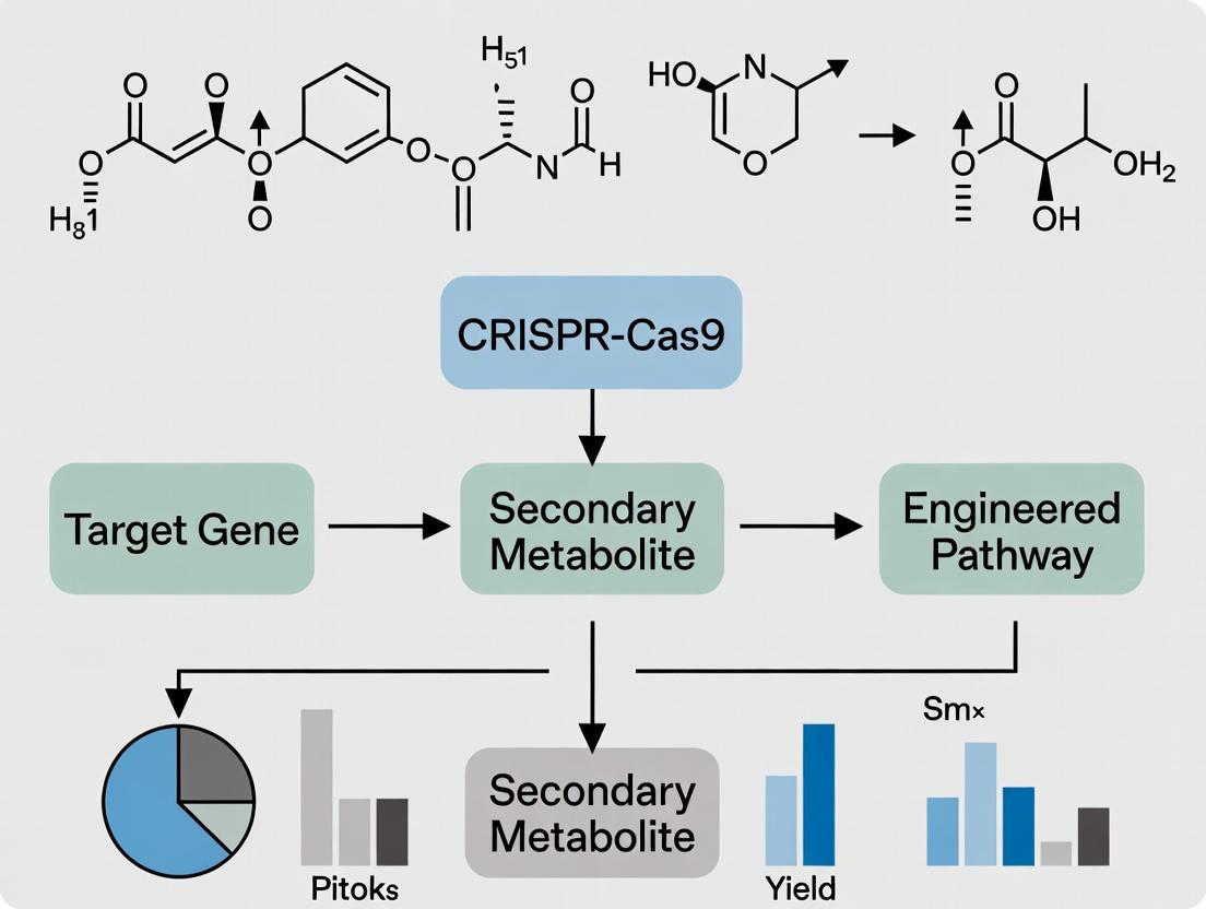
CRISPR-Cas9 Pathway Engineering: Revolutionizing Secondary Metabolite Production for Next-Gen Therapeutics
This comprehensive article explores the transformative role of CRISPR-Cas9 in engineering microbial and plant secondary metabolite pathways for drug discovery and development.
CRISPR-Cas9 Pathway Engineering: Revolutionizing Secondary Metabolite Production for Next-Gen Therapeutics
Abstract
This comprehensive article explores the transformative role of CRISPR-Cas9 in engineering microbial and plant secondary metabolite pathways for drug discovery and development. Targeting researchers and industry professionals, it covers foundational principles of pathway architecture and CRISPR mechanisms, then details cutting-edge methodologies for gene knockout, activation (CRISPRa), and repression (CRISPRi). We address common experimental hurdles and optimization strategies for efficiency and specificity. The analysis extends to validation frameworks and comparative assessments against traditional engineering tools like homologous recombination and RNAi. Finally, we synthesize key takeaways and project future directions, highlighting CRISPR's potential to unlock novel bioactive compounds and streamline therapeutic pipeline development.
CRISPR-Cas9 and Secondary Metabolites 101: Building the Blueprint for Pathway Engineering
Secondary metabolites (SMs) are organic compounds produced by plants, fungi, bacteria, and other organisms that are not directly essential for primary growth, development, or reproduction. They often serve ecological roles (e.g., defense, signaling). Their diverse chemical structures make them a vital "treasure trove" for pharmaceuticals, agrochemicals, and industrially useful compounds. Pathway engineering, particularly using CRISPR-Cas9, aims to optimize or redirect cellular machinery to overproduce target SMs or create novel analogs.
Table 1: Major Classes of Secondary Metabolites and Their Impact
| Class | Core Structure | Key Examples | Primary Sources | Major Applications/Value |
|---|---|---|---|---|
| Alkaloids | Nitrogen-containing heterocycles | Morphine, Vinblastine, Nicotine | Plants, Fungi | Analgesics, Anticancer drugs; Global plant alkaloid market ~$7.5B (2023) |
| Polyketides | Complex chains from acetyl-CoA | Erythromycin, Doxorubicin, Lovastatin | Bacteria, Fungi | Antibiotics, Statins; >20% of top-selling pharmaceuticals |
| Terpenoids/Isoprenoids | Isoprene (C5) units | Artemisinin, Taxol, Carotenoids | Plants, Microbes | Antimalarial, Anticancer, Nutraceuticals; Global terpenoid market ~$10B+ |
| Phenylpropanoids & Flavonoids | C6-C3 phenylpropane | Resveratrol, Quercetin, Lignin | Plants | Antioxidants, Anti-inflammatory, Dietary supplements |
| Non-Ribosomal Peptides (NRPs) | Amino acid derivatives | Penicillin, Cyclosporine, Vancomycin | Bacteria, Fungi | Antibiotics, Immunosuppressants |
The Rationale for Engineering Secondary Metabolite Pathways
Engineering is driven by the "supply problem": low native yield, complex extraction, and environmental pressure on natural sources. CRISPR-Cas9 enables precise, multiplex genome editing to:
- Knock-out pathway repressors or competing pathways.
- Knock-in or activate silent biosynthetic gene clusters (BGCs).
- Tune expression of key enzymes via promoter engineering.
- Introduce heterologous pathways into optimized microbial chassis (e.g., S. cerevisiae, E. coli).
Experimental Protocols for CRISPR-Cas9 Mediated SM Pathway Engineering
Protocol 4.1: Activation of a Silent Biosynthetic Gene Cluster inStreptomyces
Objective:To activate the silent 'cryptic' BGC for a novel polyketide inStreptomyces coelicolorvia CRISPRa.
Materials:
- S. coelicolor M145 strain.
- Plasmid pCRISPomyces-2 (or similar Streptomyces-optimized CRISPR-Cas9 vector).
- sgRNA designed to target promoter region of cluster transcriptional activator.
- Conjugation donor E. coli ET12567/pUZ8002.
- MS agar with appropriate antibiotics (apramycin, thiostrepton).
- HPLC-MS for metabolite analysis.
Method:
- sgRNA Design & Cloning: Design a 20-nt sgRNA sequence complementary to the -10/-35 region of the putative activator gene promoter. Clone into the BsaI site of pCRISPomyces-2.
- Conjugative Transfer:
- Prepare S. coelicolor spores (heat-shock at 50°C for 10 min).
- Mix heated spores with donor E. coli carrying the CRISPR plasmid.
- Plate on MS agar, incubate at 30°C for ~16-20h.
- Overlay plate with 1 mL water containing nalidixic acid (to counter-select E. coli) and apramycin (for plasmid selection). Incubate 3-5 days.
- Exconjugant Screening: Pick apramycin-resistant colonies. Validate via colony PCR and sequencing of the target locus.
- Metabolite Production & Analysis:
- Inoculate validated exconjugants in liquid YES medium. Shake at 30°C for 5-7 days.
- Extract culture with equal volume ethyl acetate. Dry extract under nitrogen.
- Resuspend in methanol and analyze by HPLC-MS. Compare chromatograms to wild-type control for new peaks.
Protocol 4.2: Multiplex Knockout of Competing Pathways inS. cerevisiaefor Terpenoid Production
Objective:To enhance flux towards the target terpenoid (e.g., β-carotene) by knocking out genes involved in competing sterol synthesis.
Materials:
- S. cerevisiae strain engineered with β-carotene pathway.
- CRISPR-Cas9 plasmid (e.g., pCAS-YL with LEU2 marker).
- PCR reagents for repair template (RT) assembly.
- ERG9 and ERG28 gene-specific sgRNA oligonucleotides.
- YPD and Synthetic Dropout (-Leu) media.
Method:
- Multiplex sgRNA Assembly: Clone two sgRNA expression cassettes (targeting ERG9 and ERG28) into the plasmid using Golden Gate assembly.
- Repair Template Design: Design ~80-bp single-stranded DNA oligos as RTs for each target. They contain stop codons and frameshifts near the Cas9 cut site (PAM site NGG).
- Yeast Transformation: Use the high-efficiency LiAc/SS carrier DNA/PEG method. Co-transform ~1 µg of CRISPR plasmid and 200 pmol of each RT.
- Selection and Validation: Plate on -Leu agar. Screen colonies by diagnostic PCR and Sanger sequencing to confirm biallelic disruptions.
- Flux Analysis: Ferment engineered and parent strains in controlled bioreactors. Quantify β-carotene (HPLC, OD450) and ergosterol (GC-MS) to demonstrate redirected flux.
Visualizing Pathways and Workflows
Title: CRISPR-Cas9 Secondary Metabolite Engineering Workflow
Title: Metabolic Precursor Flow to Major SM Classes
The Scientist's Toolkit: Research Reagent Solutions
Table 2: Essential Reagents for CRISPR-Cas9 SM Pathway Engineering
| Reagent/Material | Function/Description | Example Vendor/Product |
|---|---|---|
| CRISPR-Cas9 System Vectors | Host-specific plasmids for expression of Cas9 and sgRNA. | pCRISPomyces-2 (Streptomyces); pCAS-YL (Yeast); Addgene. |
| sgRNA Synthesis Oligos | Ultramer oligonucleotides for cloning or direct delivery of sgRNA. | IDT, Thermo Fisher. |
| HDR Repair Templates | Single-stranded DNA oligos or double-stranded PCR fragments for precise editing. | IDT, Genewiz. |
| Chassis Strain | Optimized microbial host for heterologous expression (e.g., high precursor flux). | S. cerevisiae CEN.PK2, E. coli BAP1, Streptomyces chassis strains. |
| Metabolite Standards | Analytical standards for quantifying target SMs via HPLC/LC-MS. | Sigma-Aldrich, Cayman Chemical. |
| HPLC-MS/MS System | For sensitive detection, quantification, and structural characterization of SMs. | Agilent, Waters, Thermo Fisher systems. |
| Bioinformatics Tools | To identify BGCs and design sgRNAs. | antiSMASH, CRISPRdirect, SnapGene. |
| Specialized Growth Media | For selection, conjugation, and optimal SM production (e.g., R5, YPD, TB). | Formulated per lab protocol or from vendors like HiMedia. |
Biosynthetic Gene Clusters (BGCs) are co-localized sets of genes in microbial genomes that encode the machinery for synthesizing a secondary metabolite. In the context of CRISPR-Cas9 pathway engineering, understanding BGC architecture is paramount for targeted genome editing, heterologous expression, and yield optimization of pharmaceutically relevant compounds.
Core Architectural Domains of a Typical BGC
A canonical BGC comprises several functional modules. Quantitative data on the average size and gene count for major classes are summarized below.
Table 1: Common BGC Classes and Their Structural Features
| BGC Class | Avg. Cluster Size (kb) | Avg. Gene Count | Core Biosynthetic Genes | Common Regulatory Elements | Example Metabolite |
|---|---|---|---|---|---|
| Non-Ribosomal Peptide Synthetase (NRPS) | 30 - 80 | 10 - 20 | NRPS genes (A, T, C domains) | LuxR-type, SARP | Penicillin, Vancomycin |
| Polyketide Synthase (PKS) | 50 - 150 | 15 - 30 | PKS genes (KS, AT, ACP domains) | TetR-family | Erythromycin, Doxorubicin |
| Terpene | 10 - 20 | 3 - 8 | Terpene synthase/cyclase | - | Geosmin, Artemisinin |
| Hybrid (e.g., NRPS-PKS) | 70 - 200 | 25 - 50 | Combined NRPS/PKS genes | Complex, pathway-specific | Rapamycin, Bleomycin |
| Ribosomally synthesized and post-translationally modified peptides (RiPPs) | 5 - 15 | 2 - 10 | Precursor peptide gene, Modification enzymes | - | Nisin, Thiostrepton |
Experimental Protocol: BGC Identification and Analysis for CRISPR Targeting
This protocol outlines steps to identify and analyze a BGC prior to CRISPR-Cas9 engineering.
Protocol 1: In silico BGC Identification and Target Design
- Genome Sequence Acquisition: Obtain the complete genome sequence of the producer organism from NCBI GenBank or via whole-genome sequencing.
- BGC Prediction: Use antiSMASH (version 7.0+) to scan the genome. Input the genome file in FASTA or GenBank format. Run with default parameters plus "--clusterhmmer" and "--asf" for detailed annotation.
- Architecture Mapping: Within the antiSMASH results, note the physical map: core biosynthetic genes, tailoring enzymes (e.g., oxidoreductases, methyltransferases), resistance genes, and putative regulatory genes.
- CRISPR Target Selection: Identify protospacers (20-nt sequences) adjacent to a 5'-NGG PAM within non-essential, permissive regions of the BGC (e.g., promoter regions, specific tailoring enzyme genes) using tools like CRISPRdirect or CHOPCHOP.
- Specificity Check: BLAST the selected protospacer sequences against the host genome to ensure single, on-target location within the BGC.
Protocol 2: CRISPR-Cas9 Mediated BGC Activation via Promoter Insertion Objective: Activate a silent BGC by replacing its native promoter with a strong, constitutive promoter. Materials:
- pCRISPR-Cas9 plasmid (with sgRNA scaffold).
- Donor DNA template containing the strong promoter (e.g., ermEp) flanked by 1-kb homology arms matching sequences upstream and downstream of the native BGC promoter.
- Electrocompetent cells of the producer strain (e.g., Streptomyces coelicolor).
- Recovery medium (e.g., LB with 10% sucrose).
- Selection antibiotics.
Method:
- sgRNA Cloning: Clone the designed protospacer targeting the sequence immediately upstream of the native BGC start codon into the pCRISPR-Cas9 plasmid using BsaI Golden Gate assembly. Transform into E. coli, isolate, and sequence-verify the plasmid.
- Donor DNA Preparation: Synthesize or PCR-amplify the donor DNA fragment.
- Co-transformation: Introduce 500 ng of the validated pCRISPR-Cas9 plasmid and 1 µg of donor DNA into electrocompetent producer strain cells via electroporation (e.g., 1.8 kV, 5 ms).
- Recovery & Selection: Recover cells in 1 mL of recovery medium at 28°C for 12-16 hours. Plate onto selective medium containing the appropriate antibiotic and incubate for 3-7 days.
- Screening: Screen colonies by colony PCR using one primer within the inserted promoter and one outside the homology arm to verify correct promoter swap. Confirm via sequencing.
- Metabolite Analysis: Culture positive mutants and analyze metabolite production via LC-MS compared to the wild-type strain.
Key Diagrams
Diagram 1: BGC Domains & CRISPR Engineering
Diagram 2: BGC Engineering Protocol Flow
The Scientist's Toolkit: Essential Research Reagents and Materials
Table 2: Key Research Reagent Solutions for BGC Engineering
| Item | Function in BGC/CRISPR Work | Example/Supplier Note |
|---|---|---|
| antiSMASH Software | Identifies and annotates BGCs in genomic data. Essential for initial architecture overview. | Public web server or standalone version (v7.0+). |
| CRISPR-Cas9 Plasmid System | Delivers Cas9 and sgRNA expression cassettes to the host cell. | pCRISPR-Cas9 vectors for Streptomyces (e.g., pCRISPomyces-2). |
| Donor DNA Fragment | Template for HDR. Contains desired edit (e.g., promoter, gene deletion) flanked by homology arms. | Synthesized as gBlocks (IDT) or amplified via PCR. |
| Electrocompetent Cells | Genetically tractable host strain prepared for efficient DNA uptake via electroporation. | High-efficiency S. coelicolor or E. coli ET12567/pUZ8002 for conjugation. |
| HDR Enhancer (e.g., RecET) | Proteins that promote homologous recombination, increasing editing efficiency in some hosts. | Plasmid co-expression or encoded on the CRISPR plasmid. |
| LC-MS/MS System | Analyzes secondary metabolite profiles pre- and post-engineering to assess product yield/change. | Agilent, Thermo Fisher, or Waters systems with reverse-phase columns. |
| Selection Antibiotics | Maintains plasmid(s) and selects for successfully edited clones. | Apramycin, Thiostrepton, Kanamycin (concentration strain-dependent). |
| PCR Reagents for Screening | Verifies correct genomic integration of the edit via colony PCR. | High-fidelity polymerase (e.g., Q5, Phusion) and specific primers. |
CRISPR-Cas9 has revolutionized metabolic engineering by enabling precise, multiplexed editing of biosynthetic gene clusters (BGCs) in microbial hosts. For researchers engineering pathways for secondary metabolites (e.g., antibiotics, anticancer agents), the system allows for targeted gene knock-outs, knock-ins, and transcriptional regulation to optimize precursor flux, eliminate competitive pathways, and enhance titers. This primer details the core mechanisms and provides actionable protocols for pathway editing applications.
Core Mechanisms: From DNA Cleavage to Pathway Modulation
The Cas9-sgRNA Ribonucleoprotein Complex
The Streptococcus pyogenes Cas9 endonuclease is guided by a single guide RNA (sgRNA), a chimeric RNA containing a user-defined 20-nucleotide spacer sequence (for target DNA recognition) and a scaffold sequence. The sgRNA spacer base-pairs with the target DNA adjacent to a Protospacer Adjacent Motif (PAM; 5'-NGG-3'), enabling Cas9 to induce a double-strand break (DSB).
DNA Repair Pathways: Harnessing for Engineering
Cellular repair of the DSB dictates the editing outcome, critical for pathway engineering.
- Non-Homologous End Joining (NHEJ): An error-prone repair pathway often used for gene knock-outs. Small insertions or deletions (indels) can disrupt coding sequences of repressor genes or non-essential pathway enzymes.
- Homology-Directed Repair (HDR): Uses a donor DNA template for precise gene knock-in or point mutation. Essential for inserting strong promoters, epitope tags, or heterologous genes into a BGC.
Table 1: Quantitative Overview of CRISPR-Cas9 Editing Outcomes in Common Hosts
| Host Organism | Typical Delivery Method | NHEJ Efficiency Range (%) | HDR Efficiency Range (%) (with donor) | Key Applications in Pathway Engineering |
|---|---|---|---|---|
| S. cerevisiae | Plasmid or RNP | 70-90 | 10-30 | Engineering of fungal polyketide pathways. |
| E. coli | Plasmid or RNP | 20-60 | <1-5 (low) | Precursor pathway optimization. |
| Streptomyces spp. | Conjugative Plasmid | 50-80 | 5-20 (with ssDNA) | Activation or refactoring of silent BGCs. |
| Aspergillus nidulans | AMA1-based Plasmid | 60-95 | 15-40 | Fungal secondary metabolite overproduction. |
| Mammalian Cells | Lentivirus or RNP | 20-50 | 1-20 | Engineering of plant metabolite pathways in cell lines. |
CRISPRi/a for Transcriptional Control
A catalytically "dead" Cas9 (dCas9) can be fused to repressor (KRAB) or activator (VP64) domains. When targeted to promoter regions, this enables CRISPR interference (CRISPRi) or activation (CRISPRa) without altering the DNA sequence. This is invaluable for fine-tuning expression levels of multiple pathway genes simultaneously.
CRISPR-Cas9 Workflow for Pathway Editing
Application Notes & Protocols for Pathway Engineering
Protocol: Multiplexed Knock-out inS. cerevisiaefor Precursor Pool Enhancement
Aim: Disrupt three genes (ERG9, ARO10, PDC5) competing for acetyl-CoA and aromatic precursors to redirect flux toward a target polyketide.
Reagents & Materials: Table 2: Research Reagent Solutions - Yeast Multiplex Editing
| Item | Function/Description | Example Supplier/Catalog |
|---|---|---|
| pCAS-SP plasmid system | Expresses Cas9, sgRNA(s), and selectable marker. | Addgene #60847 |
| sgRNA cloning oligos | 20nt target-specific sequences for Golden Gate assembly. | IDT, Custom DNA Oligos |
| BsaI-HFv2 | Restriction enzyme for Golden Gate assembly of sgRNAs. | NEB #R3733 |
| Yeast Donor Oligos (80-120nt) | Homology templates for NHEJ-driven repair, contain stop codons/frame-shifts. | IDT, Ultramer Oligos |
| YPD & SC-Selective Media | For yeast cultivation and transformation selection. | Formedium |
| Zymolyase | Digest cell wall for efficient transformation. | AMS Biotechnology |
Methodology:
- Design: For each target gene, design a sgRNA targeting an early exon using a validated tool (e.g., CHOPCHOP). Design donor oligos with 40bp homology arms flanking a STOP cassette.
- Cloning: Use BsaI-mediated Golden Gate assembly to clone three sgRNA expression cassettes into the pCAS vector.
- Transformation: Co-transform S. cerevisiae with the pCAS multiplex plasmid and the pool of donor oligos using the LiAc/SS carrier DNA/PEG method.
- Selection & Screening: Plate on appropriate selective media. Screen colonies by colony PCR and Sanger sequencing to confirm triple knock-out.
- Curing: Passage colonies on non-selective media to lose the pCAS plasmid.
Protocol: HDR-Mediated Promoter Swap inStreptomyces coelicolor
Aim: Replace the native promoter of the actII-ORF4 pathway-specific regulator with a constitutive strong promoter (ermEp) to overactivate actinorhodin production.
Reagents & Materials: Table 3: Research Reagent Solutions - Streptomyces Promoter Swap
| Item | Function/Description | Example Supplier/Catalog |
|---|---|---|
| pCRISPomyces-2 plasmid | Cas9 + sgRNA expression for Streptomyces. | Addgene #61737 |
| dsDNA Donor Fragment | Contains ermEp flanked by >1kb homology arms. | Gibson Assembly or Gene Synthesis |
| E. coli ET12567/pUZ8002 | Non-methylating donor strain for conjugation. | Lab Stock or CGSC |
| Apramycin & Thiostrepton | Antibiotics for selection in E. coli and Streptomyces. | Sigma-Aldrich |
| TSBS Medium | For Streptomyces conjugation and sporulation. | Formedium |
Methodology:
- Donor Construction: Synthesize or Gibson-assemble the ermEp sequence with 1-1.5 kb homology arms upstream and downstream of the native promoter's genomic locus.
- sgRNA Cloning: Clone sgRNA targeting the cut site within the native promoter region into pCRISPomyces-2.
- Conjugation: Transform the pCRISPomyces-2 plasmid and the donor plasmid (or fragment) into E. coli ET12567/pUZ8002. Perform intergeneric conjugation with S. coelicolor spores.
- Selection: Plate on selective media containing apramycin (plasmid) and thiostrepton (counter-selection for E. coli). Incubate at 30°C.
- Screening & Validation: Screen exconjugants by PCR for correct promoter replacement. Cure the plasmid via passaging and quantify actinorhodin via HPLC.
Streptomyces HDR Promoter Swap Workflow
The Scientist's Toolkit: Key Reagents for CRISPR Pathway Editing
Table 4: Essential Toolkit for CRISPR-based Metabolic Pathway Engineering
| Category | Item | Critical Function in Pathway Editing |
|---|---|---|
| Nucleases & Variants | Wild-Type SpCas9 | Standard DSB induction for knock-out/knock-in. |
| High-Fidelity SpCas9 (e.g., SpCas9-HF1) | Reduces off-target effects when editing large gene clusters. | |
| dCas9 (D10A, H840A) | Catalytic null base for CRISPRi/a transcriptional tuning. | |
| Delivery Vectors | AMA1-based fungal plasmids | High-copy, self-replicating plasmids for Aspergillus/Penicillium. |
| Integrative plasmids (e.g., pSET152-based) | Stable chromosomal integration in Actinomycetes. | |
| RNP complexes (Cas9 protein + sgRNA) | Direct delivery, rapid degradation, reduces off-targets in delicate hosts. | |
| Donor Templates | ssDNA Oligos (80-200nt) | For precise point mutations or short insertions via HDR. |
| dsDNA fragments (PCR/gene synthesis) | For large insertions (e.g., promoter, gene) with long homology arms. | |
| Specialized Modules | dCas9-KRAB repression domain | Strong transcriptional repression (CRISPRi) of competitive genes. |
| dCas9-VP64 activation domain | Transcriptional activation (CRISPRa) of silent/sleeping BGCs. | |
| MS2-MCP recruiting systems | For enhanced activation (dCas9-VP64-p65-Rta) or base editing fusions. | |
| Host-Specific Reagents | Zymolyase (Yeast) | Cell wall digestion for efficient transformation. |
| Thiostrepton (Streptomyces) | Selective antibiotic and potential inducer for some promoters. | |
| Polyethylene Glycol (PEG)-mediated protoplast transformation | Standard for many fungal and some bacterial hosts. |
Introduction Within a broader thesis investigating CRISPR-Cas9 for secondary metabolite pathway engineering, it is critical to understand the foundational—and limited—methodologies that preceded it. Pre-CRISPR metabolic engineering for natural product discovery and optimization was a slow, iterative process hampered by a lack of precise, multiplex genetic tools. This document outlines the key experimental approaches, their inherent limitations, and the specific protocols that defined the era, providing context for the revolutionary impact of CRISPR-based genome editing.
1. Key Pre-CRISPR Techniques and Their Quantitative Limitations The engineering of microbial hosts (e.g., Streptomyces, E. coli, S. cerevisiae) for enhanced secondary metabolite production relied on a suite of imprecise genetic tools. The table below summarizes the efficiency, throughput, and typical outcomes of these methods.
Table 1: Comparison of Pre-CRISPR Metabolic Engineering Tools
| Technique | Typical Target | Max Efficiency (Strain Modification) | Timeframe for Multiplex (3-5 loci) | Key Limitation for Pathway Engineering |
|---|---|---|---|---|
| Random Mutagenesis (UV/Chemical) | Genome-wide | 0.01-0.1% beneficial mutation | Months to years | Requires high-throughput screening; mutations are unmarked and pleiotropic. |
| Homologous Recombination (HR) via Suicide Vector | Single locus | 10^-3 to 10^-6 (non-recombineering) | 6-12 months | Extremely low efficiency in wild-type strains; laborious counter-selection required. |
| λ-Red/ET Recombineering (in E. coli) | Single locus | >10^4 recombinants/μg DNA | 1-2 months | Limited host range; often requires subsequent conjugation into producer strain. |
| PCR-Targeting (e.g., Redirect in Streptomyces) | Single locus | ~10^-2 to 10^-3 | 3-6 months | Dependent on pre-constructed cosmid libraries; leaves antibiotic resistance cassettes. |
| Site-Specific Recombination (Cre-loxP, FLP-FRT) | Marker excision | >90% excision | Adds 1-2 months per cycle | Only for removing markers; does not enable de novo insertion. |
| RNAi/Antisense RNA Knockdown | Gene expression | Variable, 30-80% knockdown | 1-2 months | Silencing is titratable but transient and incomplete; polar effects common. |
2. Detailed Protocol: Classical Homologous Recombination for Gene Knockout in Streptomyces This protocol exemplifies the complexity of pre-CRISPR, multi-step genome editing.
Objective: To disrupt a specific gene (actII-ORF4) within the actinorhodin biosynthetic gene cluster in Streptomyces coelicolor.
Materials:
- Bacterial Strains: S. coelicolor A3(2), E. coli ET12567/pUZ8002 (non-methylating, conjugation donor).
- Vectors: pKC1139 (or similar suicide vector with oriT, aac(3)IV apramycin resistance, temperature-sensitive replicon).
- Reagents: Apramycin, kanamycin, chloramphenicol, nalidixic acid, isobutyryladenosine (ISP4) media, agar.
Procedure: A. Vector Construction (2-3 weeks):
- Amplify ~1.5 kb DNA fragments upstream (UP) and downstream (DOWN) of the actII-ORF4 coding sequence via PCR.
- Ligate the UP and DOWN fragments into the multiple cloning site of pKC1139, flanking the apramycin resistance (aac(3)IV) cassette, using a three-fragment Gibson Assembly or traditional restriction/ligation.
- Sequence-verify the final construct, pKC1139-ΔactII.
B. Conjugal Transfer from E. coli to Streptomyces (1 week):
- Introduce pKC1139-ΔactII into E. coli ET12567/pUZ8002 via transformation.
- Grow donor E. coli and recipient S. coelicolor spores separately. Mix, pellet, and resuspend to spot onto ISP4 agar plates.
- Incubate at 30°C for 16-20 hours.
- Overlay plate with 1 mL water containing apramycin (50 μg/mL) and nalidixic acid (25 μg/mL) to select for Streptomyces exconjugants (which have integrated the plasmid via single-crossover) while counterselecting E. coli.
C. Selection for Double-Crossover Events (2-3 weeks):
- Pick exconjugants and streak for single colonies under apramycin selection at 30°C (permissive temperature for plasmid replication).
- Inoculate single colonies into liquid media without antibiotics and incubate at 37°C (non-permissive temperature) for 2-3 rounds of growth to promote plasmid loss.
- Plate dilutions onto non-selective agar. Replica-plate individual colonies onto plates with and without apramycin.
- Screen for Apramycin-Sensitive (Apra^S) colonies, which have undergone a second crossover and lost the vector backbone.
- Verify gene knockout via colony PCR using primers external to the UP/DOWN homology arms.
3. Pathway Engineering Workflow & Limitations
Diagram Title: Pre-CRISPR Iterative Engineering Cycle
4. The Scientist's Toolkit: Essential Reagents for Pre-CRISPR Engineering
Table 2: Key Research Reagent Solutions
| Item | Function in Pre-CRISPR Engineering |
|---|---|
| Temperature-Sensitive Suicide Vectors (e.g., pKC1139, pIJ790) | Contains oriT for conjugation, antibiotic marker, and replicon that fails at elevated temperatures, allowing for selection of double-crossover events. |
| E. coli ET12567/pUZ8002 Strain | Non-methylating dam/dmr host carrying the conjugation helper plasmid pUZ8002. Essential for mobilizing vectors from E. coli into actinomycetes. |
| cosmid/BAC Genomic Library | Large-insert clone library covering the entire biosynthetic gene cluster of interest. Served as the template for PCR-targeting or subcloning. |
| λ-Red/ET Recombineering Plasmid (e.g., pKD46, pSC101-BAD-ETγ) | Expresses phage-derived recombinases in E. coli to enable high-efficiency, PCR-based modification of cloned DNA on plasmids or BACs. |
| I-SceI Meganuclease Vector | Rare-cutting endonuclease used in conjunction with a conditionally replicating vector to stimulate double-strand break repair and increase homologous recombination efficiency. |
| Gateway or Gibson Assembly Cloning Kits | Enabled faster, more reliable in vitro assembly of multiple homology arms and markers for vector construction, but did not simplify in vivo genome integration. |
Conclusion The protocols and tools detailed here underscore the technically demanding and time-intensive nature of metabolic engineering prior to CRISPR-Cas9. The reliance on homologous recombination with low native efficiency, the necessity for selectable markers, and the near-impossibility of coordinated multiplex editing constituted fundamental barriers. This historical context directly informs the thesis that CRISPR-Cas9, with its precision, multiplexability, and marker-free editing, represents a paradigm shift in the rational redesign of secondary metabolite pathways.
This application note details the use of key model organisms in CRISPR-Cas9-mediated secondary metabolite pathway engineering, a core component of modern drug discovery research. Streptomyces species are prolific producers of clinically relevant antibiotics and other bioactive compounds, while plant cell cultures offer a sustainable platform for producing complex plant-derived pharmaceuticals. Engineering these hosts using CRISPR-Cas9 allows for precise manipulation of biosynthetic gene clusters (BGCs) to enhance yield, produce novel analogs, or activate silent pathways.
Application Notes
CRISPR-Cas9 Engineering inStreptomycesspp.
Streptomyces coelicolor and Streptomyces avermitilis are the primary model organisms for actinobacterial genetics and natural product discovery. Recent advances have established efficient CRISPR-Cas9 tools for these high-GC Gram-positive bacteria, enabling targeted gene knockouts, transcriptional activation (CRISPRa), and large-scale genomic deletions to remove competing pathways.
Key Quantitative Data: CRISPR-Cas9 Efficiency in Streptomyces
| Strain | Target Gene/Operation | Efficiency (%) | Delivery Method | Reference (Year) |
|---|---|---|---|---|
| S. coelicolor M145 | actII-ORF4 knockout | 90-100 | Conjugative plasmid | [Cobb et al., 2015] |
| S. avermitilis | 1.8 Mb genomic deletion | ~100 | Conjugative plasmid + φC31 integrase | [Tao et al., 2022] |
| S. albus J1074 | Multiplexed (3 genes) knockout | 85 | PEG-mediated protoplast transformation | [Alberti & Corre, 2019] |
| S. venezuelae | CRISPRi repression of bldD | 70-80 | Conjugative plasmid | [Roh et al., 2019] |
Pathway Engineering in Plant Cell Cultures
Plant cell cultures (e.g., from Nicotiana benthamiana, Catharanthus roseus) are emerging as controllable hosts for producing terpenoids, alkaloids, and flavonoids. Transient CRISPR-Cas9 delivery via Agrobacterium tumefaciens (agroinfiltration) or protoplast transfection allows for the knockout of competing pathway genes or repressors of biosynthesis.
Key Quantitative Data: CRISPR Outcomes in Plant Cell Cultures
| Plant Species/Culture Type | Target Pathway | Modification | Metabolite Yield Change | Transformation Method |
|---|---|---|---|---|
| N. benthamiana suspension | Monoterpenoid indole alkaloid | Knockout of strictosidine glucosidase | 60% reduction in strictosidine degradation | Agroinfiltration |
| C. roseus hairy roots | Catharanthine/vindoline | CRISPRa of ORCA3 transcriptional activator | 2.5-fold increase in terpenoid indole alkaloids | A. rhizogenes |
| Medicago truncatula cell suspension | Triterpenoid saponins | Knockout of β-amyrin synthase | Knockout confirmed; novel saponins detected | PEG-mediated protoplast transfection |
Detailed Protocols
Protocol 1: CRISPR-Cas9 Mediated Gene Knockout inStreptomyces coelicolorvia Conjugation fromE. coli
Objective: To disrupt a target gene within a biosynthetic gene cluster to elucidate function or redirect metabolic flux.
Research Reagent Solutions & Materials:
| Reagent/Material | Function/Description |
|---|---|
| pCRISPomyces-2 plasmid | A Streptomyces shuttle vector containing cas9, a traceless sgRNA cassette, and apramycin resistance. |
| ET12567(pUZ8002) E. coli donor strain | DAM-/DEM-* strain with helper plasmid for mobilizing oriT-containing plasmids via conjugation. |
| MS agar with 10 mM MgCl₂ | Solid medium for Streptomyces conjugation and sporulation. |
| Apramycin (50 µg/mL) + Nalidixic Acid (25 µg/mL) | Selection antibiotics for exconjugants (Streptomyces resistance + counter-selection against E. coli). |
| HR Repair Template DNA | Double-stranded DNA fragment containing homologous arms (≥500 bp each) flanking the desired deletion. |
| SSC Buffer (0.3 M sodium citrate, 3 M NaCl) | Used to spread on plates after overlay to promote Streptomyces growth. |
Procedure:
- sgRNA Design & Cloning: Design a 20-nt spacer sequence targeting the desired gene using a validated online tool. Clone the spacer into the BsaI site of pCRISPomyces-2 via Golden Gate assembly.
- Repair Template Construction: PCR-amplify or synthesize a linear DNA fragment containing ~1 kb homology arms upstream and downstream of the Cas9 cut site. The fragment should omit the sequence to be deleted.
- Donor Strain Preparation: Transform the assembled pCRISPomyces-2 plasmid into chemically competent E. coli ET12567(pUZ8002). Select on LB agar with apramycin (50 µg/mL) and kanamycin (50 µg/mL).
- Conjugation: a. Inoculate a single E. coli donor colony and grow in LB with antibiotics to an OD₆₀₀ of ~0.6. b. Wash cells 3x with LB to remove antibiotics. c. Prepare S. coelicolor spores by harvesting from a fresh plate and heat-shocking at 50°C for 10 minutes. d. Mix donor cells (100 µL), spores (100 µL), and repair template DNA (500 ng). Plate onto MS agar (no antibiotics). Incubate at 30°C for 16-20 hours.
- Selection & Screening: a. Overlay the plate with 1 mL of sterile water containing apramycin (final 50 µg/mL) and nalidixic acid (final 25 µg/mL). Add 1 mL of 2X SSC buffer. b. Incubate at 30°C for 3-5 days until exconjugant colonies appear. c. Patch colonies onto selective plates. Screen for successful gene deletion via PCR using primers outside the homology region.
- Curing the Plasmid: Streak positive clones on non-selective medium for several rounds to allow loss of the temperature-sensitive plasmid. Verify plasmid loss by patching onto apramycin-containing and antibiotic-free plates.
Protocol 2: Transient CRISPR-Cas9 Knockout inNicotiana benthamianaSuspension Cells via Agroinfiltration
Objective: To transiently disrupt a gene in a plant biosynthetic pathway for functional genomics or metabolic engineering.
Research Reagent Solutions & Materials:
| Reagent/Material | Function/Description |
|---|---|
| pORE-Cas9 binary vector | Plant expression vector containing a plant-codon-optimized cas9 and a kanamycin resistance marker. |
| pHEE401E sgRNA vector | Contains the AtU6-26 promoter for sgRNA expression and a spectinomycin resistance marker. |
| Agrobacterium tumefaciens strain GV3101(pMP90) | Disarmed strain with helper plasmid for T-DNA transfer, suitable for transient transformation. |
| LB Medium with appropriate antibiotics | For growth of A. tumefaciens. |
| Acetosyringone (200 µM) | Phenolic compound that induces the Agrobacterium vir genes for T-DNA transfer. |
| MS Liquid Medium (pH 5.6) | Maintenance medium for N. benthamiana suspension cells. |
| CTAB DNA Extraction Buffer | For genomic DNA extraction from plant cells for genotyping. |
Procedure:
- Vector Assembly: a. Clone a target-specific 20-nt spacer into the BsaI site of the pHEE401E sgRNA vector. b. Transform the assembled sgRNA vector and the pORE-Cas9 vector separately into electrocompetent A. tumefaciens GV3101.
- Agrobacterium Culture Preparation: a. Grow individual cultures (+ antibiotics) of the Cas9 and sgRNA strains overnight at 28°C. b. Sub-culture to an OD₆₀₀ of 0.5-0.8. Pellet cells and resuspend in MS liquid medium supplemented with 200 µM acetosyringone. c. Mix the Cas9 and sgRNA cultures in a 1:1 ratio. Let the mixture incubate at room temperature for 2-4 hours.
- Agroinfiltration of Suspension Cells: a. Use 5-7 day old N. benthamiana suspension cultures. b. Add the Agrobacterium mixture to the plant cells at a final OD₆₀₀ of ~0.2. Gently mix. c. Co-cultivate in the dark at 25°C with slow shaking (80 rpm) for 48-72 hours.
- Sampling and Analysis: a. Harvest cells by vacuum filtration. Rinse with sterile water to remove excess Agrobacterium. b. Extract genomic DNA using CTAB method. Assess editing efficiency via T7 Endonuclease I (T7EI) assay or Sanger sequencing followed by tracking of indels by decomposition (TIDE) analysis. c. For metabolite analysis, flash-freeze cell pellets in liquid nitrogen and perform HPLC-MS on extracts.
Visualizations
(Diagram: CRISPR-Cas9 Workflow for Streptomyces Gene Knockout)
(Diagram: Transient CRISPR in Plant Cells via Agroinfiltration)
Precision Editing in Action: CRISPR-Cas9 Strategies for Pathway Manipulation
1. Introduction: Within the Thesis Context This protocol details a standardized workflow for applying CRISPR-Cas9 to engineer Biosynthetic Gene Clusters (BGCs) for secondary metabolite production. It supports the broader thesis that precision genome editing, specifically via multiplexed sgRNA strategies, is a transformative tool for activating silent BGCs, refactoring complex pathways, and optimizing titers in both native and heterologous hosts for drug discovery pipelines.
2. Application Notes & Protocols
2.1. Phase I: In Silico sgRNA Design for BGC Targets
- Objective: Design specific, efficient, and multiplex-compatible sgRNAs targeting regulatory genes, pathway repressors, or specific biosynthetic modules within a BGC.
- Protocol:
- BGC Sequence Retrieval: Obtain the complete nucleotide sequence of the target BGC from databases (e.g., MIBiG, antiSMASH output).
- Protospacer Adjacent Motif (PAM) Identification: Scan the sequence for the appropriate PAM site (e.g., 5'-NGG-3' for Streptococcus pyogenes Cas9).
- sgRNA Candidate Selection: For each target site, extract the 20-nt sequence immediately 5' to the PAM as the spacer.
- Specificity Check (BLAST): Perform a BLASTn search of each spacer sequence against the host genome to minimize off-target effects. Prioritize sgRNAs with zero or minimal off-targets with ≤3 mismatches.
- Efficiency Prediction: Score candidates using validated algorithms (e.g., Doench '16 score from Broad Institute, CFD specificity score). Select the top 3-5 per target.
- Multiplexing Design: For simultaneous editing, ensure selected sgRNAs do not have significant cross-homology to avoid interference.
Quantitative Data Summary: sgRNA Design Parameters Table 1: Key Parameters for Optimal sgRNA Selection
| Parameter | Optimal Target Value | Purpose |
|---|---|---|
| GC Content | 40-60% | Enhances stability and efficiency. |
| Doench Efficiency Score | > 0.5 (Higher is better) | Predicts on-target cutting activity. |
| CFD Specificity Score | < 0.2 (Lower is better) | Predicts off-target potential. |
| Off-Target Matches | 0 with perfect seed region | Minimizes unintended genomic edits. |
2.2. Phase II: sgRNA Expression Construct Assembly
- Objective: Clone validated sgRNA spacer sequences into an appropriate CRISPR-Cas9 expression plasmid for the host organism (e.g., Streptomyces, Aspergillus, E. coli).
- Protocol (Golden Gate Assembly):
- Oligonucleotide Annealing: Synthesize complementary DNA oligos for each spacer (Forward: 5'-CACC[G]+spacer-3', Reverse: 5'-AAAC[revcomp spacer]+C-3'). Resuspend in annealing buffer (10 mM Tris, 50 mM NaCl, pH 7.5).
- Annealing Reaction: Mix oligos (1:1 ratio, 100 µM each), heat to 95°C for 5 min, and cool slowly to 25°C (~1°C/min).
- Ligation into Vector: Set up a Golden Gate reaction (e.g., using BsaI-HFv2 enzyme):
- Annealed oligo duplex (1 µL, 1:10 dilution)
- Destination plasmid (e.g., pCRISPR-Cas9, 50 ng)
- T4 DNA Ligase Buffer (1X)
- BsaI-HFv2 (1 µL)
- T4 DNA Ligase (1 µL)
- H₂O to 20 µL. Cycle: (37°C for 5 min, 20°C for 5 min) x 10 cycles, then 80°C for 5 min.
- Transformation: Transform 5 µL of reaction into competent E. coli DH5α, plate on selective media, and sequence-verify colonies.
2.3. Phase III: Host Transformation/Transfection & Screening
- Objective: Deliver the CRISPR-Cas9-sgRNA construct into the production host and identify successful editing events.
- Protocol (Protoplast Transformation for Filamentous Fungi/Actinomycetes):
- Protoplast Preparation: Grow mycelia to mid-exponential phase. Harvest and digest cell wall using lysozyme (bacteria) or lysing enzymes (e.g., Novozym 234 for fungi) in an osmotic stabilizer (0.8 M sucrose).
- Transformation: Mix 10⁷ protoplasts with 5-10 µg of plasmid DNA in 30% PEG 4000 solution. Incubate at room temperature for 20 min.
- Regeneration: Plate on regeneration agar (osmotic stabilizer + selective antibiotics) for 5-7 days.
- Primary Screening (Colony PCR): Pick regenerated colonies. Use primer sets flanking the target site to amplify the genomic region. Analyze PCR products via agarose gel electrophoresis for size shifts.
- Secondary Screening (Sequencing Validation): Sanger sequence the PCR products from primary hits. Use alignment software (e.g., SnapGene) to confirm insertions/deletions (indels) or precise edits at the target locus.
- Phenotypic Screening: Ferment validated mutants in small-scale culture. Analyze metabolite profiles via LC-MS/MS and compare to wild-type to assess pathway engineering impact (e.g., novel peak emergence, titer increase).
Quantitative Data Summary: Transformation & Screening Metrics Table 2: Typical Experimental Metrics for Protoplast-Based Editing
| Step | Key Metric | Expected Range / Target |
|---|---|---|
| Protopast Viability | Viable count per mL | 10⁷ - 10⁸ /mL |
| Transformation Efficiency | CFU per µg DNA | 10² - 10⁴ for many actinomycetes |
| Editing Efficiency | % of screened colonies with indels | 10% - 80% (host & construct dependent) |
| Validation | Sanger sequencing confirmation | >95% sequence clarity at target locus |
3. The Scientist's Toolkit: Research Reagent Solutions Table 3: Essential Materials for CRISPR-Cas9 BGC Engineering
| Item | Function / Application | Example Product/Catalog |
|---|---|---|
| CRISPR-Cas9 Expression Vector | All-in-one plasmid for Cas9 and sgRNA expression in the target host. | pCRISPR-Cas9 (host-specific variants), pKCcas9dO for Streptomyces. |
| High-Fidelity DNA Polymerase | Accurate amplification of target BGC loci for screening. | Q5 High-Fidelity DNA Polymerase (NEB). |
| Type IIS Restriction Enzyme | Enzymatic digestion for Golden Gate assembly of sgRNA arrays. | BsaI-HFv2, Esp3I (Thermo Scientific). |
| T4 DNA Ligase | Ligation of annealed oligos into the sgRNA expression scaffold. | T4 DNA Ligase (Rapid) (Thermo Scientific). |
| Protoplasting Enzymes | Digestion of microbial cell walls for DNA delivery. | Lysozyme (for bacteria), Lysing Enzymes from Trichoderma (Sigma-Aldrich). |
| Polyethylene Glycol (PEG) | Facilitates DNA uptake during protoplast transformation. | PEG 4000, 30-40% (w/v) solution. |
| Osmotic Stabilizer | Maintains protoplast integrity during processing. | Sucrose (0.8 M) or Sorbitol (0.8 M) solution. |
| LC-MS Grade Solvents | High-purity solvents for metabolite extraction and analysis. | Methanol, Acetonitrile (Fisher Chemical). |
4. Visualized Workflows & Pathways
CRISPR-Cas9 BGC Engineering Workflow
Gene Editing Outcome from Cas9-Induced DSB
Within CRISPR-Cas9-driven secondary metabolite pathway engineering, a core challenge is maximizing titers of target compounds. Native host organisms possess complex regulatory networks and competing metabolic pathways that divert flux away from the desired biosynthetic route. This application note details strategies for employing CRISPR-Cas9 knockout (KO) to silence these competing pathways and negative regulatory genes, thereby rewiring cellular metabolism for enhanced metabolite production. These protocols are framed within a thesis investigating the combinatorial optimization of polyketide synthase (PKS) clusters in Streptomyces species.
Key Target Categories for Knockout
Table 1: Common Knockout Targets in Metabolic Pathway Engineering
| Target Category | Example Genes | Rationale for Knockout | Expected Outcome |
|---|---|---|---|
| Competing Pathways | sgn (stigmatellin), red (undecylprodigiosin), act (actinorhodin) biosynthetic gene clusters | Eliminate production of endogenous secondary metabolites that consume shared precursors (e.g., acetyl-CoA, malonyl-CoA). | Increased precursor pool availability for target pathway. |
| Global Neg. Regulators | afsA, nsdA, wblA | Disrupt pleiotropic regulatory genes that repress antibiotic biosynthesis. | Derepression of multiple biosynthetic gene clusters, including target. |
| Pathway-Specific Repressors | scbR (for actinorhodin), rap genes | Remove direct transcriptional repression of target gene cluster. | Enhanced transcription of target biosynthetic genes. |
| Proteolytic/Degradation | lon, clp proteases | Reduce turnover of key biosynthetic enzymes. | Increased stability and half-life of pathway enzymes. |
| Alternative Terminal Enzymes | shc (squalene-hopene cyclase) | Block diversion of isoprenoid flux to non-target products. | Channeling of metabolic flux (e.g., farnesyl pyrophosphate) toward target terpenoid. |
Detailed Protocol: Multiplexed KO inStreptomyces coelicolor
Aim: To concurrently knockout the competing red and act pigment pathways and the regulatory gene wblA.
I. sgRNA Design and Vector Construction
- Target Identification: Select protospacer sequences (20-nt) adjacent to a 5'-NGG PAM for early essential genes within actII-ORF4 (activating regulator), redD (regulator), and wblA.
- Oligonucleotide Design: Design primers for each target:
actKO_F: 5'-CACCG[TargetSequence]-3'actKO_R: 5'-AAAC[ReverseCompTargetSequence]C-3' - Cloning into pCRISPR-Cas9: Digest the Streptomyces-optimized plasmid (containing cas9, ermE promoter, and sgRNA scaffold) with BsaI. Ligate annealed oligos into the vector. Perform triple-parental mating or PEG-mediated protoplast transformation to generate three separate plasmids.
II. CRISPR-Cas9 Delivery and Screening
- Conjugal Transfer: From E. coli ET12567/pUZ8002, mobilize each plasmid into S. coelicolor M145 spores. Plate on MS agar with apramycin (selection for plasmid) and nalidixxic acid (counter-selection against E. coli).
- Initial Phenotypic Screen: Incubate at 30°C for 5-7 days. Look for loss of blue (actinorhodin) and red (undecylprodigiosin) pigmentation in transformants.
- Genotype Validation (PCR & Sequencing): Isolate genomic DNA. Perform PCR amplification flanking each target site (primers ~500bp upstream/downstream). Analyze products by gel electrophoresis. Successful KO results in larger amplicons (due to NHEJ-mediated indels) compared to wild-type. Sequence to confirm frameshift mutations.
- Curing the Plasmid: Passage positive clones on non-selective media for several generations, then replica plate to confirm loss of apramycin resistance.
III. Metabolite Analysis
- Extraction: Ferment validated KO strains in R5 liquid medium for 120h. Acidify culture broth and extract with ethyl acetate.
- Quantification: Analyze extracts via HPLC-MS. Compare peak areas of target metabolite (e.g., a specific polyketide) against internal standard. Use UV-vis spectroscopy to quantify residual pigment production.
Table 2: Example Quantitative Output from a Triple KO Experiment
| Strain (S. coelicolor) | Target Metabolite Yield (mg/L) | Actinorhodin (% of WT) | Undecylprodigiosin (% of WT) | Final Titer Improvement |
|---|---|---|---|---|
| Wild-Type (M145) | 10.2 ± 1.5 | 100% | 100% | 1x (Baseline) |
| Δact | 18.5 ± 2.1 | <5% | 110% | 1.8x |
| Δact Δred | 35.7 ± 3.8 | <5% | <5% | 3.5x |
| Δact Δred ΔwblA | 72.4 ± 6.3 | <5% | <5% | 7.1x |
The Scientist's Toolkit
Table 3: Essential Research Reagents & Materials
| Item | Function & Application |
|---|---|
| pCRISPR-Cas9 (Streptomyces optimized) | Shuttle vector containing Cas9, sgRNA scaffold, and temperature-sensitive origin for curing. |
| BsaI-HFv2 Restriction Enzyme | High-fidelity enzyme for golden-gate assembly of sgRNA oligos into the plasmid. |
| E. coli ET12567/pUZ8002 | Non-methylating E. coli donor strain for intergeneric conjugation with Streptomyces. |
| MS Agar with Apramycin/Nalidixxic Acid | Selective medium for isolating exconjugants post-conjugation. |
| Mycelial Lysis Buffer (Lysozyme/Proteinase K) | For efficient genomic DNA extraction from thick Streptomyces mycelia. |
| Phire Plant Direct PCR Master Mix | Enables rapid PCR screening directly from mycelial or spore samples. |
| HPLC-MS System with C18 Column | For separation, identification, and quantification of secondary metabolites. |
Visualizations
Title: Knockout Strategy Logic Flow
Title: Experimental Workflow for KO Strain Generation
The engineering of microbial and fungal hosts for the overproduction of high-value secondary metabolites (e.g., antibiotics, anticancer agents) is a cornerstone of modern pharmaceutical research. A persistent challenge in this field, central to broader thesis research on CRISPR-Cas9 pathway engineering, is the precise, tunable, and simultaneous regulation of multiple biosynthetic gene cluster (BGC) genes. Traditional knock-out/knock-in strategies are binary and limited. CRISPR-mediated transcriptional regulation—CRISPR activation (CRISPRa) and interference (CRISPRi)—provides a dynamic, programmable solution. By fusing a catalytically "dead" Cas9 (dCas9) to transcriptional effector domains, researchers can upregulate (activate) or downregulate (repress) target genes without altering the genomic sequence, enabling the fine-tuning of metabolic flux for optimized metabolite yield.
Core Mechanisms and System Components
CRISPRa: Employs dCas9 fused to transcriptional activators (e.g., VP64, p65, Rta) or recruiter proteins (e.g., SunTag, SAM system). The sgRNA guides the complex to a promoter or enhancer region, recruiting RNA polymerase and co-activators to initiate transcription.
CRISPRi: Utilizes dCas9 fused to transcriptional repressors like the KRAB (Krüppel-associated box) domain. The dCas9-KRAB complex binds to a target site within or near a promoter, inducing heterochromatin formation and blocking transcriptional initiation or elongation.
Key Design Parameters:
- sgRNA Target Site: For CRISPRi, targeting the non-template strand near the transcription start site (TSS) is most effective. For CRISPRa, targeting upstream of the TSS (e.g., -50 to -400 bp) is typical.
- Effector Strength: Multi-domain systems (e.g., VPR, SAM) offer stronger activation than single domains (e.g., VP64).
- Delivery: Systems can be delivered via plasmid, viral vector, or stable integration into the host genome.
Application Notes for Pathway Engineering
- Multiplexed Regulation: Co-expression of multiple sgRNAs allows for simultaneous activation of rate-limiting enzymes and repression of competing pathways, a critical strategy for redirecting metabolic flux.
- Tunability: Expression levels can be tuned by modulating sgRNA expression (using promoters of varying strength), effector domain dosage, or the use of chemically inducible systems.
- Dynamic Control: Inducible promoters (e.g., Tet-On/Off) for dCas9 or sgRNA expression allow for temporal control over gene regulation, aligning pathway activation with growth phases.
- Screening: Pooled CRISPRa/i sgRNA libraries enable high-throughput screening for genes that, when regulated, enhance metabolite production.
Table 1: Quantitative Comparison of Common CRISPRa/i Systems
| System | Core dCas9 Fusion | Key Effector Domains | Typical Fold Change (Activation/Repression) | Best For |
|---|---|---|---|---|
| CRISPRi (Basic) | dCas9-KRAB | KRAB | Repression: 5-100x (80-95% knockdown) | Strong, consistent repression of individual genes. |
| CRISPRa (VP64) | dCas9-VP64 | VP64 (x4) | Activation: 2-10x | Moderate, reliable activation. |
| CRISPRa (SAM) | dCas9-VP64 | MS2-p65-HSF1 (recruited via MS2 RNA loops) | Activation: 10-1000x+ | High-level activation, sensitive to sgRNA design. |
| CRISPRa (VPR) | dCas9-VPR | VP64, p65, Rta | Activation: 5-300x | Robust, single-vector activation with broad cell type utility. |
| CRISPRa (SunTag) | dCas9-SunTag | scFv-GCN4-VP64 (x10) | Activation: 5-200x | Very high activation via antibody-peptide recruitment. |
Experimental Protocols
Protocol 1: Design and Cloning of CRISPRa/i sgRNAs for a Bacterial BGC
Objective: To construct sgRNA expression plasmids targeting key genes in a secondary metabolite pathway (e.g., Streptomyces).
Materials: See "The Scientist's Toolkit" below.
Methodology:
- Target Identification: Identify target genes (e.g., pathway-specific positive regulator, competing pathway enzyme). Use bioinformatics (e.g., AntiSMASH) to define the BGC.
- sgRNA Design: For CRISPRi, design 20-nt sgRNAs complementary to the non-template strand within 50 bp downstream of the TSS. For CRISPRa, design sgRNAs targeting regions 50-400 bp upstream of the TSS. Use tools like CHOPCHOP or CRISPick. Include an NGG PAM (for SpCas9).
- Oligo Annealing: Synthesize oligos: Forward: 5'-CACCG[20-nt GUIDE SEQUENCE]-3', Reverse: 5'-AAAC[20-nt GUIDE RC SEQUENCE]C-3'. Resuspend in annealing buffer (10 mM Tris, 50 mM NaCl, 1 mM EDTA, pH 7.5). Mix 1 µL of each oligo (100 µM), heat to 95°C for 5 min, then slowly cool to 25°C.
- Golden Gate Cloning: Digest and ligate the annealed oligo duplex into a BsaI-linearized sgRNA expression plasmid (e.g., pCRISPRi or pCRISPRa-v2) using T4 DNA Ligase. The plasmid typically contains a constitutive promoter (e.g., J23119) driving the sgRNA.
- Transformation: Transform ligation into E. coli DH5α, plate on selective agar, and incubate overnight.
- Verification: Screen colonies by colony PCR or restriction digest, followed by Sanger sequencing of the sgRNA insert.
Protocol 2: Co-transformation and Screening in a Fungal Host (e.g.,Aspergillus nidulans)
Objective: To introduce dCas9-effector and sgRNA plasmids into a fungal host and screen for altered metabolite production.
Methodology:
- Strain Preparation: Grow the fungal strain in appropriate medium to prepare protoplasts using lysing enzymes (e.g., Driselase, Novozyme).
- DNA Preparation: Prepare the dCas9-effector expression plasmid (constitutively expressed, e.g., under gpdA promoter) and the sgRNA plasmid(s) (under a RNA Pol III promoter like tRNA).
- Protoplast Co-transformation: Mix 5 µg of each plasmid with 100 µL of protoplasts (10^7 cells/mL) in STC buffer. Incubate on ice for 30 min. Add 1 mL of PEG solution (60% PEG 4000, 50 mM CaCl₂, 10 mM Tris-HCl, pH 7.5), mix gently, and incubate at room temperature for 20 min.
- Regeneration and Selection: Plate the transformation mix onto regeneration agar containing appropriate selective agents (e.g., hygromycin for dCas9, phleomycin for sgRNA). Incubate at 30°C for 3-5 days.
- Phenotypic Screening: Pick transformants to multi-well plates with production medium. After growth, extract metabolites (e.g., with ethyl acetate) and analyze via HPLC or LC-MS.
- Validation: Quantify target gene expression changes in high- and low-producing strains via RT-qPCR to correlate phenotype with CRISPRa/i activity.
Visualization
CRISPRa/i Tunes Metabolic Flux for Yield
CRISPRa/i Strain Engineering Workflow
The Scientist's Toolkit
Table 2: Essential Research Reagents for CRISPRa/i in Microbial Engineering
| Reagent/Material | Function & Explanation |
|---|---|
| dCas9-Effector Plasmids | Core vectors expressing dCas9 fused to activator (VPR, SunTag) or repressor (KRAB) domains under a host-specific promoter. |
| sgRNA Cloning Backbone | Plasmid with a BsaI site for Golden Gate assembly of sgRNA sequences, driven by a Pol III (e.g., U6, tRNA) or constitutive promoter. |
| BsaI-HF v2 (NEB) | High-fidelity restriction enzyme for Type IIS digestion in Golden Gate assembly of sgRNA oligos. |
| T4 DNA Ligase | Ligates annealed sgRNA oligo duplex into the BsaI-digested backbone with high efficiency. |
| Chemically Competent E. coli (e.g., DH5α, NEB Stable) | For plasmid amplification and library construction. |
| Host-Specific Transformation Kit | e.g., Fungal protoplasting enzymes (Driselase), electroporation kits for actinomycetes. |
| Selective Antibiotics/Antimetabolites | For stable maintenance of plasmids in the engineered host (e.g., hygromycin, phleomycin, apramycin). |
| RT-qPCR Master Mix & Primers | For validation of transcriptional changes in target genes post-CRISPRa/i application. |
| Metabolite Analysis Standards | Authentic chemical standards of the target secondary metabolite for HPLC/LC-MS quantification. |
1. Introduction and Application Notes Within the broader thesis of CRISPR-Cas9 engineering of secondary metabolite pathways, multiplexed editing of gene clusters represents a pivotal strategy. Polyketide, non-ribosomal peptide, and terpene clusters often contain multiple, sequentially acting genes. Simultaneous targeting of several loci within such a cluster enables rapid combinatorial knockout, activation, or refactoring to elucidate pathway logic, eliminate competing branches, or optimize production titers. This approach accelerates the design-build-test-learn cycle compared to sequential editing, reducing screening time and enabling complex pathway remodeling in a single transformation.
2. Data Presentation: Key Quantitative Outcomes from Recent Studies Table 1: Summary of Recent Multiplexed Editing Applications in Metabolic Clusters
| Organism (Cluster) | Target Loci (#) | Editing Goal | Efficiency (All Modifications) | Key Outcome | Citation (Year) |
|---|---|---|---|---|---|
| Streptomyces coelicolor (Actinorhodin) | 3 | Combinatorial Knockout | 65% (in triple transformant) | Defined essential tailoring steps | Wang et al. (2023) |
| Aspergillus nidulans (Sterigmatocystin) | 4 | Promoter Swap & Knockout | 42% (quadruple edit) | 8.5x titer increase | Zhang et al. (2024) |
| Bacillus subtilis (Surfactin) | 5 | NRPS Module Excision | 28% (penta-edit) | Produced novel lipopeptide variants | Chen & Li (2023) |
| Saccharomyces cerevisiae (β-Carotene) | 3 (Integrated Cluster) | Tuning Enzyme Expression | 91% (triple integration) | Optimized flux, 2.3x yield | Park et al. (2024) |
3. Experimental Protocols
Protocol 1: Design and Assembly of a Multiplex sgRNA/Cas9 Plasmid for a Bacterial Gene Cluster Objective: Construct a single plasmid expressing Cas9 and up to five sgRNAs targeting distinct loci within a biosynthetic gene cluster. Materials: pCRISPomyces-2 backbone, BsaI-HFv2, T4 DNA Ligase, oligonucleotides for sgRNA scaffolds, PCR reagents, Gibson Assembly Master Mix. Procedure:
- sgRNA Design: For each target gene (e.g., actI, actIII, actIV), design a 20-nt spacer sequence using CRISPR design tools (e.g., CHOPCHOP). Avoid off-targets within the cluster.
- Oligo Annealing: Synthesize complementary oligonucleotides for each spacer, flanked by BsaI overhangs. Anneal by heating to 95°C for 5 min and cooling slowly.
- *Golden Gate Assembly: Set up a reaction with BsaI-digested pCRISPomyces-2, all annealed oligo duplexes, BsaI-HFv2, and T4 DNA Ligase. Cycle: 37°C (5 min), 16°C (10 min), 30x; then 50°C (5 min), 80°C (10 min).
- Transformation & Verification: Transform into E. coli DH5α, select on apramycin. Verify by colony PCR and Sanger sequencing across the array.
Protocol 2: High-Efficiency Multiplex Editing in Streptomyces via Conjugation Objective: Deliver the multiplex CRISPR plasmid and a repair template (if needed) to achieve simultaneous knockouts. Materials: Assembled plasmid, E. coli ET12567/pUZ8002, Streptomyces spores, apramycin, nalidixic acid, Thiostrepton. Procedure:
- Prepare Donor: Transform the multiplex plasmid into E. coli ET12567/pUZ8002. Grow in LB with apramycin, kanamycin, chloramphenicol.
- Conjugation: Mix donor E. coli (washed) with Streptomyces spores (heat-shocked). Plate on MS agar, incubate 37°C for 16-20h. Overlay with apramycin (for plasmid selection) and nalidixic acid (to kill E. coli).
- Screening & Curing: Incubate until exconjugants appear. Patch colonies onto selective and non-selective media. Screen for desired edits by multiplex colony PCR. Cure the plasmid by passaging without antibiotic.
- Genotype Validation: Perform diagnostic PCR and sequencing of all target loci on cured strains to confirm mutations.
4. Visualization
Multiplexed Gene Cluster Editing Workflow
Multiplex Editing Streamlines a Metabolic Pathway
5. The Scientist's Toolkit: Research Reagent Solutions Table 2: Essential Materials for Multiplexed Cluster Editing
| Item | Function & Application | Example Product/Catalog |
|---|---|---|
| Modular CRISPR Plasmid Backbone | All-in-one vector for expressing Cas9 and assembling sgRNA arrays. | pCRISPomyces-2 (Addgene #79872) |
| Type IIS Restriction Enzyme | Enables Golden Gate assembly of multiple sgRNA expression cassettes. | BsaI-HFv2 (NEB #R3733) |
| Gibson Assembly Master Mix | For seamless assembly of large HDR repair templates. | NEBuilder HiFi DNA Assembly (NEB #E5520) |
| E. coli Donor Strain | Facilitates intergeneric conjugation for delivery into actinomycetes. | ET12567/pUZ8002 |
| High-Fidelity Polymerase | Accurate amplification of verification amplicons and repair templates. | Q5 Hot Start (NEB #M0493) |
| sgRNA Design Software | Identifies specific, high-efficiency targets with minimal off-targets. | CHOPCHOP, CRISPRdirect |
| Next-Gen Sequencing Kit | Validates complex, multiplexed edits across entire clusters. | Illumina MiSeq Reagent Kit v3 |
Application Notes
This document details the application of CRISPR-Cas9 for engineering secondary metabolite pathways in Actinobacteria and fungi to produce novel or optimized antibiotics and anti-cancer agents. These case studies are framed within a thesis investigating the precision and multiplexing capabilities of CRISPR-based tools for polyketide synthase (PKS) and non-ribosomal peptide synthetase (NRPS) pathway refactoring.
Case Study 1: Engineering Streptomyces for Novel Polyketide Antibiotics CRISPR-Cas9 was utilized to perform double-strand breaks (DSBs) in the genome of Streptomyces coelicolor, targeting the actinorhodin (ACT) PKS gene cluster. A homology-directed repair (HDR) template introduced modified acyltransferase (AT) domains from the stambomycin gene cluster, altering the extender unit specificity. This led to the production of "actino-stambomycins," novel hybrid polyketides with demonstrated enhanced activity against methicillin-resistant Staphylococcus aureus (MRSA).
Case Study 2: Refactoring the Epothilone Pathway in Sorangium cellulosum Epothilones are microtubule-stabilizing anti-cancer agents. To improve titers, a multiplexed CRISPR-Cas9 protocol was applied to replace native promoters of the 8-gene epothilone (epo) cluster with a set of strong, constitutive synthetic promoters. This derepressed pathway expression and eliminated a key transcriptional bottleneck, resulting in a 12-fold increase in Epothilone B yield in a heterologous Myxococcus xanthus host.
Case Study 3: Generating Novel β-Lactam Derivatives in Penicillium chrysogenum To explore novel β-lactam scaffolds, CRISPR-Cas9 was used to target the isopenicillin N synthase (ipns) and expandase (cefEF) genes in the penicillin/cephalosporin pathway. Donor DNA encoding engineered, broad-substrate-spectrum synthase variants was co-transformed. The strategy yielded novel isopenicillin N analogs with altered side chains, which were subsequently modified by the downstream pathway, producing a small library of cephalosporin-like compounds with activity against resistant strains.
Quantitative Data Summary
Table 1: Summary of CRISPR-Cas9 Engineering Outcomes in Case Studies
| Case Study | Target Organism | Target Pathway/Genes | Primary Engineering Goal | Key Quantitative Outcome |
|---|---|---|---|---|
| 1. Novel Polyketide | Streptomyces coelicolor | Actinorhodin PKS AT domains | AT domain swapping via HDR | 3 novel compounds isolated; Lead compound MIC vs. MRSA: 0.5 µg/mL (vs. 8 µg/mL for parent ACT) |
| 2. Epothilone Yield | Myxococcus xanthus (heterologous host) | Epothilone (epoA-epoK) promoter regions | Promoter replacement via NHEJ/HDR | Epothilone B titer increased from 0.8 mg/L to 9.6 mg/L (12-fold increase) in shake-flask culture. |
| 3. β-Lactam Derivatives | Penicillium chrysogenum | ipns, cefEF genes | Gene replacement with engineered variants | 15 stable transformants; 8 produced detectable novel compounds; 1 analog showed a 4-fold reduction in MIC for an ESBL E. coli strain. |
Experimental Protocols
Protocol 1: Multiplexed CRISPR-Cas9 Promoter Replacement in Actinobacteria
Objective: To replace native promoters of a target biosynthetic gene cluster (BGC) with synthetic constitutive promoters.
Materials: See "Research Reagent Solutions" below. Duration: 4-5 weeks.
Procedure:
- sgRNA Design & Plasmid Construction:
- Design two sgRNAs per promoter target, flanking the ~200 bp native promoter region. Use CRISPR design tools (e.g., CHOPCHOP).
- Clone sgRNA expression cassettes (using a U6 or J23119 promoter) into the E. coli-Streptomyces shuttle vector pCRISPomyces-2.
- For each target, synthesize a linear HDR donor DNA containing the synthetic promoter (e.g., ermEp*) flanked by 1 kb homology arms corresponding to sequences upstream and downstream of the cut sites.
Transformation & Screening:
- Transform the constructed CRISPR plasmid and the pooled HDR donor fragments into the competent E. coli ET12567/pUZ8002 strain via electroporation.
- Conjugate this E. coli strain with the target Streptomyces strain on MS agar plates with 10 mM MgCl2. After 8-12h, overlay with apramycin (for plasmid selection) and nalidixic acid (to counter-select E. coli).
- Incubate at 30°C for 5-7 days until exconjugant colonies appear.
Genotype Validation:
- Isolate genomic DNA from exconjugants.
- Perform PCR screening using a primer pair annealing outside the homology arms to confirm correct promoter swap (size change). Sequence the PCR product.
- Streak candidate clones on plates without antibiotic to facilitate plasmid curing. Screen for apramycin-sensitive colonies.
Phenotype Analysis (Metabolite Production):
- Inoculate validated mutants in liquid culture medium (e.g., TSB).
- Extract metabolites from the supernatant and mycelium with ethyl acetate.
- Analyze extracts via HPLC-MS. Compare chromatograms and product titers (using a standard curve) to the wild-type strain.
Protocol 2: CRISPR-Cas9-Mediated Gene Knock-in for Fungal Pathway Engineering
Objective: To replace a native gene in a fungal BGC with an engineered variant.
Materials: See "Research Reagent Solutions" below. Duration: 6-8 weeks.
Procedure:
- CRISPR RNP Complex Preparation:
- In vitro transcribe and purify the target sgRNA (or purchase synthetic sgRNA).
- Form Ribonucleoprotein (RNP) complexes by incubating 10 µg of purified Cas9 nuclease with a 1.2x molar ratio of sgRNA in NEBuffer 3.1 at 25°C for 10 min.
Donor DNA Preparation:
- Prepare a linear donor DNA fragment containing the engineered gene variant, flanked by at least 1 kb homology arms. Include a selectable marker (e.g., hph for hygromycin resistance) for initial screening, flanked by loxP sites for subsequent Cre-mediated excision.
Protoplast Transformation:
- Grow the target fungus (e.g., Penicillium) in high-osmolarity medium. Digest the cell wall with lytic enzymes (e.g., Lysing Enzymes from Trichoderma harzianum) to generate protoplasts.
- Mix 10^7 protoplasts with the pre-formed RNP complex and 2-3 µg of the linear donor DNA in a PEG-CaCl2 solution. Incubate on ice for 30 min.
- Add PEG solution, incubate at room temperature for 20 min, then dilute and plate onto regeneration agar containing hygromycin B.
Selection and Marker Excision:
- After 3-5 days, pick resistant transformants. Validate correct integration by diagnostic PCR across both homology junctions.
- Transform positive clones with a plasmid expressing Cre recombinase to remove the hph marker. Screen for hygromycin-sensitive, PCR-positive clones.
Metabolite Analysis:
- Culture marker-free engineered strains and the wild-type in production medium.
- Perform LC-HRMS on culture extracts to identify novel compounds based on predicted mass shifts and fragmentation patterns.
Visualizations
Title: CRISPR Workflow for Actinobacteria Engineering
Title: CRISPR Promoter Swap to Boost Metabolite Titer
Research Reagent Solutions
Table 2: Essential Research Reagents and Materials
| Item Name | Category | Function in Protocol | Example/Supplier Note |
|---|---|---|---|
| pCRISPomyces-2 Plasmid | CRISPR Vector | All-in-one E. coli-Streptomyces shuttle vector expressing Cas9 and sgRNA(s). Contains apramycin resistance. | Widely used toolkit for actinobacteria; enables multiplexing. |
| High-Fidelity DNA Polymerase | Molecular Biology | Accurate amplification of homology arms and donor DNA fragments for HDR. | Phusion or Q5 polymerase to avoid mutations in donor DNA. |
| ET12567/pUZ8002 E. coli | Bacterial Strain | Non-methylating, conjugation-proficient donor strain for delivering plasmids to actinobacteria. | Essential for intergeneric conjugation from E. coli to Streptomyces. |
| Cas9 Nuclease (Purified) | Protein | For fungal RNP protocols. Creates double-strand breaks at genomic DNA sites specified by sgRNA. | Commercial suppliers (e.g., NEB, IDT). Used for protoplast co-transformation. |
| In vitro Transcription Kit | Molecular Biology | For generating sgRNA for RNP complex formation in fungal protocols. | T7 or SP6 polymerase-based kits. Alternatively, use synthetic sgRNA. |
| Lysing Enzymes from T. harzianum | Cell Biology | Digest fungal cell walls to generate protoplasts for transformation. | Sigma-Aldrich L1412. Concentration and time must be optimized per fungus. |
| Polyethylene Glycol (PEG) 4000 | Transformation Reagent | Facilitates the uptake of DNA and RNP complexes into fungal and actinobacterial protoplasts. | Critical component of transformation mix. |
| Hygromycin B | Selection Antibiotic | Selective agent for fungal transformants containing the hph marker gene in the donor DNA. | Common dominant selection marker in fungi. |
| Cre Recombinase Expression Plasmid | Molecular Biology | For removing selection markers flanked by loxP sites after initial screening (marker recycling). | Allows creation of marker-free, clean engineered strains. |
| HPLC-MS System | Analytical Chemistry | For metabolite profiling, titer quantification, and novel compound identification. | Requires reversed-phase C18 column and electrospray ionization (ESI) source. |
Navigating Experimental Hurdles: Optimizing CRISPR-Cas9 for Complex Metabolic Networks
Improving Editing Efficiency in GC-Rich and Hard-to-Transform Hosts
Context: Within a broader thesis on CRISPR-Cas9 secondary metabolite pathway engineering, this application note addresses the critical bottleneck of low editing efficiency in industrially relevant but genetically recalcitrant hosts, such as Streptomyces and other high-GC Actinobacteria. These organisms are prolific producers of secondary metabolites but are notoriously difficult to engineer, hampering pathway optimization and novel drug discovery.
Key Challenges and Recent Quantitative Insights
Recent studies have systematically quantified the barriers to efficient editing in GC-rich, hard-to-transform hosts. The primary factors include inefficient DNA delivery, poor Cas9 expression/splicing, high endogenous nuclease activity, and inefficient homology-directed repair (HDR). The table below summarizes recent quantitative findings and their implications.
Table 1: Quantified Barriers and Solutions for Editing in Recalcitrant Hosts
| Challenge Category | Quantitative Impact (Reported Data) | Proposed/Validated Solution | Resultant Efficiency Improvement |
|---|---|---|---|
| DNA Delivery | Classical PEG-mediated protoplast transformation in Streptomyces: ~10³ – 10⁴ CFU/µg DNA. Conjugation often <1% exconjugants. | Electroporation of pre-germinated spores (M. Tao et al., 2022). Optimized intergeneric conjugation using methyltransferase-deficient E. coli donors. | Electroporation: 10⁵ – 10⁶ CFU/µg DNA. Conjugation: ~10-100x increase in exconjugant yield. |
| Cas9 Expression & Toxicity | Constitutive cas9 expression reduces transformation efficiency by >90% in some Streptomyces spp. | Inducible promoters (tipAp, ermE), tRNA-spliced *cas9, or transient delivery of pre-assembled RNP complexes. | 5-10x higher transformation efficiency vs. constitutive expression. RNP methods yield >80% editing efficiency in some cases. |
| Host Nuclease Activity | Extracellular nuclease activity in Streptomyces degrades >95% of exogenous dsDNA within hours. | Use of host strains lacking major nucleases (e.g., Δrec3) or plasmid donor DNA protected by phosphorothioate linkages. | ~3-5x increase in DNA recovery and editing efficiency. |
| HDR Efficiency | In high-GC hosts, HDR using standard dsDNA donors is often <1%. Single-stranded oligonucleotide (ssODN) donors are rapidly degraded. | Long (~1 kb) GC-balanced homology arms. Use of ssDNA donors from phagemid systems or chemical protection (PEgylated). Coupling with NHEJ inhibitors (e.g., SCR7). | 10-50% precise editing with optimized ssDNA donors vs. <1% with standard dsDNA. |
| GC-Rich Protospacer Adjacent Motif (PAM) Limitation | Canonical SpCas9 NGG PAM is suboptimal for targeting GC-rich regions (potential bias). | Use of Cas9 variants with relaxed PAMs (e.g., SpCas9-NG, xCas9, SpRY). | Expands targetable genomic space by >4x in GC-rich genomes, enabling targeting of previously inaccessible pathway genes. |
Detailed Experimental Protocol: CRISPR-Cas9 RNP Electroporation forStreptomyces
This protocol details an efficient method for gene knockout in Streptomyces species using pre-assembled Ribonucleoprotein (RNP) complexes delivered via electroporation, bypassing transcription and translation barriers.
Materials & Reagents:
- Streptomyces sp. strain of interest.
- T4 DNA Ligase Buffer (or Cas9-specific buffer): For RNP complex assembly.
- Alt-R S.p. Cas9 Nuclease V3 (or similar high-activity Cas9).
- Chemically synthesized crRNA and tracrRNA (or synthetic sgRNA).
- Electrocompetent cells: Prepared from pre-germinated spores.
- Recovery medium: Sucrose-containing broth.
- Donor DNA (optional): For HDR, use single-stranded DNA (ssDNA) with 80-bp homology arms.
- PCR reagents for screening.
Procedure:
- crRNA:tracrRNA Complex Formation: Resuspend synthetic crRNA and tracrRNA to 100 µM in nuclease-free buffer. Mix equimolar ratios (e.g., 2 µL each), heat to 95°C for 5 min, and cool slowly to room temperature.
- RNP Complex Assembly: In a 1.5 mL tube, combine the following on ice:
- 5 µL (62.5 pmol) Cas9 nuclease (from 10 µM stock).
- 2.5 µL (62.5 pmol) annealed guide RNA complex.
- 1.5 µL T4 DNA Ligase Buffer (10X).
- 6 µL nuclease-free water. Incubate at 25°C for 10-20 min.
- Preparation of Electrocompetent Streptomyces Cells:
- Harvest spores and heat-shock (55°C, 10 min) in sterile water.
- Incubate spores in rich broth for 6-8 hours to initiate germination.
- Harvest germinated spores by centrifugation, wash 3x with ice-cold 10% glycerol, and concentrate 100x. Keep on ice.
- Electroporation: Mix 20-50 µL of competent cells with 5 µL of assembled RNP complex (and 1-2 µL of 10 µM ssDNA donor if performing HDR). Transfer to a 2-mm electroporation cuvette. Apply a pulse (typical parameters: 2.5 kV, 400 Ω, 25 µF for E. coli; optimize for Streptomyces, e.g., 1.8-2.2 kV).
- Recovery and Outgrowth: Immediately add 1 mL of ice-cold recovery medium with 0.5M sucrose. Transfer to a tube and incubate with shaking at 30°C for 24-48 hours.
- Plating and Screening: Plate outgrowth on selective agar plates. After 3-5 days, screen colonies by colony PCR and sequencing of the target locus.
The Scientist's Toolkit: Key Research Reagent Solutions
Table 2: Essential Reagents for Advanced Editing in Recalcitrant Hosts
| Reagent / Material | Function / Rationale | Example Product / Specification |
|---|---|---|
| Cas9 Nuclease, HiFi or V3 | High-specificity, high-activity enzyme for RNP assembly. Reduces off-target effects crucial for clean pathway engineering. | Integrated DNA Technologies (IDT) Alt-R S.p. Cas9 Nuclease V3. |
| Chemically Modified sgRNA | 2'-O-methyl 3' phosphorothioate modifications increase stability against host nucleases, improving RNP half-life and efficacy. | Synthesized sgRNA with 3-5 terminal modifications. |
| Single-Stranded DNA (ssDNA) Donor | For HDR in hosts with low dsDNA recombination. Phagemid-produced or chemically synthesized (ultramer). | IDT Ultramer DNA Oligos (up to 200 nt). |
| NHEJ Repair Inhibitor | Shifts repair balance towards HDR by inhibiting the non-homologous end joining pathway. | SCR7 pyrazine (small molecule inhibitor of DNA Ligase IV). |
| Broad-Host-Range Conjugative Vector | Enables plasmid delivery from E. coli to hard-to-transform hosts via conjugation. | pSET152-based vectors, or oriT-containing shuttle vectors. |
| Methyltransferase-Deficient E. coli Donor Strain | Prevents methylation-based restriction of introduced DNA in Streptomyces, drastically improving conjugation efficiency. | E. coli ET12567/pUZ8002. |
| Tunable Inducible Promoter Systems | Controls toxic Cas9 expression temporally. Leaky expression is minimized. | tipAp (thiostrepton-inducible), ermE (a strong, constitutive promoter that can be used in a repressed host). |
Visualized Workflows and Pathways
Title: Dual-Delivery CRISPR Workflow for Recalcitrant Hosts
Title: Molecular Pathway to Improved HDR Efficiency
Within the context of CRISPR-Cas9 secondary metabolite pathway engineering, a central challenge is the inherent metabolic burden imposed by heterologous gene expression. This burden—comprising resource competition, energy drain, and stress responses—can reduce host cell fitness and growth, ultimately diminishing the yield of the target high-value compound. This application note provides detailed protocols and analytical frameworks for quantifying and balancing this trade-off, enabling the optimization of microbial cell factories for drug development.
Table 1: Key Metrics for Assessing Metabolic Burden and Yield
| Metric | Measurement Method | Typical Impact on Fitness | Typical Impact on Yield | Ideal Target Range |
|---|---|---|---|---|
| Specific Growth Rate (μ) | OD600 over time | Direct indicator. High burden reduces μ. | Inverse correlation; low μ often precedes high yield. | >70% of wild-type rate. |
| Heterologous Protein Load | Fluorescence (GFP/RFP), proteomics | High load decreases ATP pools, slows growth. | Necessary for pathway enzymes; requires optimization. | Pathway-specific; minimize non-essential expression. |
| ATP/ADP Ratio | Luminescent assay kits | Low ratio indicates energy deficit, growth arrest. | Very low ratio halts biosynthesis. | >50% of wild-type level. |
| Plasmid Copy Number | qPCR of origin vs. genome | High copy number increases burden. | May increase pathway enzyme dosage. | Tune via origin and repressor systems. |
| Target Metabolite Titer | HPLC-MS/MS | Indirect; high titer often coincides with low fitness at harvest. | Primary success metric. | Maximize while maintaining viable culture. |
| ROS (Reactive Oxygen Species) Levels | Fluorescent probes (e.g., H2DCFDA) | High ROS causes oxidative stress, cell damage. | Inhibits enzyme activity, degrades metabolites. | Minimize increase vs. control. |
Table 2: Common CRISPR-Cas9 Toolkit Elements for Burden Mitigation
| Genetic Tool | Primary Function | Expected Fitness Change | Expected Yield Change |
|---|---|---|---|
| T7 Promoter (Inducible) | Strong, controlled expression. | ++ if induced late-log phase. | +++ with optimized induction timing. |
| CRISPRi (dCas9) | Tunable repression of native/competing pathways. | + (redirects flux, may relieve burden). | ++ (increases precursor availability). |
| Terminator Library | Fine-tune transcription levels. | + (avoids excessive RNA load). | + (optimizes enzyme stoichiometry). |
| Genomic Integration | Eliminates plasmid maintenance burden. | ++ (removes antibiotic, replication cost). | ++ (improves genetic stability). |
| Ribosome Binding Site (RBS) Libraries | Fine-tune translation efficiency. | + (balances enzyme expression). | ++ (optimizes metabolic flux). |
| Two-vector Systems (Separation) | Separates Cas9 from pathway expression. | ++ (reduces burden during fermentation). | + (maintains editing capability). |
Experimental Protocols
Protocol 1: Quantifying Metabolic Burden via Growth Kinetics and Fluorescent Reporters
Objective: To correlate heterologous pathway expression with host cell fitness. Materials: Microplate reader, spectrophotometer, fluorescent protein reporter plasmid, M9/minimal media, shaking incubator. Procedure:
- Strain Preparation: Transform the production host (e.g., E. coli BL21(DE3)) with two plasmids: (a) the secondary metabolite pathway plasmid, and (b) a constitutive GFP reporter plasmid. Create a control with only the GFP plasmid.
- Cultivation: Inoculate 96-well deep-well plates with colonies in 1 mL of appropriate media with antibiotics. Incubate at desired temperature with shaking.
- High-throughput Monitoring: Use a microplate reader to take periodic measurements (every 30 min) over 24 hours.
- OD600: Measures cell density/growth.
- GFP Fluorescence (Ex/Em 485/520 nm): Serves as a proxy for general cellular translational capacity and health.
- Data Analysis: Calculate specific growth rate (μ) from the exponential phase of the OD600 curve. Normalize GFP fluorescence to OD600. Compare the normalized fluorescence and μ of the engineering strain to the control. A significant drop in both indicates high metabolic burden.
Protocol 2: CRISPRi-Mediated Flux Rebalancing to Alleviate Burden
Objective: To repress competing native pathways and redirect resources toward product synthesis. Materials: dCas9 protein expression vector, sgRNA library targeting genes in competing pathways (e.g., TCA cycle, lactate production), qPCR reagents, metabolite extraction kit. Procedure:
- sgRNA Design & Library Construction: Design 3-5 sgRNAs per target gene (e.g., gltA, ldhA). Clone into an inducible sgRNA expression vector.
- Strain Engineering: Co-transform the production pathway strain with the dCas9 vector and the sgRNA library. Screen colonies on selective plates.
- Screening for Fitness:
- Inoculate transformants in 96-well plates.
- Induce dCas9 and sgRNA expression at mid-exponential phase.
- Monitor growth kinetics (OD600) for 8-12 hours post-induction. Select strains with growth rates closest to the wild-type.
- Yield Validation: Cultivate selected strains in shake flasks, induce pathway expression, and quantify target metabolite titer via HPLC at stationary phase. The optimal strain shows minimal growth penalty with maximal titer increase.
Protocol 3: Multi-omics Correlation Analysis for Burden Diagnosis
Objective: To identify the molecular sources of metabolic burden (transcriptomic, proteomic). Materials: RNA sequencing kit, LC-MS/MS for proteomics, data analysis software (e.g., Python/R). Procedure:
- Sample Collection: Harvest cells from the engineering strain and control at two key timepoints: mid-exponential phase and early stationary phase. Immediately snap-freeze in liquid N2.
- RNA-seq: Extract total RNA, prepare libraries, and sequence. Map reads to the host genome and plasmid sequences.
- Proteomics: Lyse cells, digest proteins with trypsin, and analyze peptides by LC-MS/MS.
- Data Integration:
- Identify significantly upregulated/downregulated native host genes (especially in stress responses, ribosome biogenesis).
- Quantify absolute abundance of heterologous pathway enzymes.
- Correlate enzyme abundance with precursor pool depletion (from metabolomics data if available) and growth defects. This pinpoints the most burdensome pathway nodes.
Visualizations
Title: Core Trade-off: Burden vs. Yield
Title: Burden Balancing Iterative Workflow
The Scientist's Toolkit: Research Reagent Solutions
| Item | Function & Application in Burden Analysis |
|---|---|
| pET Duet-1 Vector | Allows co-expression of two pathway genes from a single plasmid, reducing total plasmid number and associated burden. |
| dCas9-KRAB (CRISPRi) Plasmid | For tunable transcriptional repression of competing host genes to redirect metabolic flux and reduce burden. |
| BacTiter-Glo Assay | Luminescent assay to quantify ATP levels in vivo, a direct readout of cellular energy burden. |
| ROS Detection Kit (e.g., CellROX) | Fluorescent probes to measure reactive oxygen species, indicating oxidative stress from heterologous expression. |
| RNaseq Library Prep Kit (Illumina) | For transcriptomic profiling to identify host stress responses and resource reallocation upon pathway expression. |
| Tunable Promoter Libraries (e.g., Anderson) | To systematically vary the expression strength of each pathway gene and find the burden-minimizing combination. |
| Genomic Integration Kits (Lambda Red) | For moving pathway genes from plasmids to the chromosome to eliminate plasmid replication/antibiotic burden. |
| Microplate Reader with Gasper | Enables high-throughput, parallel monitoring of growth (OD600) and fluorescent reporter signals for burden screening. |
Application Notes
Within CRISPR-Cas9 secondary metabolite pathway engineering research, the rapid identification of high-producer strains is the critical bottleneck following genome editing. High-throughput screening (HTS) and selection methods bridge the gap between genetic perturbation and measurable industrial yield. This document details contemporary methodologies for evaluating strains engineered for compounds such as polyketides, non-ribosomal peptides, and terpenes.
Key Challenges Addressed:
- Genetic Heterogeneity: Following CRISPR-Cas9 multiplex editing, clonal populations exhibit varying production phenotypes.
- Pathway Complexity: Engineered pathways often involve multiple regulatory nodes and competing metabolic fluxes.
- Throughput vs. Sensitivity: Balancing the screening of thousands of clones with accurate, quantitative detection of target molecules.
Modern Solutions: Current trends integrate biosensors, microfluidics, and label-free spectroscopic techniques with automated analytics, moving beyond traditional plate-based assays.
Table 1: Comparison of High-Throughput Screening & Selection Modalities
| Method | Throughput (Clones/Day) | Quantification | Key Advantage | Primary Limitation |
|---|---|---|---|---|
| Microtiter Plate Assay | 10^2 - 10^4 | End-point, bulk culture | Standardized, compatible with HPLC/MS validation | Low spatial resolution, bulk averaging |
| Fluorescence-Activated Cell Sorting (FACS) | 10^7 - 10^8 | Single-cell, fluorescence intensity | Ultra-high-speed, single-cell resolution | Requires a fluorescence reporter (biosensor or labeled antibody) |
| Microfluidic Droplet Sorting | 10^6 - 10^7 | Single-cell, fluorescence/absorbance | Compartmentalization, minimal cross-talk, low reagent use | Device complexity, potential for droplet coalescence |
| Raman-Activated Cell Sorting (RACS) | 10^3 - 10^5 | Single-cell, chemical fingerprint | Label-free, direct chemical phenotype measurement | Lower throughput, complex data interpretation |
| Nanowell Array/MALDI-TOF | 10^3 - 10^4 | Single-cell, mass spectrometry | Direct metabolite detection, high molecular specificity | Low throughput, expensive instrumentation |
Table 2: Performance Metrics of Representative Biosensors for Selection
| Biosensor Type | Target Compound Class | Dynamic Range | Response Time (min) | Reference (Recent Example) |
|---|---|---|---|---|
| Transcription Factor-Based | Tetracyclines, Macrolides | 10 - 1000 µM | 30-120 | ACS Synth. Biol. 2023, 12, 5 |
| FRET-Based Peptide | Non-Ribosomal Peptides | 1 - 100 µM | 5-15 | Nat. Commun. 2022, 13, 233 |
| Riboswitch (GFP reporter) | Flavins, Thiamine | 0.1 - 10 µM | 10-30 | Nucleic Acids Res. 2024, 52, gkae001 |
Detailed Experimental Protocols
Protocol 3.1: FACS-Based Screening Using a Transcription Factor Biosensor
This protocol is for isolating high-producing strains following CRISPR-Cas9 engineering of a pathway where the product (e.g., an antibiotic) activates a transcription factor linked to GFP.
I. Materials & Strain Preparation
- Biosensor Strain: Engineered host containing:
- CRISPR-Cas9-modified secondary metabolite gene cluster.
- Chromosomally integrated biosensor construct: Product-responsive promoter (P_resp) driving GFPmut3.
- Growth Media: Appropriate production medium (e.g., R5A for Streptomyces, defined minimal medium for E. coli).
- Controls: A known high-producer (positive control) and a biosensor strain with a non-functional cluster (negative control).
- Equipment: Flow cytometer/cell sorter (e.g., Sony SH800, BD FACSymphony), 96-well deep-well plates, microplate shaker.
II. Procedure
- Culture Induction: From frozen glycerol stocks, inoculate clones in 200 µL of production medium in 96-deep-well plates. Incubate with shaking (e.g., 300 rpm, 30°C) for 24-48 hours to reach mid-late exponential phase.
- Sample Preparation: Dilute cultures 1:100 in sterile PBS or Tris-EDTA buffer. Filter through a 40 µm cell strainer to remove aggregates.
- FACS Instrument Setup:
- Use a 488 nm laser for excitation.
- Set detection filters: 530/30 nm bandpass (GFP), 585/30 nm bandpass (autofluorescence control).
- Gating Strategy: a. Forward-scatter (FSC) vs. Side-scatter (SSC) to gate on single cells. b. SSC-H vs. SSC-W to exclude doublets. c. Plot GFP vs. autofluorescence. Define a sorting gate based on the top 0.5-1% of GFP signal from the negative control population.
- Sorting: Sort 10,000-100,000 cells from the high-GFP gate directly into 1 mL of fresh medium in a 24-well plate. Set instrument for "Single-Cell" or "1-Cell Per Well" mode if sorting into 96-well plates for clonal isolation.
- Recovery & Validation: Incubate sorted pools or clonal plates for 2-3 days. Transfer to production media in secondary plates for metabolite extraction and validation via LC-MS.
Protocol 3.2: Microtiter Plate Screening with High-Content Imaging
This protocol uses in-situ staining and automated imaging to quantify intracellular product accumulation in a 96- or 384-well format.
I. Materials
- Strains: Arrayed clones in 384-well microtiter plates with optically clear bottoms.
- Stain: Nile Red (for lipid/polyketide droplets) or a product-specific fluorescent dye/probe (if available). Prepare 1 µg/mL stock in DMSO.
- Equipment: High-content imaging system (e.g., ImageXpress Micro Confocal, Opera Phenix), plate washer.
II. Procedure
- Growth: Culture clones in 50 µL production medium for desired period (e.g., 72h).
- Fixation & Staining: Add 10 µL of 10% formaldehyde to each well, incubate 15 min at RT. Wash 2x with PBS. Add 20 µL of PBS containing Nile Red (final conc. 100 ng/mL). Incubate in dark for 30 min.
- Image Acquisition: Using a 40x air objective, acquire 9 fields per well. Excitation/Emission: 488 nm / 570-650 nm for Nile Red. Include brightfield.
- Image Analysis (Typical Pipeline):
- Cell Segmentation: Use brightfield or nuclear stain (if used) to identify individual cells.
- Fluorescence Quantification: Measure mean fluorescence intensity (MFI) of Nile Red within the cytoplasmic mask for each cell.
- Data Aggregation: Calculate the median per-cell MFI for each well. Normalize to the plate median.
- Hit Selection: Select clones from the top 2% of normalized MFI for expansion and LC-MS/MS validation.
Diagrams
Diagram 1 Title: High-Throughput Screening Workflow for Engineered Strains
Diagram 2 Title: Linking Pathway Engineering to Screening via Biosensor
The Scientist's Toolkit: Research Reagent Solutions
Table 3: Essential Materials for High-Throughput Strain Screening
| Item | Function & Application | Example Product/Supplier |
|---|---|---|
| Fluorescent Biosensor Plasmids | Provide a genetic circuit that converts product concentration into fluorescence for FACS or plate readers. | "pSenSpec" kits (e.g., for acyl-homoserine lactones, tetracyclines); Addgene repositories. |
| Live-Cell Compatible Stains | Label intracellular structures or products without killing cells for time-course imaging. | Nile Red (lipid droplets), Fura-2 AM (calcium), CellROX (ROS). (Thermo Fisher, Sigma). |
| Microfluidic Droplet Generation Oil | Forms stable, monodisperse water-in-oil emulsions for single-cell compartmentalization. | Bio-Rad Droplet Generation Oil for Probes; QX200 Droplet Generation Oil (Bio-Rad). |
| Cell Sorting Collection Media | Sterile, recovery-optimized medium to maintain viability of sorted cells. | BD CytoSort Collection Tubes with Serum; SONY SH800 Collection Tubes. |
| 384-Well Black/Clear Bottom Plates | Optimal for cell culture, fluorescence assays, and high-content imaging with minimal cross-talk. | Corning #3760 (black, clear bottom); Greiner #781090. |
| Lysis & Metabolite Extraction Buffer | Quench metabolism and extract intracellular metabolites for miniaturized LC-MS validation. | 40:40:20 Methanol:Acetonitrile:Water with 0.1% Formic Acid (v/v/v), chilled to -20°C. |
| Data Analysis Software | Analyze flow cytometry or high-content imaging data for population statistics and hit picking. | FlowJo (BD), FCS Express; MetaXpress (Molecular Devices), CellProfiler (Open Source). |
Application Notes
Within CRISPR-Cas9 pathway engineering for secondary metabolite production, delivery system selection is paramount. The host organism—ranging from prokaryotic bacteria to eukaryotic fungi, plants, and mammalian cells—dictates the requisite vector architecture and transformation methodology. Successful editing necessitates not only Cas9 and gRNA delivery but also the efficient introduction of donor DNA templates for precise pathway refactoring. Key considerations include cargo capacity, transient vs. stable genomic integration, host-specific promoters, and compatibility with the organism's DNA repair machinery.
Current State Data & Quantitative Comparisons
Table 1: Vector Systems for Diverse Hosts in Metabolic Engineering
| Host Organism | Preferred Vector Type | Typical Cargo Capacity | Key Selection Markers | Primary Transformation Efficiency Range | Integration Type |
|---|---|---|---|---|---|
| E. coli | Plasmid (high-copy) | Up to 15 kb | Ampᵣ, Kanᵣ | 10⁸ – 10⁹ CFU/µg DNA | Episomal |
| Streptomyces spp. | Shuttle Cosmid/BAC | 30 – 50 kb | Thiostreptonᵣ, Apramycinᵣ | 10⁴ – 10⁶ CFU/µg DNA | Episomal / Integrative |
| S. cerevisiae | Episomal/Integrative Plasmid | 5 – 20 kb | URA3, LEU2, Hygromycinᵣ | 10⁵ – 10⁷ transformants/µg DNA | Both |
| Filamentous Fungi | AMA1-based Plasmids | > 20 kb | Hygromycinᵣ, Phleomycinᵣ | 10¹ – 10³ transformants/µg DNA | Mostly Ectopic |
| Plant Protoplasts | T-DNA Binary Vector | Virtually unlimited | Kanamycinᵣ, Hygromycinᵣ | 10-40% transient transfection rate | Random Integration |
| Mammalian Cells | Lentiviral Vector | ~8 kb | Puromycinᵣ, Blasticidinᵣ | MOI-dependent; near 100% with selection | Random Integration |
Table 2: Physical Transformation Technique Efficiencies
| Technique | Applicable Hosts | Key Parameter | Typical Efficiency (Viable Cells) | Optimal For |
|---|---|---|---|---|
| Heat Shock | E. coli, Yeast, Protoplasts | Field Strength (kV/cm) | Varies by host (see Table 1) | High-throughput plasmid delivery |
| Electroporation | Bacteria, Yeast, Protoplasts, Mammalian cells | Pulse Length (ms) | 10-50% (mammalian cells) | Hard-to-transform cells |
| Agrobacterium-mediated (ATMT) | Plants, Fungi | Acetosyringone concentration | 10²-10⁴ transformants/co-culture | Stable genomic integration |
| PEG-Mediated | Protoplasts (Fungal, Plant) | PEG Molecular Weight | 0.01-1% | Cells lacking cell walls |
| Lipofection | Mammalian cells, Plant protoplasts | Lipid:DNA Ratio | 70-90% transient (mammalian) | Transient delivery, sensitive cells |
| Particle Bombardment | Plants, Fungi, Mammalian cells | Helium Pressure (psi) | 10⁻³ - 10⁻² (stable integration) | Organisms with tough cell walls |
Detailed Protocols
Protocol 1: CRISPR-Cas9 Plasmid Delivery via Agrobacterium tumefaciens-Mediated Transformation (ATMT) for Filamentous Fungi Objective: Achieve stable integration of Cas9, gRNA, and homology-directed repair (HDR) template for pathway gene knockout/editing in Aspergillus nidulans. Materials: A. tumefaciens strain (e.g., AGL1), fungal spores, binary T-DNA vector with Cas9 (fungal codon-optimized), sgRNA (U6 promoter), and HDR template (flanked by >1kb homology arms), induction medium (IM) with acetosyringone, co-cultivation medium, selection plates (e.g., containing hygromycin B and cefotaxime). Procedure:
- Vector Construction: Clone your pathway-specific gRNA and HDR template into a binary vector containing a fungal Cas9 expression cassette and a selectable marker (e.g., hph for hygromycin resistance).
- Transform Agrobacterium: Introduce the recombinant binary vector into competent A. tumefaciens cells via electroporation. Select on appropriate antibiotics.
- Co-cultivation Preparation: Grow Agrobacterium overnight in LB with antibiotics. Pellet and resuspend to OD₆₀₀=0.5 in IM supplemented with 200 µM acetosyringone. Induce for 6 hrs at 28°C. Harvest fresh fungal spores (10⁶ spores/mL).
- Co-cultivation: Mix 100 µL induced Agrobacterium with 100 µL fungal spore suspension. Spread onto sterile nitrocellulose filters placed on IM agar plates with acetosyringone. Incubate at 24°C for 48-72 hrs.
- Selection: Transfer filters to selection plates containing hygromycin B (for fungal transformants) and cefotaxime (to kill Agrobacterium). Incubate at 37°C for 3-5 days until fungal transformants appear.
- Screening: Purify putative transformants through successive re-streaking. Confirm gene editing via PCR genotyping and sequencing of the targeted metabolite pathway locus.
Protocol 2: Multiplexed gRNA Delivery via Golden Gate Assembly into a Lentiviral Vector for Mammalian Cell Line Engineering Objective: Create a stable mammalian cell line (e.g., HEK293) with multiple knockouts in regulatory genes of a targeted metabolite biosynthetic cluster. Materials: Lentiviral backbone plasmid (e.g., pLenti-CRISPRv2), BsmBI-v2 restriction enzyme, T4 DNA Ligase, Golden Gate Assembly reaction mix, HEK293T packaging cells, transfection reagent (e.g., PEI), packaging plasmids (psPAX2, pMD2.G), polybrene, puromycin. Procedure:
- gRNA Oligo Design & Cloning: Design 3-4 gRNA sequences targeting your genes of interest. Order oligos as forward/reverse pairs with BsmBI overhangs.
- Golden Gate Assembly: Set up a one-pot reaction: 50 ng BsmBI-digested lentiviral backbone, 1:3 molar ratio of each annealed gRNA oligo duplex, 10 U BsmBI-v2, 400 U T4 Ligase, 1x T4 Ligase buffer. Cycle: (37°C 5 min, 16°C 10 min) x 30 cycles; 50°C 5 min; 80°C 5 min.
- Lentivirus Production: Transform assembled plasmid into E. coli and sequence-verify. Co-transfect HEK293T cells with the verified plasmid, psPAX2, and pMD2.G using PEI. Harvest virus-containing supernatant at 48 and 72 hrs post-transfection.
- Transduction & Selection: Filter supernatant (0.45 µm) and add to target HEK293 cells in the presence of 8 µg/mL polybrene. Spinoculate (1000 x g, 90 min, 32°C) to enhance infection. After 48 hrs, begin selection with 2 µg/mL puromycin for 7 days.
- Validation: Harvest genomic DNA from polyclonal or single-cell-derived populations. Assess editing efficiency at each target locus via T7E1 assay or next-generation sequencing. Analyze metabolite profile changes via LC-MS.
The Scientist's Toolkit: Research Reagent Solutions
Table 3: Essential Reagents for Delivery & Transformation
| Reagent/Material | Function & Application Note |
|---|---|
| pCas9-Guide Plasmid Backbones | Pre-cloned, host-optimized vectors (e.g., pFC332 for E. coli, pFC900 for yeast) speeding assembly. |
| 2x Hifi Assembly Master Mix | Enables rapid, seamless cloning of large HDR templates and multigene constructs into any vector. |
| S. cerevisiae Spheroplasting Kit | Enzymatically removes cell wall for efficient PEG-mediated transformation of large DNA. |
| Lipofectamine 3000 | High-efficiency lipid nanoparticle for transient/stable delivery of CRISPR ribonucleoproteins into mammalian cells. |
| Hygromycin B (Analytical Grade) | Selective antibiotic for fungi, plants, and mammalian cells when used with corresponding resistance markers. |
| Acetosyringone (100 mM Stock) | Phenolic inducer of Agrobacterium vir genes, critical for efficient T-DNA transfer in ATMT. |
| Polybrene (Hexadimethrine Bromide) | Cationic polymer that enhances viral transduction efficiency by neutralizing charge repulsion. |
| RNP Complex (Cas9 + sgRNA) | Pre-formed Ribonucleoprotein for delivery via electroporation, eliminating need for DNA vectors and reducing off-target integration. |
Visualizations
Title: ATMT Workflow for Fungal CRISPR Editing
Title: Decision Flow for Delivery System Selection
Benchmarking Success: Validating and Comparing CRISPR to Traditional Engineering Tools
Within CRISPR-Cas9 engineering of secondary metabolite pathways, robust validation is paramount. This article details integrated application notes and protocols for metabolomic, transcriptomic, and genotypic analyses, forming a cohesive validation framework to confirm intended edits and characterize unintended effects in engineered microbial or plant systems.
Metabolomic Analysis: Targeted LC-MS/MS for Pathway Product Quantification
Objective: Quantify changes in secondary metabolite titers and related pathway intermediates following CRISPR intervention.
Protocol 1.1: Sample Preparation for Microbial Cultures
- Culture & Quenching: Harvest 10 mL of culture at mid-log and stationary phases by rapid vacuum filtration onto a 0.45 μm nylon filter. Immediately quench metabolism by submerging the filter in 5 mL of -20°C quenching solution (60% methanol, 40% PBS).
- Metabolite Extraction: Transfer cells to a tube with 1 mL of -20°C extraction solvent (80% methanol, 20% water with 0.1% formic acid). Vortex for 30 seconds.
- Cell Disruption: Subject tubes to three cycles of freeze-thaw (liquid nitrogen, 37°C water bath) followed by 10 minutes of sonication in an ice-water bath.
- Pellet Removal: Centrifuge at 16,000 x g for 15 minutes at 4°C.
- Sample Clean-up: Pass supernatant through a 3 kDa molecular weight cutoff filter. Dry the filtrate in a vacuum concentrator.
- Reconstitution: Reconstitute dried metabolites in 100 μL of LC-MS grade 10% methanol for LC-MS/MS analysis.
Protocol 1.2: Targeted LC-MS/MS Analysis
- Column: BEH C18 (2.1 x 100 mm, 1.7 μm)
- Mobile Phase A: 0.1% Formic acid in water
- Mobile Phase B: 0.1% Formic acid in acetonitrile
- Gradient: 5% B to 95% B over 12 minutes, hold 2 min.
- Flow Rate: 0.35 mL/min
- Mass Spectrometer: Triple quadrupole operated in Multiple Reaction Monitoring (MRM) mode.
- Data Analysis: Integrate peak areas for metabolite-specific MRM transitions. Quantify using external calibration curves (6-point) for each target metabolite.
Table 1: Representative Metabolomic Data from Engineered Streptomyces sp.
| Metabolite (Target) | Control Titer (mg/L) | Engineered Strain Titer (mg/L) | Fold Change | p-value |
|---|---|---|---|---|
| Polyketide A | 15.2 ± 1.8 | 142.5 ± 12.7 | 9.4 | <0.001 |
| Intermediate B | 5.5 ± 0.9 | 3.1 ± 0.5 | 0.56 | 0.012 |
| Byproduct C | 22.7 ± 3.1 | 58.3 ± 6.8 | 2.6 | <0.001 |
Transcriptomic Analysis: RNA-seq for Pathway Gene Expression Profiling
Objective: Assess genome-wide expression changes to validate CRISPR-mediated modulation of pathway regulators and identify off-target transcriptional effects.
Protocol 2.1: RNA Extraction & Library Prep
- RNA Isolation: Extract total RNA from triplicate biological samples using a kit with on-column DNase I digestion. Assess integrity (RIN > 8.5) via Bioanalyzer.
- Library Preparation: Using 1 μg total RNA, perform poly-A selection, fragmentation, first and second strand cDNA synthesis, end repair, A-tailing, and adapter ligation per standard Illumina protocols.
- Library Amplification: Amplify libraries with 12 cycles of PCR. Clean up with SPRI beads.
- QC & Sequencing: Quantify libraries by qPCR, pool equimolar amounts, and sequence on an Illumina platform (e.g., NovaSeq) for ≥ 25 million 150 bp paired-end reads per sample.
Protocol 2.2: Bioinformatic Analysis Workflow
- Quality Control: Use FastQC and Trimmomatic to assess and trim adapter/low-quality sequences.
- Alignment: Map reads to the reference genome using STAR aligner.
- Quantification: Generate gene-level read counts using featureCounts.
- Differential Expression: Perform analysis with DESeq2 in R (adjusted p-value < 0.05, |log2FoldChange| > 1).
- Pathway Enrichment: Conduct Gene Ontology (GO) and KEGG pathway enrichment analysis using clusterProfiler.
Table 2: Key Transcriptomic Changes in Engineered Strain
| Gene ID | Annotation | Log2(FC) | Adjusted p-value | Associated Pathway |
|---|---|---|---|---|
| SM01G_12345 | Pathway-Specific Regulator | +4.78 | 2.5E-12 | Target Polyketide |
| SM01G_23456 | Key Biosynthetic Enzyme | +3.21 | 8.7E-09 | Target Polyketide |
| SM01G_34567 | Global Regulator | -1.85 | 0.0034 | Primary Metabolism |
| SM01G_45678 | Putative Off-Target Gene | -2.15 | 0.0011 | Unknown |
Genotypic Analysis: NGS for On-Target and Off-Target Editing Verification
Objective: Confirm precision of CRISPR-Cas9 edits and identify potential off-target mutations via whole-genome sequencing (WGS).
Protocol 3.1: Amplicon & Whole Genome Sequencing
- On-Target Validation:
- PCR Amplification: Design primers flanking the target edit site (~500 bp amplicon). Perform high-fidelity PCR.
- Library Prep & Sequencing: Clean amplicons, prepare libraries, and sequence deeply (>10,000x coverage) on a MiSeq.
- Off-Target Screening (WGS):
- Genomic DNA Extraction: Use a kit for high-molecular-weight gDNA.
- Library Preparation: Prepare PCR-free, 350 bp insert libraries.
- Sequencing: Sequence to a minimum coverage of 100x on an Illumina NovaSeq platform.
Protocol 3.2: Sequencing Data Analysis
- Read Processing: Trim adapters with Trimmomatic.
- Alignment: Map reads to the reference genome using BWA-MEM.
- Variant Calling: For on-target sites, use CRISPResso2 for precise quantification of editing efficiency and outcomes (indels, HDR). For WGS, call variants with GATK HaplotypeCaller.
- Off-Target Filtering: Filter WGS variants against the parental strain. Compare remaining variants to in silico predicted off-target sites from Cas-OFFinder.
Table 3: Genotypic Validation Results
| Analysis Type | Target Locus | Editing Efficiency | Predominant Edit | Observed Off-Targets |
|---|---|---|---|---|
| Amplicon-Seq | pksR | 92% | 15 bp deletion | N/A |
| Whole-Genome Seq | Genome-wide | N/A | N/A | 1 (in intergenic region) |
The Scientist's Toolkit
Table 4: Essential Research Reagent Solutions
| Item/Category | Example Product/Kit | Function in Validation Framework |
|---|---|---|
| Metabolite Standards | Custom Synthetic Compounds | Absolute quantification of target secondary metabolites via LC-MS/MS calibration. |
| Quenching/Extraction Kit | Biocrates solvent systems | Reproducible, rapid quenching and extraction of intracellular metabolites. |
| RNA Preservation & Kit | RNAlater & RNeasy Mini Kit | Stabilizes RNA in situ and provides high-integrity, DNA-free total RNA. |
| Stranded mRNA Seq Kit | Illumina Stranded mRNA Prep | Converts RNA to sequencing libraries, preserving strand information. |
| High-Fidelity PCR Mix | Q5 Hot-Start (NEB) | Accurate amplification of target loci for sequencing-based genotypic validation. |
| PCR-Free WGS Kit | Illumina DNA PCR-Free Prep | Prevents PCR bias during whole-genome library construction for off-target detection. |
| NGS Validation Software | CRISPResso2, DESeq2, GATK | Specialized bioinformatic tools for analyzing editing outcomes and omics data. |
LC-MS Metabolomic Validation Workflow
Transcriptional Regulation Post-CRISPR
Integrated Genotypic Validation Strategy
In the pursuit of engineering microbial secondary metabolite pathways for novel drug discovery, the selection of a genomic editing tool is paramount. This analysis compares the modern CRISPR-Cas9 system with the established techniques of Homologous Recombination (HR) and λ-Red Recombineering.
Quantitative Comparison of Key Features
Table 1: Tool Comparison for Pathway Engineering
| Feature | CRISPR-Cas9 | HR (with selection) | λ-Red Recombineering |
|---|---|---|---|
| Editing Efficiency | High (can exceed 80% in optimized strains) | Very Low (<0.1%) without selection | High (~10⁴–10⁸ CFU/µg DNA) |
| Time to Isolate Mutant | Days to a week | Weeks to months (due to screening) | Days |
| Multiplexing Ability | High (simultaneous multi-locus editing) | None | Low (sequential edits) |
| Requires Selection Marker? | No (enables marker-free editing) | Yes (typically mandatory) | No (for simple edits) |
| Insert Size Limit | Large (∼10s of kb) with careful design | Large (∼10s of kb) | Limited (<3-5 kb optimal) |
| Primary Best Use | Knock-ins, knock-outs, multiplexed pathway refactoring | Large, precise insertions (e.g., entire pathway) | Rapid, oligo-mediated point mutations & knock-ins |
Application Notes in Pathway Engineering
CRISPR-Cas9 excels in rapid, iterative strain engineering. It is ideal for knocking out regulatory genes, activating silent clusters via promoter swaps, and simultaneously deleting multiple competing pathway genes. The ability to perform markerless edits is crucial for stacking multiple modifications.
Homologous Recombination (HR), while inefficient, remains the gold standard for introducing large, precise DNA constructs. It is indispensable for inserting entire heterologous biosynthetic gene clusters (BGCs) into a defined genomic locus, often coupled with selectable markers like antibiotic resistance.
λ-Red Recombineering is a specialist tool for E. coli and related strains. It is unparalleled for making quick, precise point mutations in regulatory elements (e.g., promoter regions) or for rapidly assembling pathway components via in vivo recombination, serving as a bridge between in vitro DNA assembly and chromosomal integration.
Detailed Experimental Protocols
Protocol 1: CRISPR-Cas9 for Gene Knock-in in Streptomyces
- Design: Design a 20-nt guide RNA (gRNA) sequence targeting the desired genomic locus. Design a donor DNA template containing your gene of interest flanked by ∼1 kb homology arms.
- Plasmid Assembly: Clone the gRNA into a Streptomyces CRISPR-Cas9 plasmid (e.g., pCRISPomyces-2). Clone the donor DNA into a separate, or the same, plasmid.
- Transformation: Introduce both plasmids into the host Streptomyces strain via intergeneric conjugation from E. coli.
- Selection & Curing: Select exconjugants with apramycin (plasmid selection). Screen for successful edits via colony PCR. Subculture positive colonies at elevated temperature without antibiotic to cure the Cas9/gRNA plasmid.
- Verification: Perform final genomic DNA extraction and PCR sequencing to confirm precise integration.
Protocol 2: λ-Red Recombineering for Promoter Replacement in E. coli
- Strain Preparation: Transform an E. coli strain (e.g., BW25113) harboring a λ-Red plasmid (pKD46 or similar) with arabinose-inducible gam, bet, exo genes. Grow at 30°C.
- Induction: Dilute an overnight culture 1:100 and grow to an OD₆₀₀ of ∼0.4-0.6. Add L-arabinose (0.1% final) to induce λ-Red proteins. Incubate 1 hour.
- Electrocompetent Cell Preparation: Chill cells on ice, wash extensively with ice-cold 10% glycerol.
- Transformation: Electroporate with 100-200 ng of a linear dsDNA fragment or a single-stranded oligo. The DNA should contain the new promoter sequence flanked by 50-nt homology arms matching the target region.
- Recovery & Screening: Recover cells in SOC medium for 2-3 hours at 37°C. Plate on LB agar. Screen colonies by colony PCR.
Pathway Engineering Workflow Diagram
Title: Tool Selection Workflow for Metabolic Pathway Engineering
The Scientist's Toolkit: Essential Reagent Solutions
Table 2: Key Research Reagents & Materials
| Reagent/Material | Function in Experiments |
|---|---|
| CRISPR-Cas9 Plasmid System (e.g., pCRISPomyces) | All-in-one vector expressing Cas9, gRNA, and often containing a temperature-sensitive origin for curing. |
| λ-Red Plasmid (e.g., pKD46, pSIM series) | Conditionally expresses Gam, Bet, Exo proteins in E. coli to enable recombineering. |
| Linear Donor DNA / ssDNA Oligos | Repair templates for HR or recombineering; must contain homology arms for targeted integration. |
| Gateway or Gibson Assembly Cloning Kits | For rapid construction of donor plasmids or gRNA expression cassettes. |
| HPLC-MS Grade Solvents (Acetonitrile, Methanol) | Essential for extracting and analyzing secondary metabolites from engineered cultures. |
| Agarose for PFGE | Pulsed-field gel electrophoresis verifies large genomic insertions/deletions of BGCs. |
| T4 DNA Ligase & High-Fidelity Polymerase | Critical for all molecular cloning steps in constructing editing vectors. |
| Anhydrotetracycline (aTc) / Arabinose | Small molecule inducers for tightly regulated Cas9 or λ-Red protein expression, respectively. |
This application note, framed within a thesis on CRISPR-Cas9 engineering of secondary metabolite pathways, provides a systematic comparison of three primary gene silencing/editing technologies: CRISPR-Cas9 (clustered regularly interspaced short palindromic repeats-associated protein 9), RNA interference (RNAi), and Antisense RNA (asRNA). For researchers in natural product and drug development, selecting the optimal tool depends on the required efficiency, speed, durability, and mechanism of action. CRISPR-Cas9 mediates permanent DNA-level knockouts, while RNAi and asRNA offer transient transcriptional or post-transcriptional silencing.
Quantitative Comparison Table
Table 1: Comparative Overview of Key Parameters
| Parameter | CRISPR-Cas9 | RNAi (shRNA/siRNA) | Antisense RNA (Gapmers) |
|---|---|---|---|
| Molecular Target | Genomic DNA | Cytoplasmic mRNA (post-transcriptional) | Pre-mRNA/mRNA (nuclear/cytoplasmic) |
| Primary Mechanism | Double-strand break → NHEJ/HDR | RISC-mediated mRNA cleavage & translational inhibition | RNase H-mediated mRNA cleavage |
| Typical Efficiency | High (70-95% knockout efficiency in clonal populations) | Moderate to High (70-90% knockdown at mRNA level, variable protein depletion) | Moderate (50-80% knockdown, highly sequence-dependent) |
| Onset of Action | Slow (Requires DNA repair and cell division; effects manifest in ~24-72h) | Fast (mRNA degradation within hours, peak at 24-72h) | Fast (mRNA degradation within hours) |
| Duration of Effect | Permanent, heritable | Transient (days to a week in dividing cells) | Transient (days) |
| Key Off-Target Risk | DNA off-target cleavage; mitigated by high-fidelity Cas variants | Seed region-mediated miRNA-like off-target mRNA repression | RNase H off-target cleavage; mitigated by careful design |
| Speed from Design to Data | Slower (Clonal validation needed: 2-4 weeks) | Faster (Transfection & assay in 3-5 days) | Fast (Transfection & assay in 3-5 days) |
| Therapeutic Development | In vivo gene therapy (clinical trials) | siRNA therapeutics (FDA-approved, e.g., Patisiran) | Antisense oligonucleotides (FDA-approved, e.g., Nusinersen) |
| Best for Pathway Engineering | Permanent knockout of competing pathway genes; precise knock-in of regulators. | Rapid, multiplexed knockdown of pathway bottleneck genes for screening. | Rapid knockdown in systems hard to transfect with large plasmids (e.g., some fungi). |
Detailed Protocols for Secondary Metabolite Pathway Perturbation
Protocol 3.1: CRISPR-Cas9 Knockout for a Competing Gene in a Fungal Metabolite Pathway
Objective: Generate a stable knockout of a repressor gene (e.g., creA in Aspergillus) to derepress a target secondary metabolite gene cluster.
Materials:
- Fungal strain (e.g., Aspergillus nidulans)
- Cas9 expression plasmid (AMAI-containing for fungi)
- gRNA expression plasmid (with fungal promoter like SNR52)
- Donor DNA template for homology-directed repair (HDR) (if performing knock-in/point mutation)
- Protoplasting solution (VinoTaste Pro, Novozymes)
- PEG-mediated transformation reagents
- Selective media (containing hygromycin or pyrithiamine)
Procedure:
- Design: Identify a 20-nt target sequence (5'-NGG PAM) in the early exon of the target gene using design tools (CHOPCHOP). Synthesize oligonucleotides, clone into the gRNA plasmid.
- Co-transformation: Prepare fungal protoplasts. Co-transform 5 µg of linearized Cas9 plasmid and 3 µg of gRNA plasmid (and 5 µg donor DNA if applicable) using PEG/CaCl₂.
- Selection & Screening: Plate on selective media. Isolate transformants after 3-5 days.
- Genotype Validation: Perform colony PCR across the target locus and sequence to confirm indel mutations (NHEJ) or precise editing (HDR).
- Metabolite Analysis: Culture validated knockouts in production medium. Extract metabolites and analyze via LC-MS for increased titers of the target compound.
Protocol 3.2: RNAi-mediated Knockdown of a Key Pathway Enzyme in Plant Cell Suspension Culture
Objective: Rapidly silence a rate-limiting enzyme (e.g., cytochrome P450) in a plant alkaloid pathway to study flux redirection.
Materials:
- Plant cell suspension (e.g., Catharanthus roseus)
- Agrobacterium strain LBA4400
- RNAi binary vector (e.g., pHELLSGATE)
- Target gene-specific PCR product (300-500 bp)
- Acetosyringone
- LB agar plates with appropriate antibiotics
Procedure:
- Construct: Gateway clone an inverted repeat of the target gene fragment into the RNAi binary vector.
- Transform: Introduce the vector into Agrobacterium via electroporation.
- Co-cultivation: Mix late-log Agrobacterium culture (OD₆₀₀=0.5) with plant cells in the presence of 100 µM acetosyringone. Co-cultivate for 48h.
- Selection & Induction: Wash and transfer plant cells to selection media with antibiotic and 400 mg/L cefotaxime. Subculture weekly.
- Analysis: Harvest cells at 7, 14, 21 days. Extract RNA for qRT-PCR confirmation of knockdown. Analyze metabolite profiles via HPLC.
Protocol 3.3: Antisense Oligonucleotide (Gapmer) Inhibition in Mammalian Cell Factory Lines
Objective: Transiently inhibit a negative regulator of a polyketide synthase (PKS) expression unit in a engineered CHO cell line.
Materials:
- CHO-S cells expressing a heterologous PKS pathway
- Chemically modified antisense gapmers (e.g., 2'-MOE, phosphorothioate backbone) targeting the regulator's mRNA
- Lipofectamine RNAiMAX transfection reagent
- Serum-free Opti-MEM medium
Procedure:
- Design & Obtain: Design 16-20 nt gapmers with a central DNA region flanked by modified RNA. Order from commercial suppliers.
- Transfection: Seed cells in 6-well plates. Dilute gapmer (50 nM final) in Opti-MEM. Mix with RNAiMAX. Incubate 20 min, add to cells.
- Incubation: Assay at 24, 48, and 72 hours post-transfection.
- Validation: Isolate RNA for target mRNA quantification by qPCR. Perform western blot for target protein if antibody available.
- Pathway Output: Quantify polyketide titer in supernatant using LC-MS/MS.
Visualization: Mechanisms and Workflows
The Scientist's Toolkit: Essential Research Reagent Solutions
Table 2: Key Reagents and Materials for Implementation
| Reagent/Material | Supplier Examples | Function & Application Note |
|---|---|---|
| High-Fidelity Cas9 Nuclease | Integrated DNA Technologies, Thermo Fisher | Minimizes DNA off-target effects. Critical for generating clean knockouts in pathway engineering to avoid confounding phenotypes. |
| Custom sgRNA Synthesis Kit | Synthego, ToolGen | Enables rapid, arrayed gRNA production for multiplexed knockout screens of entire secondary metabolite gene clusters. |
| RNase H-enhanced Antisense Gapmers | Qiagen, Sigma-Aldrich | Chemically modified (2'-MOE/PS) for stability and potent mRNA cleavage. Ideal for acute, dose-responsive knockdowns in mammalian cell factories. |
| Lipofectamine RNAiMAX | Thermo Fisher | Low-cytotoxicity transfection reagent optimized for siRNA/gapmer delivery into adherent and suspension cells, including CHO and HEK293. |
| VinoTaste Pro Enzymes | Novozymes | Reliable fungal protoplasting enzyme mix for efficient transformation of filamentous fungi (e.g., Aspergillus, Penicillium) with CRISPR components. |
| Gateway-compatible RNAi Vectors | Invitrogen, Addgene | Facilitates quick cloning of hairpin constructs for stable RNAi in plant and fungal systems, allowing long-term pathway modulation studies. |
| HPLC-MS/MS Systems | Agilent, Waters | For quantitative analysis of secondary metabolite titers. Essential for measuring the functional output of genetic perturbations. |
| NGS-based Off-Target Analysis Kit | Illumina, IDT | Enables CIRCLE-seq or GUIDE-seq to empirically profile CRISPR off-target sites, a crucial step before deploying edits in production strains. |
Assessing Genetic Stability and Long-Term Production in Engineered Strains.
This application note details protocols for evaluating the genetic stability and long-term production fidelity of microbial strains engineered via CRISPR-Cas9 for secondary metabolite pathway optimization. Within the broader thesis on CRISPR-Cas9 pathway engineering, ensuring that engineered genetic constructs remain stable over serial passages is critical for translating laboratory-scale production to industrially viable bioprocesses and reliable drug development pipelines.
Key Assessment Protocols
Protocol 2.1: Serial Passaging for Stability Assessment
Objective: To simulate long-term cultivation and assess the genetic and phenotypic stability of engineered strains over generations.
- Inoculation: Initiate cultures from a single colony of the engineered strain in appropriate production medium.
- Growth Cycles: Grow the culture to late exponential/early stationary phase. This constitutes one passage.
- Dilution and Transfer: Aseptically dilute the culture (typically 1:100 to 1:1000) into fresh medium to initiate the next passage. Repeat for a predetermined number of passages (e.g., 50-100).
- Sampling and Storage: At regular intervals (e.g., every 10 passages), sample and cryopreserve culture aliquots for downstream analysis.
- Monitoring: Record growth kinetics (OD600) and, if applicable, production phase metrics for each sampled passage.
Protocol 2.2: Targeted Amplicon Sequencing for Mutation Detection
Objective: To screen for mutations or indels within the integrated CRISPR-Cas9-edited loci over time.
- DNA Isolation: Extract genomic DNA from sampled passages (Protocol 2.1, Step 4).
- PCR Amplification: Design primers flanking the edited genomic region(s). Perform high-fidelity PCR.
- Library Preparation & Sequencing: Purify amplicons and prepare libraries for deep sequencing on platforms like Illumina MiSeq. Target a minimum coverage of 10,000x per amplicon.
- Bioinformatic Analysis: Align sequences to the reference (unedited) genome. Use tools like CRISPResso2 to quantify the percentage of reads with perfect edits, indels, or other mutations.
Protocol 2.3: HPLC/LC-MS Quantification of Secondary Metabolite Yield
Objective: To quantify the target secondary metabolite production titers across passages.
- Standardized Production: Inoculate cryopreserved samples from different passages into production medium under identical conditions.
- Metabolite Extraction: At peak production time, harvest culture, separate biomass, and extract metabolites using a solvent system optimized for the target compound (e.g., ethyl acetate for polyketides).
- Analysis: Separate and quantify the target metabolite using High-Performance Liquid Chromatography (HPLC) with UV/Vis or mass spectrometry (LC-MS) detection. Use a purified standard for calibration.
- Calculation: Normalize titer to optical density (OD) or dry cell weight (DCW).
Data Presentation
Table 1: Genetic and Production Stability Over Serial Passaging
| Passage Number | Perfect Edit Frequency (%)* | Total Mutation Frequency (%)* | Specific Productivity (mg/L/OD600) | Relative Titer (% of Passage 1) |
|---|---|---|---|---|
| 1 (Baseline) | 98.5 ± 0.5 | 1.5 ± 0.5 | 15.2 ± 0.8 | 100 |
| 20 | 97.1 ± 0.7 | 2.9 ± 0.7 | 14.9 ± 0.9 | 98.0 |
| 40 | 95.3 ± 1.2 | 4.7 ± 1.2 | 13.5 ± 1.1 | 88.8 |
| 60 | 91.8 ± 1.8 | 8.2 ± 1.8 | 11.2 ± 1.5 | 73.7 |
| 80 | 87.4 ± 2.1 | 12.6 ± 2.1 | 9.5 ± 1.8 | 62.5 |
Data from Protocol 2.2 (mean ± SD, n=3). *Data from Protocol 2.3 for target metabolite (mean ± SD, n=3).
Table 2: Research Reagent Solutions Toolkit
| Item | Function & Application |
|---|---|
| High-Fidelity PCR Master Mix | Ensures accurate amplification of target loci for sequencing with minimal polymerase errors. |
| CRISPResso2 Software | Quantifies genome editing outcomes from next-generation sequencing data. |
| Certified Metabolite Standard | Provides reference for accurate quantification and identification via HPLC/LC-MS. |
| Stabilized Production Medium | Chemically defined medium optimized for consistent secondary metabolite yield across batches. |
| Next-Gen Sequencing Library Prep Kit | Facilitates preparation of amplicon libraries for deep sequencing on platforms like Illumina. |
| Genomic DNA Purification Kit (Microbe) | Reliable isolation of high-quality, shearing-free genomic DNA from microbial cultures. |
Visualization
Title: Workflow for Long-Term Stability Assessment
Title: Causes of Instability in Engineered Pathways
1. Introduction Within CRISPR-Cas9-mediated secondary metabolite pathway engineering, scaling engineered microbial strains from shake flasks to bioreactors presents critical challenges. This protocol outlines a systematic assessment to evaluate and mitigate scale-up risks, ensuring titers, yields, and productivities (TYPs) are maintained or improved under controlled, scalable conditions. The workflow is integral to translating laboratory discoveries into industrially viable bioprocesses for novel drug candidates.
2. Key Scalability Parameters & Assessment Protocol
2.1. Pre-Bioreactor Shake Flask Screening
- Objective: Identify top-performing engineered strains under simulated bioreactor conditions.
- Protocol:
- Inoculate 50 mL of defined production medium in 250 mL baffled shake flasks with single colonies of CRISPR-engineed strains and the parental control.
- Incubate at the target process temperature (e.g., 30°C), 80% humidity, with shaking at 250 rpm (orbital diameter: 50 mm).
- Monitor growth (OD600) and substrate (e.g., glucose) concentration hourly using an automated cell density meter and HPLC/Rapid Assay.
- At stationary phase, induce pathway-specific promoters as per the genetic construct design.
- Harvest samples at 12, 24, 48, and 72 hours post-induction for analysis of target metabolite titers via LC-MS/MS.
- Calculate specific productivity (mg product/g DCW/h) for each strain.
2.2. Bioreactor Feasibility Run in Bench-Top Fermenter
- Objective: Validate strain performance under controlled, scalable parameters.
- Protocol:
- Setup: Configure a 5 L bench-top bioreactor with 3 L working volume of the same defined medium. Calibrate pH and dissolved oxygen (DO) probes.
- Inoculum: Prepare a 10% v/v inoculum from a shake flask culture in mid-exponential phase (OD600 ~10).
- Process Parameters: Set initial conditions: Temperature = 30°C, Agitation = 500 rpm, Aeration = 1.0 vvm (air), pH = 6.8 (controlled with NH4OH and H2SO4), DO = 40% saturation (maintained by cascading agitation up to 1200 rpm and then blending in O2).
- Fed-Batch Initiation: Allow batch growth until the initial carbon source is depleted (indicated by a DO spike). Initiate exponential glucose feed (μ_set = 0.15 h⁻¹) to control growth rate and minimize overflow metabolism.
- Induction & Production Phase: At the target cell density (e.g., DCW = 30 g/L), induce metabolite pathway via temperature shift or inducer addition into the feed medium.
- Monitoring: Record online data (pH, DO, temperature, agitation, off-gas O2/CO2) continuously. Take offline samples every 3-4 hours for OD600, DCW, substrate/metabolite analysis, and HPLC for product titer.
3. Data Analysis & Comparison Tables
Table 1: Comparative Performance Metrics of Engineered Strain A vs. Control
| Metric | Shake Flask (Batch) | 5 L Bioreactor (Fed-Batch) | Scale Factor (Bioreactor/Flask) | Acceptable Range |
|---|---|---|---|---|
| Max OD600 | 45.2 ± 3.1 | 125.5 ± 8.4 | 2.78 | >1.5 |
| Final Titer (mg/L) | 850 ± 65 | 2450 ± 210 | 2.88 | >2.0 |
| Yield (mg/g Glc) | 22.1 ± 1.8 | 35.4 ± 2.5 | 1.60 | >1.2 |
| Peak Specific Productivity (mg/g/h) | 4.1 ± 0.3 | 5.8 ± 0.4 | 1.41 | >1.0 |
| Process Time (h) | 72 | 96 | 1.33 | <1.5 |
Table 2: Critical Bioreactor Process Parameters & Their Impact
| Parameter | Setpoint/Target | Observed Impact on Metabolite Titer | Engineering Target |
|---|---|---|---|
| Dissolved O2 | >40% saturation | Drop <20% reduced titer by 60% | Cascade control (Agit → O2 blend) |
| pH | 6.8 ± 0.1 | Variation >0.3 reduced yield by 15% | Tight PID control |
| Growth Rate (μ) | 0.15 h⁻¹ (feed phase) | μ > 0.18 led to acetate accumulation | Exponential feed algorithm |
| Induction Cell Density | 30 g/L DCW | Induction at 20 g/L reduced final titer by 30% | In-line capacitance probe |
4. The Scientist's Toolkit: Key Research Reagent Solutions
| Item | Function & Relevance to Scalability Assessment |
|---|---|
| Defined Chemical Medium | Eliminates batch-to-batch variability from complex ingredients (yeast extract, peptone), essential for reproducible fed-batch process development. |
| CRISPR-Cas9 Plasmid System | Enables precise knock-in/knock-out of pathway genes in the host chromosome, ensuring genetic stability without antibiotic selection at scale. |
| LC-MS/MS Standards | Isotopically labeled internal standards for the target secondary metabolite enable absolute quantification and accurate titer comparison across scales. |
| DO & pH Probes (Sterilizable) | Provide real-time, in-situ data on critical process parameters (CPPs) that directly impact cell physiology and product formation. |
| Antifoam Agent (Structured Silicone) | Controls foam formation in aerated bioreactors, preventing probe fouling and volume loss, which is negligible at flask scale. |
| Exponential Feed Controller | Software/hardware that dynamically calculates nutrient feed rate to maintain a constant, optimal growth rate, maximizing biomass and productivity. |
5. Visualization of Workflows & Pathways
Title: Scalability Assessment Workflow from Lab to Bioreactor
Title: CRISPR-Engineered Secondary Metabolite Pathway Logic
Conclusion
CRISPR-Cas9 has unequivocally established itself as a paradigm-shifting tool for secondary metabolite pathway engineering, offering unprecedented precision, speed, and multiplexing capability. By moving beyond simple knockouts to sophisticated transcriptional control, it enables the rational redesign of metabolic networks to overproduce known therapeutics and discover novel chemical entities. While challenges in delivery, specificity, and host fitness remain, ongoing advancements in base editing, prime editing, and synthetic biology are poised to address these limitations. The integration of CRISPR engineering with AI-driven pathway prediction and automation will further accelerate the drug discovery pipeline, promising a new era of bioengineered medicines for treating antibiotic-resistant infections, cancers, and other complex diseases. The future lies in harnessing these tools to unlock the full, untapped potential of microbial and plant genomes for human health.