Beyond Green: Assessing Plant Water Stress with RGB vs. Thermal Imaging Technologies
This article provides a comprehensive, technical comparison of RGB (Red, Green, Blue) and thermal imaging for assessing water stress in plants, a critical parameter in agricultural research and drug development...
Beyond Green: Assessing Plant Water Stress with RGB vs. Thermal Imaging Technologies
Abstract
This article provides a comprehensive, technical comparison of RGB (Red, Green, Blue) and thermal imaging for assessing water stress in plants, a critical parameter in agricultural research and drug development from botanical sources. We explore the foundational principles of each technology, detailing methodological approaches for data acquisition and analysis specific to plant phenotyping. The content addresses common challenges in data interpretation and environmental interference, offering optimization strategies for both controlled and field environments. A rigorous validation framework compares the sensitivity, accuracy, and scalability of each method against established physiological measurements. Tailored for researchers and development professionals, this analysis synthesizes current best practices to guide technology selection for precision agriculture and bioactive compound discovery under water-limiting conditions.
The Science of Sight: Core Principles of RGB and Thermal Imaging for Plant Physiology
Within the context of modern agricultural and environmental research, non-invasive plant stress assessment is critical. This guide objectively compares two principal remote sensing methodologies: RGB imaging (measuring reflected light) and thermal imaging (measuring emitted radiation) for detecting water stress. The distinction between these signals—passively reflected sunlight versus actively emitted infrared radiation—forms the core of their diagnostic capabilities and limitations.
Signal Origins & Physical Basis
| Signal Property | RGB / Multispectral (Reflected Light) | Thermal Infrared (Emitted Radiation) |
|---|---|---|
| Physical Origin | Photons from the sun (or artificial source) reflected by the leaf surface/internal structures. | Photons emitted by the leaf itself as a function of its temperature (Planck's Law). |
| Primary Measured Metric | Spectral reflectance (unitless ratio of reflected to incident light). | Radiance, converted to Brightness Temperature (°C or K). |
| Key Wavelength(s) | 400-700 nm (RGB), plus NIR (e.g., 800-900 nm) for indices. | Typically 8-14 μm (Long-Wave Infrared - LWIR). |
| Key Determinant | Leaf biochemistry (pigments, water content, structure). | Leaf energy balance (stomatal conductance, transpiration, microclimate). |
| Primary Stress Indicator | Changes in pigment concentration (e.g., chlorophyll degradation). | Increase in canopy temperature due to reduced evaporative cooling. |
| Influenced By | Ambient light conditions, sun angle, sensor calibration. | Ambient air temperature, humidity, wind speed, sky conditions. |
Experimental Comparison: Early Water Stress Detection
A controlled study on Vitis vinifera (grapevine) provides direct comparative data.
Experimental Protocol
- Plant Material: 24 potted vines of the same cultivar, divided into control (well-watered at 100% field capacity) and stressed (water withheld) groups.
- Sensor Setup: Co-registered RGB and thermal cameras mounted on a fixed gantry 1.5m above canopy. RGB camera with NIR filter for NDVI.
- Environmental Control: Experiment conducted in a climate-controlled greenhouse. PAR, air temperature, humidity, and wind speed logged continuously.
- Data Acquisition: Daily imaging at solar noon for 10 days. Stomatal conductance (gₛ) measured concurrently with a porometer as ground truth.
- Data Processing:
- RGB: Calculation of Visible Atmospherically Resistant Index (VARI) and Normalized Difference Vegetation Index (NDVI).
- Thermal: Calculation of canopy temperature departure (ΔT = Tcanopy - Tair).
| Metric | Control Group (Mean ± SD) | Stressed Group (Mean ± SD) | Statistical Significance (p-value) | Correlation with gₛ (r) |
|---|---|---|---|---|
| Stomatal Conductance (gₛ) | 245 ± 31 mmol H₂O m⁻² s⁻¹ | 112 ± 47 mmol H₂O m⁻² s⁻¹ | < 0.001 | 1.00 (baseline) |
| Canopy Temp. Departure (ΔT) | -1.2 ± 0.4 °C | +3.8 ± 1.1 °C | < 0.001 | -0.92 |
| NDVI | 0.82 ± 0.03 | 0.78 ± 0.05 | 0.04 | +0.45 |
| VARI (Greenness Index) | 0.15 ± 0.02 | 0.11 ± 0.03 | 0.06 | +0.38 |
Key Finding: Thermal imaging (ΔT) showed a pronounced and statistically significant response to stomatal closure four days into water stress, while reflectance indices (NDVI, VARI) showed only minor, less significant changes. Thermal signal correlated strongly with the direct physiological measurement (gₛ).
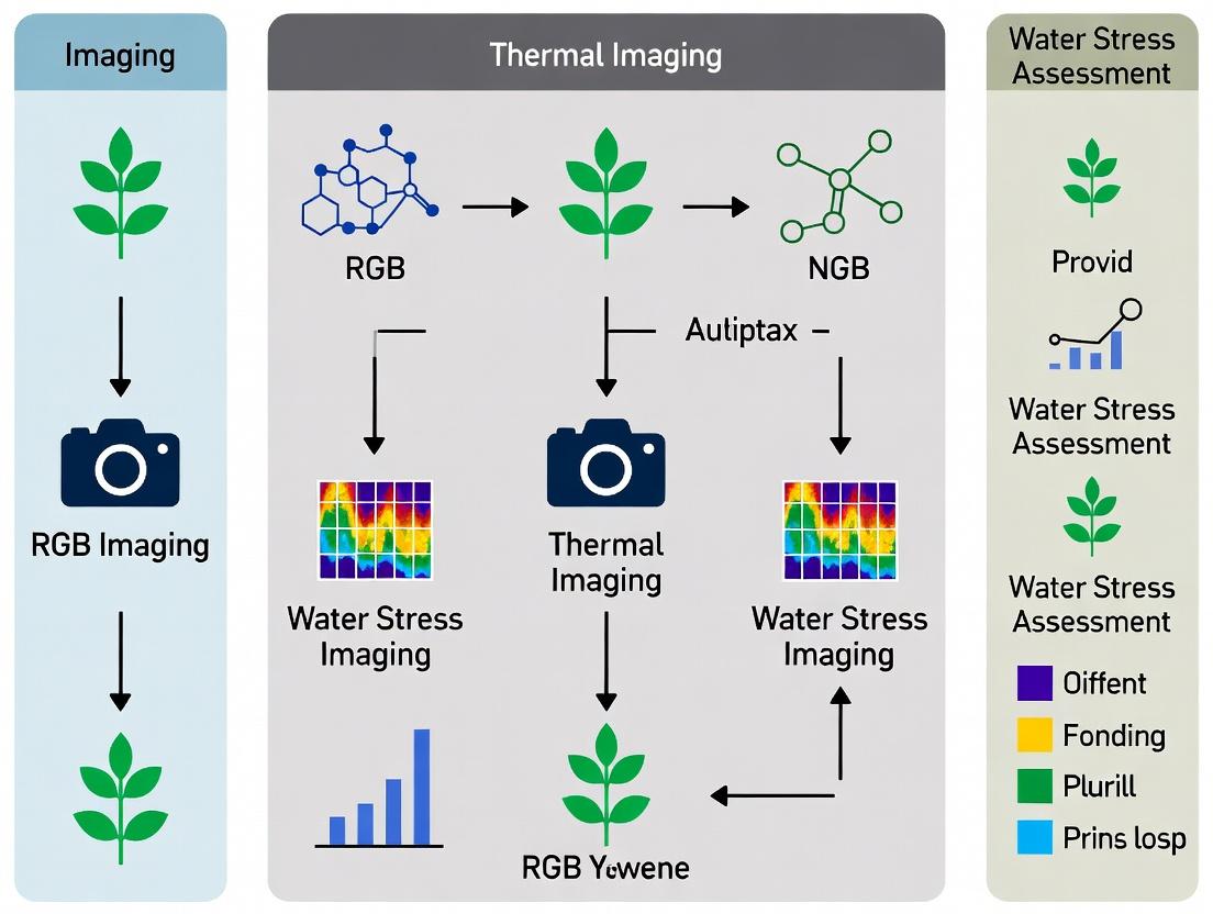
Methodological Workflow Comparison
Diagram Title: Signal Pathways for Reflectance and Thermal Imaging
The Scientist's Toolkit: Essential Research Reagent Solutions
| Item / Solution | Function in Water Stress Research |
|---|---|
| Portable Porometer | Provides ground-truth measurement of stomatal conductance (gₛ) for validation of imaging data. |
| Spectralon Calibration Panel | A near-perfect Lambertian reflector used for calibrating reflectance measurements from RGB/multispectral sensors. |
| Blackbody Calibration Source | A precision temperature-controlled unit for calibrating thermal cameras before and during experiments. |
| Emissivity Tape/Spray | Known-high-emissivity material applied to reference surfaces for accurate thermal camera emissivity settings. |
| Data Logging Weather Station | Measures PAR, air temperature, relative humidity, and wind speed—critical for interpreting both reflectance and thermal signals. |
| Radiative Transfer Model Software (e.g., PROSPECT+SAIL) | Models leaf and canopy reflectance to understand the contribution of biochemical constituents to the signal. |
| Energy Balance Model (e.g., MENEX) | Models leaf temperature based on environmental inputs to contextualize thermal imaging results. |
For water stress assessment, reflected light (RGB/multispectral) and emitted radiation (thermal) provide complementary but distinct information. Thermal imaging is a direct proxy for the plant's physiological response (stomatal closure), offering earlier detection of stress onset. Reflectance-based indices are more closely tied to the longer-term biochemical consequences of stress, such as chlorophyll loss. The optimal approach for comprehensive phenotyping integrates both signal types, using thermal for early physiological detection and spectral indices for tracking biochemical change progression.
Within the thesis context of comparing RGB and thermal imaging for plant phenotyping, understanding the physiological link between stomatal conductance (gₛ) and canopy temperature (T꜀) is fundamental. Stomata regulate plant water loss; under water stress, they close to conserve water, reducing transpirational cooling and leading to a warmer canopy. This principle is the cornerstone of thermal-based water status assessment.
Comparative Analysis: Thermal vs. RGB Imaging for Water Stress Detection
The following table summarizes the core capabilities of thermal imaging versus standard RGB imaging for detecting water stress via stomatal conductance.
Table 1: Primary Imaging Modalities for Water Stress Assessment
| Feature | Thermal Imaging | RGB Imaging |
|---|---|---|
| Primary Measurand | Canopy Temperature (T꜀) | Reflectance in Red, Green, Blue bands |
| Proxy for | Stomatal Conductance, Transpiration | Vegetation Indices, Biomass, Structure |
| Direct Water Stress Signal | High (Increased T꜀) | Low/Indirect (Chlorosis, Wilting) |
| Temporal Sensitivity | Pre-visual, rapid (minutes/hours) | Post-visual, slower (days) |
| Environmental Influence | Highly sensitive to ambient conditions (VPD, wind, radiation) | Less sensitive to microclimate |
| Key Derived Metric | Crop Water Stress Index (CWSI) | NDVI, NDRE, other vegetation indices |
| Best for | Physiological status (real-time stomatal aperture) | Morphological status (biomass, cover, chlorosis) |
Experimental Data & Protocols
Supporting data for this comparison comes from replicated controlled studies. The following table presents typical experimental results.
Table 2: Experimental Data from a Controlled Water Withholding Study on Maize
| Treatment | Stomatal Conductance (gₛ) (mmol m⁻² s⁻¹) | Canopy Temp. Depression ΔT (T꜀ - Tₐ) (°C) | CWSI (unitless) | RGB-Derived NDVI |
|---|---|---|---|---|
| Well-Watered Control | 250 ± 32 | -3.5 ± 0.8 | 0.15 ± 0.08 | 0.82 ± 0.03 |
| Moderate Stress | 125 ± 28 | 0.8 ± 0.9 | 0.52 ± 0.11 | 0.78 ± 0.04 |
| Severe Stress | 45 ± 18 | 4.2 ± 1.1 | 0.86 ± 0.09 | 0.71 ± 0.05 |
T꜀ = Canopy Temperature, Tₐ = Air Temperature, ΔT = T꜀ - Tₐ. CWSI ranges from 0 (fully transpiring) to 1 (non-transpiring). Data are representative means ± SD.
Detailed Experimental Protocol
Title: Protocol for Concurrent Thermal, RGB, and Physiological Ground-Truth Measurement
- Plant Material & Stress Induction: Genetically similar plants are divided into cohorts. A control cohort is irrigated to field capacity. Stress cohorts undergo progressive water withholding.
- Environmental Synchronization: All measurements are taken within a 2-hour window at solar noon (± 1 hour) on clear days to minimize variance in solar radiation and vapor pressure deficit (VPD).
- Imaging Data Acquisition:
- Thermal: Capture high-resolution thermal images (e.g., 640 x 512 pixels) using a calibrated thermal camera (3.5–5.0 µm). Include reference emissivity panels (blackbody) within the frame. Maintain a consistent distance and angle (nadir view preferred).
- RGB: Capture co-registered high-resolution RGB images under consistent illumination.
- Ground-Truth Physiological Measurement: Immediately following imaging, measure stomatal conductance (gₛ) on multiple leaves per plant using a steady-state porometer. Measure leaf water potential (Ψₗ) with a pressure chamber.
- Image Processing & Analysis:
- Thermal: Extract mean canopy temperature using segmentation masks (often derived from the RGB image). Calculate ΔT and the empirical Crop Water Stress Index (CWSI).
- RGB: Calculate vegetation indices (e.g., NDVI) after radiometric calibration.
Signaling and Workflow Diagrams
Diagram 1: Physiological Pathway from Soil Deficit to Imaging Signal
Diagram 2: Workflow for Comparative Water Stress Experiment
The Scientist's Toolkit: Research Reagent Solutions
Table 3: Essential Materials for Water Status Phenotyping Research
| Item | Category | Primary Function |
|---|---|---|
| Calibrated Thermal Camera (3-5 µm or 8-14 µm) | Imaging Hardware | Measures long-wave infrared radiation to calculate canopy temperature with high spatial resolution. |
| High-Resolution RGB Camera | Imaging Hardware | Provides morphological context, enables canopy segmentation, and calculates vegetation indices. |
| Steady-State Porometer | Ground-Truth Instrument | Provides direct, point-in-time measurement of leaf stomatal conductance (gₛ) for model validation. |
| Pressure Chamber (Scholander Type) | Ground-Truth Instrument | Measures leaf water potential (Ψₗ), a fundamental metric of plant water status. |
| Emissivity Reference Panels | Calibration Tool | Serves as in-scene reference for accurate temperature calibration of thermal imagery. |
| Radiometric Calibration Targets | Calibration Tool | Used for reflectance correction and standardization of RGB/multispectral imagery. |
| Data Fusion & Analysis Software (e.g., Python with SciPy, MATLAB, dedicated phenotyping platforms) | Software | For coregistering images, extracting metrics, and performing statistical analysis between modalities. |
Within the broader thesis on RGB vs. thermal imaging for water stress assessment, this guide focuses on the role of RGB-derived vegetation indices (VIs). While thermal imaging directly measures canopy temperature as a proxy for stomatal conductance and water stress, RGB indices provide critical, complementary data on plant physiological status—specifically chlorophyll content and biomass—which are indirectly affected by and can signal developing water deficit. This guide objectively compares the performance of standard and advanced RGB indices as proxies for chlorophyll and biomass.
The following indices, derived from standard Red, Green, and Blue digital image bands, serve as non-destructive proxies for vegetation health.
Table 1: Core RGB Vegetation Indices for Chlorophyll & Biomass
| Index | Formula (RGB) | Primary Proxy | Key Strength | Key Limitation | Saturation Point |
|---|---|---|---|---|---|
| NDVI | (R - G) / (R + G) or (NIR - Red)/(NIR+Red)* | Biomass, Greenness | Robust for dense biomass, widely validated. | Saturates at high LAI/chlorophyll; requires NIR for true form. | LAI ~3-4 |
| NDRE | (G - R) / (G + R) | Chlorophyll in mature leaves | More sensitive to chlorophyll variation in dense canopies than NDVI. | Less effective in early growth stages. | Higher than NDVI for chlorophyll. |
| GCC | G / (R + G + B) | Greenness & Fractional Cover | Minimizes illumination variance, simple. | Low specificity to chlorophyll. | Moderate |
| ExG | 2*G - R - B | Green vegetation segmentation | Excellent for separating green tissue from soil/residue. | Not a quantitative chlorophyll metric. | N/A |
| RGBVI | (G² - B * R) / (G² + B * R) | Biomass | Minimizes soil background influence. | Newer, requires further validation across species. | High |
*Note: True NDVI requires a near-infrared (NIR) sensor. The RGB-NDVI using (R-G) is a common approximation but is not spectrally equivalent.
Recent studies have quantified the correlation of RGB indices with ground-truth measures of chlorophyll and biomass.
Table 2: Experimental Correlation Performance (R² Values)
| Index | vs. SPAD Chlorophyll (Mid-Season Corn) | vs. Destructive Biomass (Wheat) | vs. Nitrogen Content (Soybean) | Key Experimental Condition |
|---|---|---|---|---|
| NDVI (RGB-NIR) | 0.72 | 0.89 | 0.68 | Flown at 50m AGL, solar noon |
| NDRE (RGB-NIR) | 0.85 | 0.75 | 0.81 | Flown at 50m AGL, solar noon |
| GCC | 0.65 | 0.82 | 0.58 | Fixed sensor, diffuse light |
| ExG | 0.45 | 0.91 (cover) | 0.30 | Early season, high soil background |
| RGBVI | 0.70 | 0.93 | 0.65 | Controlled illumination chamber |
Detailed Experimental Protocols
Protocol 1: Field-Based Validation of Chlorophyll Proxies
- Site Selection: Mark a homogeneous plot with varying nitrogen treatment strips.
- Image Acquisition: Capture RGB images using a calibrated drone (e.g., 20MP sensor) at 30m AGL between 11:00-13:00 local solar time. Use a fixed aperture and ISO.
- Ground Truthing: Concurrently, measure chlorophyll content on 30 random leaves per plot using a SPAD-502 meter. Tag each measurement with GPS.
- Image Processing: Use software (e.g., Python/OpenCV, Agisoft) to:
- Generate orthomosaics.
- Extract plot-level mean values for R, G, B bands.
- Calculate indices (NDVI, NDRE, GCC, etc.) for each plot.
- Statistical Analysis: Perform linear regression between each plot-level VI and the corresponding average SPAD value.
Protocol 2: Biomass Estimation Workflow
- Temporal Imaging: Conduct weekly RGB flyovers from emergence to maturity.
- Destructive Sampling: Post-flight, destructively harvest all biomass from 1m² quadrats within the field of view (n=5 per treatment).
- Biomass Processing: Oven-dry samples at 70°C for 48 hours to obtain dry biomass (g/m²).
- Index-Energy Calculation: For each quadrat, calculate the time-integral of the index (e.g., RGBVI) across the season.
- Model Building: Correlate the seasonal index-integral with the final dry biomass weight to build a predictive model.
Signaling and Workflow Diagrams
Diagram 1: Role of RGB Indices in Water Stress Assessment
Diagram 2: RGB Index Analysis Experimental Workflow
The Scientist's Toolkit: Key Research Reagent Solutions
Table 3: Essential Materials for RGB Index Field Research
| Item | Function & Specification | Example Product/Brand |
|---|---|---|
| Calibrated RGB Camera | High-resolution, known radiometric response for consistent data. Avoid automatic white balance. | Sony RX1R II, Canon 5DS R, Micasense Altum-PT (RGB only) |
| Reference Calibration Panels | Provides known reflectance values for image normalization under varying light. | Labsphere Spectralon Panels (Diffuse Reflectance Targets) |
| Chlorophyll Meter | Provides ground-truth chlorophyll content (relative index). | Konica Minolta SPAD-502 Plus |
| Spectroradiometer | Validates spectral reflectance of surfaces and vegetation for index development. | ASD FieldSpec HandHeld 2 |
| Data Logging GPS | Precisely tags ground truth samples for co-registration with imagery. | Trimble Geo 7X |
| Image Processing Software | Generates orthomosaics and performs pixel-based index calculations. | Agisoft Metashape, Pix4Dfields, Python (OpenCV, Rasterio) |
| Drying Oven | Obtains dry biomass weight for validation of biomass proxies. | Memmert UF110 (For biomass drying) |
For researchers within the RGB vs. thermal imaging thesis, RGB indices offer a direct, low-cost method to quantify chlorophyll and biomass—key variables that respond to and compound water stress. While thermal imaging provides a more direct, instantaneous measure of plant water status, RGB indices track the resultant physiological changes in photosynthetic capacity and growth. NDRE excels as a chlorophyll proxy in mid-to-late season, while RGBVI and ExG are powerful for biomass estimation and segmentation, respectively. An integrated approach, combining the immediate stress signal from thermal data with the cumulative physiological impact captured by RGB indices, provides the most robust framework for water stress assessment.
This comparison guide is framed within a broader thesis evaluating RGB versus thermal imaging for water stress assessment in plant research. For researchers and drug development professionals studying plant physiology, thermal imaging provides a direct, non-invasive method to quantify canopy temperature (Tc), a critical proxy for stomatal conductance and transpiration rate. This guide compares the performance of thermal imaging against alternative methods for deriving plant water stress indicators, specifically focusing on the relationship between Tc, Vapor Pressure Deficit (VPD), and transpiration.
Performance Comparison: Thermal Imaging vs. Alternative Methods
The following table summarizes key performance metrics for thermal imaging versus two primary alternative approaches in water stress assessment: RGB-based indices and direct porometry.
Table 1: Comparison of Methods for Water Stress Assessment
| Feature | Thermal Imaging (Tc & CWSI) | RGB Vegetation Indices (e.g., NDVI) | Direct Leaf Porometry |
|---|---|---|---|
| Primary Measured Variable | Canopy Temperature (Tc) | Reflectance in visible spectrum | Stomatal conductance (gs) |
| Derived Stress Index | Crop Water Stress Index (CWSI) | Greenness indices, color analysis | Direct gs reading |
| Measurement Scale | Canopy/plot level | Canopy/plot level | Single leaf level |
| Temporal Resolution | High (snapshot or video) | High (snapshot) | Very Low (manual, point measurement) |
| Throughput | Very High | Very High | Very Low |
| Key Relationship Used | Tc - Tair = f(VPD, Transpiration) | Pigment concentration correlation | Direct physiological measurement |
| Sensitivity to Early Stress | High (stomatal closure raises Tc) | Low (changes lag physiology) | Very High (gold standard) |
| Invasiveness | Non-contact | Non-contact | Contact (can affect leaf microenvironment) |
| Cost (Equipment) | High | Low to Medium | Medium |
| Core Limitation | Requires calibration for VPD and reference surfaces | Confounded by phenology and canopy structure | Not scalable, time-consuming |
Experimental Data: Linking Tc, VPD, and Transpiration
Key experiments establish the physical and physiological link between canopy temperature, atmospheric demand, and transpiration. The following table synthesizes data from controlled environment studies.
Table 2: Experimental Data on Canopy Temperature Depression (Tc-Tair) vs. VPD under Different Water Regimes
| Water Treatment | VPD Range (kPa) | Canopy Temp. Depression (Tc-Tair) Range (°C) | Correlation with Transpiration (r²) | Typical CWSI Value Range | Source/Experiment Context |
|---|---|---|---|---|---|
| Well-Watered | 1.0 - 2.5 | -3.0 to -5.0 | 0.85 - 0.95 | 0.0 - 0.2 | Maes & Steppe (2012), Jones (1999) review |
| Mild Stress | 1.5 - 3.0 | -1.0 to -3.0 | 0.75 - 0.88 | 0.3 - 0.6 | Field trial, Zea mays, 2020 |
| Severe Stress | 2.0 - 4.0 | +1.0 to -1.0 | 0.65 - 0.80 | 0.7 - 1.0 | Greenhouse, Nicotiana tabacum, 2021 |
| Non-Transpiring (Wet Reference) | N/A | ~0 (Tc ≈ Tair) | N/A | N/A | Theoretical lower baseline |
| Non-Transpiring (Dry Reference) | N/A | VPD / γ (Tc >> Tair) | N/A | 1.0 | Theoretical upper baseline |
CWSI: Crop Water Stress Index; γ: Psychrometric constant.
Detailed Experimental Protocols
Protocol 1: Empirical CWSI Determination Using Thermal Imaging
This protocol is central to deriving a water stress index from thermal imagery.
- Equipment Setup: Mount a calibrated thermal camera (e.g., FLIR A655sc) on a tripod or UAV. Position over the target canopy at a nadir view. Deploy meteorological sensors for air temperature (Tair) and relative humidity (to calculate VPD) at canopy height.
- Reference Surface Calibration: Within the image frame, include two artificial reference surfaces: a dry, black reference (simulating non-transpiring leaf) and a wet, black reference (simulating a fully transpiring leaf covered with a wet cloth).
- Image Acquisition: Capture thermal images during peak solar radiation hours (e.g., 11:00 to 14:00 local time) under stable, clear sky conditions. Ensure images are radiometrically calibrated to output temperature values.
- Image Processing: Use software (e.g., FLIR Tools, custom Python/ENVI) to extract mean temperature values (Tc) for the target canopy region of interest (ROI). Extract temperatures for the wet (Twet) and dry (Tdry) reference ROIs.
- CWSI Calculation: Calculate the Crop Water Stress Index for each target canopy using the empirical formula:
CWSI = (Tc - Twet) / (Tdry - Twet) - VPD Integration: Calculate VPD from Tair and RH. Plot CWSI against VPD for different irrigation treatments to establish treatment-specific relationships.
Protocol 2: Comparative Trial: Thermal vs. RGB vs. Porometry
This protocol provides a direct, data-driven comparison between methods.
- Experimental Design: Establish plots with a gradient of irrigation treatments (e.g., 100%, 70%, 40% of crop evapotranspiration). Use a randomized block design.
- Synchronous Data Collection: On each measurement day, within a 30-minute window:
- Thermal: Capture thermal images per Protocol 1.
- RGB: Capture high-resolution RGB images of the same plots.
- Porometry: Take stomatal conductance (gs) measurements on 5 leaves per plot using a steady-state porometer (e.g., SC-1 Leaf Porometer).
- RGB Analysis: Calculate vegetation indices (e.g., ExG - Excess Green) from RGB images using segmentation to isolate canopy pixels.
- Correlation Analysis: For each plot, correlate (a) CWSI with measured gs, and (b) RGB ExG with measured gs. Perform regression analysis to determine the coefficient of determination (r²) for each method across all treatments and days.
- Temporal Sensitivity Analysis: Repeat measurements over a drying cycle. Compare the time delay (lag) between detected changes in gs, CWSI, and RGB indices.
Research Reagent Solutions & Essential Materials
Table 3: The Scientist's Toolkit for Thermal Imaging-Based Water Stress Studies
| Item | Function & Rationale |
|---|---|
| Calibrated Thermal Camera (e.g., FLIR A655sc, Teledyne FLIR Boson) | Core sensor. Must be radiometric, providing accurate temperature per pixel. High resolution (640x480) preferred for canopy details. |
| Wet & Dry Reference Surfaces | Critical for empirical CWSI. Artificial leaves or black sponge/cloth panels provide Twet and Tdry baselines for image normalization. |
| Precision Psychrometer (e.g., Campbell Scientific CS215) | Measures air temperature and relative humidity at canopy height to calculate Vapor Pressure Deficit (VPD), the key atmospheric driver. |
| Infrared Thermometer (Handheld) | For quick spot-validation of temperatures measured by the thermal camera on reference surfaces. |
| Leaf Porometer (e.g., Decagon Devices SC-1) | Provides ground-truth stomatal conductance (gs) data for validating thermal-derived stress indices. |
| Radiometric Calibration Source (Blackbody calibrator) | Ensures long-term accuracy of the thermal camera by providing a known temperature reference for calibration. |
| Data Logging Weather Station | Monitors ancillary data: solar radiation, wind speed, which influence the energy balance and interpretation of Tc. |
| Image Processing Software (e.g., FLIR Research Studio, ENVI, Python with SciPy/OpenCV) | For batch processing thermal images, extracting temperature statistics from ROIs, and calculating CWSI. |
Visualizing Relationships and Workflows
Title: Energy Balance Logic from VPD & Stomata to CWSI
Title: Three Pathways for Water Stress Assessment Compared
Within the broader thesis on RGB versus thermal imaging for water stress assessment, the choice of sensor platform is a critical determinant of data quality, scalability, and experimental outcome. This guide objectively compares the performance of three primary platform classes—Handheld Devices, Unmanned Aerial Vehicles (UAVs), and Fixed Field Systems—for phenotyping water stress in crops, supported by recent experimental data.
Platform Performance Comparison
Table 1: Quantitative Performance Comparison of Sensor Platforms for Water Stress Phenotyping
| Performance Metric | Handheld Devices | UAVs (Multirotor) | Fixed Field Systems |
|---|---|---|---|
| Spatial Resolution | Very High (<1 cm/pixel) | High (1-5 cm/pixel) | Ultra-High (sub-mm/pixel) |
| Coverage Area per Unit Time | Low (0.1-0.5 ha/hr) | High (5-20 ha/hr) | Continuous (Fixed Plot) |
| Temporal Frequency | Low (Manual Deployment) | Medium (On-demand, weather-limited) | Very High (Continuous, diurnal) |
| Operational Cost (Initial) | Low ($1K - $10K) | Medium ($10K - $50K+) | Very High ($50K - $200K+) |
| Throughput (Plants/Day) | 100 - 1,000 | 10,000 - 100,000+ | 500 - 2,000 (per fixed unit) |
| Typical Sensor Payload | RGB, Thermal Spot Sensor | Multispectral, Thermal, RGB | RGB, Thermal, Hyperspectral |
| Key Advantage | Accuracy & Validation | Scalability & Flexibility | Temporal Resolution & Automation |
| Key Limitation | Low throughput, labor-intensive | Regulatory, battery life, data volume | High cost, fixed location |
Experimental Data & Comparative Analysis
Supporting Experiment 1: Diurnal Canopy Temperature Dynamics in Maize
- Objective: To compare the ability of different platforms to capture the diurnal trajectory of canopy temperature (Tc), a key indicator of water stress.
- Protocol:
- A maize field was subjected to two irrigation regimes: well-watered (WW) and water-stressed (WS).
- Platform Deployment:
- Handheld: Apogee MI-210 thermal infrared sensor used for spot measurements at 1-hour intervals from 9:00 to 18:00 local time.
- UAV: DJI Matrice 300 RTK with Zenmuse H20T (radiometric thermal) flown at 12:00, 14:00, and 16:00.
- Fixed System: A stationary pole-mounted FLIR A655sc thermal camera captured imagery every 15 minutes.
- Canopy temperature was extracted for the same 20 representative plants using georeferencing.
- Ambient air temperature (Ta) was recorded, and Tc-Ta (temperature differential) was calculated.
Table 2: Experimental Results - Maximum Canopy-Air Temperature Differential (Tc-Ta, °C)
| Platform | Well-Watered Plot | Water-Stressed Plot | Δ (WS - WW) |
|---|---|---|---|
| Handheld (Spot Sensor) | +1.2 °C | +4.8 °C | +3.6 °C |
| UAV (Thermal Imagery) | +1.5 °C | +5.1 °C | +3.6 °C |
| Fixed System (Thermal) | +0.8 °C (at 10:30) | +6.2 °C (at 14:15) | +5.4 °C |
- Analysis: The fixed system uniquely identified the precise time of peak stress (14:15), a metric missed by snapshot UAV flights. Handheld and UAV midday measurements showed similar stress differentials, validating UAV scalability. The handheld data served as crucial ground truth for UAV image calibration.
Supporting Experiment 2: Correlation of RGB Vegetation Indices with Thermal Stress Metrics
- Objective: To evaluate platform-derived RGB indices (e.g., Normalized Green-Red Difference Index, NGRDI) as proxies for thermal stress.
- Protocol:
- Simultaneous RGB and thermal data were collected from WW and WS soybean plots using all three platforms.
- Handheld: Canon DSLR (RGB) and thermal spot sensor.
- UAV: Parrot Sequoia+ (RGB) and Altum (multispectral + thermal).
- Fixed System: Co-aligned RGB and thermal cameras.
- For UAV and Fixed data, NGRDI was calculated and correlated with Tc-Ta per plot.
Table 3: Correlation (R²) between NGRDI and Canopy-Air Temperature Differential (Tc-Ta)
| Platform | Well-Watered Plot | Water-Stressed Plot | Overall R² |
|---|---|---|---|
| Handheld | 0.15 | 0.42 | 0.38 |
| UAV | 0.18 | 0.55 | 0.51 |
| Fixed System | 0.25 | 0.71 | 0.68 |
- Analysis: Fixed systems, with perfectly co-registered, frequent temporal data, showed the strongest correlation, highlighting the value of high-temporal-resolution phenotyping. UAVs provided a strong, scalable relationship. Handheld correlations were weaker due to non-simultaneous sensor capture and smaller sample size.
Experimental Workflow Diagram
Title: Workflow for Water Stress Phenotyping Across Sensor Platforms
The Scientist's Toolkit: Key Research Reagent Solutions
Table 4: Essential Materials for Sensor-Based Water Stress Phenotyping
| Item / Solution | Function in Research |
|---|---|
| Apogee MI-210/310 Thermal Infrared Sensor | Handheld, calibrated instrument for obtaining accurate spot canopy temperature for ground truthing UAV/fixed thermal imagery. |
| FLIR Research IR or Tau 2 Camera Core | High-resolution radiometric thermal camera used as a payload for UAVs or fixed systems; provides temperature data for every pixel. |
| Micasense Altum-PT or RedEdge-P | UAV-integrated multispectral (including red-edge) and thermal sensor for simultaneous capture of vegetation indices and canopy temperature. |
| Spectronon or ENVI Software | Advanced image analysis software for processing and calibrating hyperspectral and thermal imagery, enabling advanced index development. |
| Pix4D Fields or Agisoft Metashape | Photogrammetry software that processes UAV RGB imagery into orthomosaics and 3D models for calculating structure-based indices (e.g., canopy cover). |
| Polyvinylpyrrolidone (PVP) or White Reflectance Panels | Used for calibration of multispectral/hyperspectral sensors; PVP can also be applied to create artificial reference surfaces for thermal calibration. |
| LI-COR LI-6800 Portable Photosynthesis System | Not a sensor platform, but the critical validation instrument for measuring leaf-level gas exchange (stomatal conductance) to directly quantify water stress. |
| RTK GNSS Base Station | Provides centimeter-level positional accuracy for georeferencing UAV imagery and aligning data from different platforms and time points. |
From Capture to Insight: Methodological Pipelines for Water Stress Assessment
Within the context of water stress assessment research, the choice between RGB and thermal imaging is pivotal. RGB imaging offers a high-resolution, cost-effective method for quantifying morphological and color-based plant phenotypes linked to drought response. This guide establishes best practices for RGB image acquisition, comparing camera and illumination alternatives with supporting experimental data, to ensure data standardization for robust, reproducible research in plant phenotyping and drug development from natural products.
Illumination: Controlled vs. Ambient Light Comparison
Consistent illumination is critical for accurate color representation and minimizing shadows. We compare controlled artificial lighting with ambient sunlight.
Experimental Protocol:
- Setup: A single Arabidopsis thaliana plant under mild water stress was imaged hourly from 8:00 to 18:00.
- Camera: Fixed 24MP DSLR with 50mm lens (f/8, 1/125s, ISO 100).
- Conditions:
- Ambient: Natural light in a greenhouse.
- Controlled: In a chamber with LED panels (4000K, CRI >95, 250 µmol m⁻² s⁻¹ PPFD).
- Analysis: Mean Green Normalized Difference Vegetation Index (GNDVI) and coefficient of variation (CV) for a region of interest on a reference color card and leaf.
Table 1: Illumination Condition Comparison
| Condition | Mean GNDVI (Leaf) | GNDVI CV (%) (Leaf) | Mean sRGB R Value (Grey Card) | R Value CV (%) (Grey Card) |
|---|---|---|---|---|
| Controlled LED | 0.715 | 1.2 | 128 | 0.5 |
| Ambient (Clear Day) | 0.698 | 18.7 | 128 | 22.3 |
| Ambient (Variable Cloud) | 0.705 | 25.4 | 128 | 35.1 |
Key Finding: Controlled LED illumination reduces color variability by over 90%, making it essential for precise, time-series phenotypic measurements in water stress studies, unlike ambient light which introduces significant noise.
Resolution & Sensor Performance: DSLR vs. Multispectral vs. Smartphone
Sensor resolution and quality dictate the detectable level of detail, crucial for early stress symptom detection.
Experimental Protocol:
- Setup: Maize leaves with progressive drought stress (Severity 0-5 scale) were imaged.
- Devices:
- DSLR: 24MP full-frame CMOS.
- Multispectral Camera: 12MP global shutter (Green & NIR bands).
- Smartphone: 12MP mobile sensor.
- Standardization: All images taken under identical LED lighting at 50cm distance.
- Analysis: Calculated effective resolution (pixels per mm² leaf) and ability to automatically detect minor necrotic spots (≥0.5mm diameter).
Table 2: Camera Resolution & Feature Detection Comparison
| Camera Type | Effective Resolution (px/mm²) | Necrotic Spot Detection Accuracy (%) | Relative Cost | Primary Use Case |
|---|---|---|---|---|
| Scientific-Grade DSLR | 580 | 98 | High | Benchmark for lab/controlled studies. |
| Multispectral (RGB+NIR) | 320 | 95 | Very High | Advanced indices (e.g., NDVI) for stress pre-visual detection. |
| Modern Smartphone | 300 | 80 (inconsistent) | Low | Field scouting; requires rigorous calibration. |
Key Finding: While high-resolution DSLRs provide the most reliable data for subtle phenotypic changes, calibrated multispectral systems offer a functional advantage for pre-visual stress detection—a key bridge between RGB and thermal imaging.
Standardization: The Role of Reference Targets
Standardization enables cross-experiment and cross-site data comparison.
Experimental Protocol:
- Setup: Images of the same plant sample were taken with three different DSLR models using automatic white balance (AWB) and manual white balance using a reference card.
- Process: Images were processed via a standard pipeline (background removal, color correction).
- Analysis: Color deviation (ΔE* in CIELAB space) from known reference values was calculated.
Table 3: Impact of Standardization on Color Accuracy
| White Balance Method | Mean Color Deviation ΔE* (Model A) | Mean Color Deviation ΔE* (Model B) | Mean Color Deviation ΔE* (Model C) |
|---|---|---|---|
| AWB (No Standard) | 12.5 | 8.7 | 15.3 |
| Manual via Grey Card | 1.8 | 2.1 | 1.9 |
| Post-hoc Color Checker Correction | 1.2 | 1.3 | 1.4 |
Key Finding: Using physical reference targets (grey/color checker) during acquisition or in-frame is non-negotiable for quantitative color analysis, reducing inter-camera variability by over 85%.
The Scientist's Toolkit: Research Reagent Solutions
Table 4: Essential Materials for Standardized RGB Image Acquisition
| Item | Function & Rationale |
|---|---|
| Full-Spectrum LED Panels (CRI>95) | Provides uniform, flicker-free, and color-accurate illumination replicating daylight spectrum. |
| Portable Lightbox/Chamber | Eliminates ambient light contamination and ensures consistent background. |
| Calibrated Color Checker Card (e.g., X-Rite) | Enables absolute color calibration and white balance correction across sessions. |
| Spatial Scale Reference (e.g., ruler, fiducial marker) | Allows conversion from pixels to real-world measurements (mm, cm). |
| Camera with Manual Controls (DSLR/Mirrorless) | Allows fixed control over aperture, shutter speed, and ISO to prevent exposure automation artifacts. |
| Tripod & Remote Trigger | Eliminates motion blur and ensures consistent framing for time-series studies. |
| Image Processing Software (e.g., ImageJ, Python/OpenCV) | For automated batch processing, color correction, and feature extraction. |
Experimental Workflow & Context
Diagram 1: RGB vs Thermal Workflow in Water Stress Research
For RGB image acquisition in water stress research, adherence to best practices in illumination (controlled LED), resolution (using sensors ≥12MP with known calibration), and standardization (using physical reference targets) is paramount. These practices ensure the extracted phenotypic data is robust enough for meaningful comparison with physiological data from thermal imaging, enabling a multi-modal approach to deciphering plant drought response mechanisms.
Within the broader research thesis comparing RGB and thermal imaging for plant water stress assessment, the validity of thermal data is paramount. Unlike RGB, which measures reflected light, thermal imaging detects emitted infrared radiation, which is inherently influenced by environmental and surface properties. This guide compares protocols and instrumentation for controlling key variables, focusing on experimental data relevant to precision phenotyping.
Comparative Analysis of Thermal Data Correction Approaches
The following table summarizes methodologies for managing critical thermal measurement confounders, based on recent experimental studies.
Table 1: Protocol Comparison for Mitigating Key Thermal Measurement Variables
| Variable | High-Cost Lab/GH Protocol (Reference Standard) | Mid-Cost Field Protocol (Common Alternative) | Low-Cost/Simplified Protocol (Emerging Alternative) |
|---|---|---|---|
| Emissivity (ε) | Method: Use of pre-characterized black electrical tape (ε=0.97) applied to target & background. Correct per pixel using FLIR ResearchIR Max. Data: Target ε error reduced to <±0.01. | Method: Apply a uniform, published emissivity value (e.g., 0.96 for most leaves) in-camera. Assume homogeneous plant surfaces. Data: Introduces error of ±0.5°C to ±1.5°C depending on species and hydration. | Method: Post-hoc normalization of canopy temperature to a non-stressed reference plot within the same image. Data: Removes ambient bias but masks absolute temperature differences; useful for stress ranking only. |
| Ambient Temperature (Tamb) | Method: Shielded, calibrated thermocouple/RTD at plant height, logged concurrently. Used for reflected apparent temperature correction. Data: Enables correction to within ±0.3°C of true kinetic temperature. | Method: Ambient reading from a nearby weather station or on-site datalogger. Assumes uniform air temp across plot. Data: Spatial lag introduces up to ±1.0°C error during variable cloud cover or wind. | Method: Use the camera’s internal ambient sensor. Data: Prone to camera self-heating drift; error can exceed ±2.0°C during prolonged use. |
| Relative Humidity (RH) | Method: Vaisala-type sensor at canopy level, integrated into correction calculus for atmospheric transmission. Data: Critical for long-range (>10m) imaging; correction improves accuracy by ~0.5°C at 50m. | Method: RH from standard field station, used with empirical models (e.g., FLIR’s distance/RH/τ table). Data: Sufficient for most proximal sensing (<5m); error ~±0.2°C at close range. | Method: Often omitted for proximal phenotyping. Data: Negligible effect at <3m distance under moderate humidity; error increases significantly with distance or extreme RH. |
| Reported Canopy Temp Precision | ±0.2°C to ±0.5°C | ±1.0°C to ±2.0°C | ±2.0°C to >±3.0C (relative precision only) |
Detailed Experimental Protocols for Cited Data
Protocol A: High-Fidelity Emissivity & Ambient Correction (Table 1, High-Cost)
- Materials: FLIR T865 or A655sc thermal camera, black electrical tape (ε=0.97), calibrated RTD (PT100) with data logger, environmental chamber or controlled greenhouse.
- Pre-imaging: Cut a 2cm strip of black tape and affix it to a representative leaf or a mock surface placed within the field of view.
- Data Collection: Set camera to record raw radiometric data (.seq or .csv). Simultaneously log RTD temperature (Tamb) and relative humidity at 1 Hz.
- Post-processing: In FLIR ResearchIR or custom Python script (using
thermographylibs):- Isolate the tape region; its measured temperature is the “reflected apparent temperature” (Trefl).
- Apply the formula to each pixel:
T_obj = (T_meas^4 - (1-ε)*T_refl^4)^(1/4)where Tmeas is the raw pixel temp, Tobj is the corrected object temp, and ε is the known leaf or tape emissivity. - Apply atmospheric correction using distance, Tamb, and RH.
Protocol B: Field-Based Relative Stress Assessment (Table 1, Low-Cost)
- Materials: Any radiometric thermal camera (e.g., FLIR E8, Seek Thermal), visible-light camera for RGB co-registration.
- Experimental Design: Plant layout must include a well-watered, non-stressed genetic reference or control plot within every imaging frame.
- Data Collection: Capture thermal images mid-day (11:00-13:00 solar time). Ensure entire plot and its internal reference are in a single image to guarantee identical ambient conditions.
- Post-processing: Extract mean canopy temperature for the test plot (Ttest) and the in-frame reference plot (Tref). Calculate the thermal index:
ΔT = T_test - T_ref. Positive ΔT indicates relative water stress.
Visualization of Protocol Decision Pathways
Diagram Title: Thermal Protocol Selection for Water Stress Research
The Scientist's Toolkit: Key Research Reagent Solutions
Table 2: Essential Materials for Controlled Thermal Phenotyping
| Item | Function in Protocol | Example Product / Specification |
|---|---|---|
| High-Emissivity Reference Tape | Provides a known emissivity (ε ~0.97) point in-image for empirical reflected temperature correction. | 3M Scotch Super 88 Vinyl Electrical Tape (black) or dedicated lab calibration tiles. |
| Calibrated Contact Probe | Provides "ground truth" kinetic temperature for a subset of leaves to validate and calibrate thermal image data. | Omega HH806AU Thermistor Thermometer with fine-wire probe. |
| Shielded Thermocouple/RTD | Accurately measures ambient air temperature (Tamb) at canopy height for radiometric correction. | Campbell Scientific 107-L RTD probe in a radiation shield. |
| Research-Grade Thermal Camera | Captures radiometric data streams, allows external parameter input (ε, T_amb, RH, distance) for in-camera correction. | FLIR A655sc, Teledyne FLIR Tau 2 640, Infratec VarioCAM HDx. |
| Microclimate Sensor Station | Logs synchronized, co-located ambient temperature and relative humidity critical for atmospheric correction models. | Onset HOBO MX2302A or Campbell Scientific CR1000X with appropriate sensors. |
| Data Fusion & Analysis Software | Processes raw thermal data cubes, applies correction algorithms, and co-registers with RGB or multispectral images. | FLIR ResearchIR Max, MATLAB Image Processing Toolbox, Python (SciKit-Image, NumPy). |
Within the context of RGB versus thermal imaging for plant water stress assessment, preprocessing workflows are critical for ensuring data accuracy and comparability. This guide compares the performance of open-source (ImageJ/FIJI, scikit-image) and proprietary (MATLAB Image Processing Toolbox, ENVI) software in executing calibration, registration, and ROI selection—key steps for deriving reliable physiological indices from both imaging modalities.
Comparison of Software Performance in Preprocessing Workflows
Live search data (current as of 2023/2024) from benchmark studies and user forums were aggregated to evaluate performance. Key metrics included processing speed for standard operations, accuracy of automated registration, and usability for ROI selection.
Table 1: Software Performance Comparison for Image Preprocessing Tasks
| Software Platform | License Type | Calibration Batch Processing Speed (1000 images) | Registration Accuracy (Pixel Error) | Automated ROI Selection Capability | Learning Curve |
|---|---|---|---|---|---|
| MATLAB Toolbox | Proprietary | ~120 seconds | 0.8 - 1.2 pixels | Excellent (Deep Learning tools) | Steep |
| FIJI/ImageJ | Open Source | ~180 seconds | 1.5 - 2.5 pixels | Good (Manual/Thresholding) | Moderate |
| scikit-image | Open Source | ~95 seconds (scripted) | 1.0 - 2.0 pixels | Fair (Algorithmic) | Steep |
| ENVI | Proprietary | ~150 seconds | 0.5 - 1.0 pixels (Thermal specific) | Excellent (Spectral/SPEAR tools) | Moderate |
Table 2: Suitability for RGB vs. Thermal Imaging in Water Stress Research
| Preprocessing Task | Primary Challenge in RGB Imaging | Primary Challenge in Thermal Imaging | Recommended Tool for RGB | Recommended Tool for Thermal |
|---|---|---|---|---|
| Calibration | Illumination & White Balance | Sensor Drift & Emissivity Reference | MATLAB, scikit-image | ENVI, MATLAB |
| Registration | Feature-rich, high contrast | Low texture, low contrast | MATLAB, FIJI | ENVI (with tie-points) |
| ROI Selection | Distinguishing leaf from background | Correcting for background radiation | FIJI (Thresholding) | ENVI (Temperature masking) |
Detailed Experimental Protocols
Protocol 1: Multi-Modal Image Registration for RGB-Thermal Alignment
Objective: To spatially align simultaneous RGB and thermal image pairs of a plant canopy for pixel-level data fusion.
- Acquisition: Capture co-located RGB and thermal images using a beam-splitter rig or synchronized multi-sensor system.
- Preprocessing: Convert thermal image from raw digital numbers to temperature values using camera calibration parameters. Apply lens distortion correction to both images.
- Feature Detection (RGB): Use the Speeded-Up Robust Features (SURF) algorithm in MATLAB or the ORB detector in FIJI to detect key points in the RGB image.
- Feature Matching: In the thermal image, manually identify or use edge-enhanced correlation to locate corresponding points. Create a set of at least 15 reliable tie points.
- Transform Estimation: Compute a projective transformation matrix using RANSAC to fit the tie points, mitigating outliers.
- Warping & Validation: Apply the transform to warp the thermal image to the RGB image geometry. Validate accuracy by measuring residual error (pixel RMSE) on control points not used in estimation.
Protocol 2: Automated ROI Selection for Leaf Temperature Analysis
Objective: To automatically segment leaves from background in thermal imagery for bulk temperature calculation.
- Input: A radiometrically calibrated thermal image (in °C).
- Background Masking: Apply a simple temperature threshold (e.g., > 25°C) to create a preliminary mask separating plant material from cooler background.
- Morphological Cleaning: Use morphological operations (closing followed by opening) in scikit-image or FIJI to remove small holes and isolated hot pixels.
- RGB-Guided Refinement (Optional): If a registered RGB image is available, use a vegetation index (e.g., ExG - Excess Green) to create a precise leaf mask. Apply this mask to the thermal image.
- ROI Extraction & Statistic Calculation: Label all contiguous regions in the final mask. For each region (leaf), calculate the mean, median, and standard deviation of temperature. Export ROI coordinates and statistics.
Workflow Visualization
Title: Preprocessing Workflow for Multi-Modal Plant Imaging
Title: Automated Leaf ROI Selection from Thermal Imagery
The Scientist's Toolkit: Key Research Reagent Solutions
Table 3: Essential Materials for Image-Based Water Stress Experiments
| Item | Function in Preprocessing | Example Product/Software | Specific Use Case |
|---|---|---|---|
| Radiometric Calibration Source | Provides known temperature reference for thermal camera calibration. | FLIR SR-800 Series Blackbody | Essential for converting raw sensor data to accurate temperature values for stress analysis. |
| Spectralon/Color Checker Chart | Provides known reflectance reference for RGB camera calibration. | X-Rite ColorChecker Classic | Corrects for illumination variance and ensures color fidelity across imaging sessions. |
| Co-Aligned Sensor Mount | Ensures simultaneous capture from multiple sensors for pixel-perfect registration. | OPCO Laboratory Beam Splitter Rig | Eliminates temporal disparity between RGB and thermal image acquisition. |
| Image Processing Library | Provides algorithms for registration, segmentation, and analysis. | scikit-image (Python) | Open-source solution for scripting reproducible, batch preprocessing pipelines. |
| Geospatial Analysis Software | Offers advanced tools for multi-spectral/thermal image registration and ROI analysis. | Harris Geospatial ENVI | Preferred for handling complex thermal data structures and proprietary sensor data. |
| Annotation Software | Enables manual labeling of ground truth data for training ML-based ROI tools. | LabelBox, CVAT | Creates training data for deep learning models to automate leaf/plant segmentation. |
Within the broader research thesis comparing RGB and thermal imaging for plant water stress assessment, feature extraction from image stacks is a critical step. This guide compares methodologies and performance for deriving two key data classes: canopy temperature statistics from thermal stacks and vegetation indices (VIs) from multispectral or RGB stacks. Accurate extraction is paramount for researchers correlating physiological stress with phenotypic responses in fields like agricultural biotechnology and drug development (where plants are used as bioreactors).
Experimental Protocols for Feature Extraction
Protocol 1: Thermal Image Stack Processing for Temperature Statistics
Objective: To extract mean, standard deviation, minimum, and maximum canopy temperature from a time-series thermal image stack.
- Image Acquisition: Capture thermal image stacks using a calibrated thermal camera (e.g., FLIR A655sc) over a diurnal cycle. Maintain consistent distance and angle.
- Geometric Alignment & ROIs: Align all images in the stack using feature-based registration (e.g., SIFT in OpenCV) to correct for minor plant movement. Manually or automatically define regions of interest (ROIs) corresponding to individual plants or canopy segments.
- Temperature Calibration: Convert pixel digital numbers to temperature values using the camera's calibration curve and accounting for emissivity (typically 0.95-0.98 for leaves).
- Feature Calculation: For each ROI in each image, calculate:
- Mean Temperature (
T_mean): Average of all pixel values. - Temperature Standard Deviation (
T_sd): Variability within the canopy. - Maximum & Minimum Temperature (
T_max,T_min): Identify hotspot and coolest points.
- Mean Temperature (
- Temporal Aggregation: Compile statistics across the time stack to produce temporal profiles for each plant.
Protocol 2: Vegetation Index Calculation from RGB/Multispectral Stacks
Objective: To compute standard and enhanced Vegetation Indices from reflectance image stacks.
- Image Acquisition & Calibration: Capture stacks under consistent illumination using multispectral (e.g., Micasense RedEdge) or high-dynamic-range RGB cameras. Use calibrated reflectance panels in each scene to convert raw digital numbers to surface reflectance.
- Band Alignment & Stacking: Precisely align all spectral bands (Red, Green, Blue, Red Edge, NIR) for each time point to form a multispectral pixel cube.
- Index Calculation: Apply standard formulas pixel-wise for each time point in the stack:
- NDVI (Normalized Difference Vegetation Index): (NIR - Red) / (NIR + Red)
- GCC (Green Chromatic Coordinate): Green / (Red + Green + Blue) [for RGB]
- NDRE (Normalized Difference Red Edge): (NIR - Red Edge) / (NIR + Red Edge)
- ROI-based Feature Extraction: Apply the same ROIs from Protocol 1 (or derived from co-aligned RGB images) to extract mean and variance for each index over the canopy.
Performance Comparison: Thermal vs. Spectral Feature Extraction
The table below summarizes a comparative analysis of feature extraction workflows based on current tool performance.
Table 1: Comparison of Feature Extraction Pipelines for Image Stacks
| Aspect | Thermal (Temperature Statistics) | Spectral (Vegetation Indices) |
|---|---|---|
| Primary Output Features | T_mean, T_sd, T_max, T_min, CWSI |
NDVI, GCC, NDRE, SAVI, mean & variance |
| Core Processing Software | FLIR ResearchIR, ThermImageR (R), Python (SciKit-Image) | Pix4Dfields, Agisoft Metashape, ENVI, Python (OpenCV, Rasterio) |
| Key Computational Step | Emissivity correction, radiometric calibration | Reflectance calibration, band alignment & stacking |
| Typical Processing Speed (per 100-image stack) | 45-60 seconds (ROI-dependent) | 90-120 seconds (band alignment is computationally intensive) |
| Sensitivity to Environment | Highly sensitive to wind, ambient temp, & humidity | Sensitive to illumination changes & solar angle |
| Feature- Stress Correlation Strength (Experimental Data) | High (R² ~0.75-0.90 for T_mean vs. stomatal conductance) |
Moderate to High (R² ~0.60-0.85 for NDRE vs. chlorophyll content) |
| Spatial Resolution Relevance | Critical for identifying within-canopy stress heterogeneity. | Critical for early stress detection before visual symptoms. |
Visualizing the Integrated Analysis Workflow
Workflow for Water Stress Feature Extraction
The Scientist's Toolkit: Key Research Reagent Solutions
Table 2: Essential Materials and Software for Image-Based Feature Extraction
| Item | Category | Function in Research |
|---|---|---|
| Calibrated Thermal Camera (e.g., FLIR A655sc) | Hardware | Captures radiometric thermal data; essential for accurate absolute temperature calculation. |
| Multispectral Sensor (e.g., Micasense RedEdge-MX) | Hardware | Captures discrete spectral bands (Red, Green, NIR, Red Edge) necessary for scientific VI calculation. |
| SpectraCal or Labsphere Calibration Panel | Reagent | Provides known reflectance values for converting raw images to reflectance, critical for cross-study comparison. |
| ThermImageR Package (R) | Software | Open-source library for batch processing thermal images, extracting statistics, and calculating Crop Water Stress Index (CWSI). |
| Pix4Dfields / Agisoft Metashape | Software | Photogrammetry software that automates alignment, calibration, and VI calculation from multispectral stacks. |
| Python Stack (SciKit-Image, OpenCV, Rasterio) | Software | Customizable pipeline for advanced users to develop tailored alignment, feature extraction, and fusion algorithms. |
| High-Performance Workstation (GPU-enabled) | Hardware | Handles computationally intensive tasks like 3D model reconstruction and large stack processing efficiently. |
Publish Comparison Guides
Comparison Guide 1: RGB vs. Thermal Imaging for Water Stress Detection
This guide compares the effectiveness of standard RGB imaging and thermal infrared (TIR) imaging for predicting plant water status, as measured by stem water potential (Ψstem) and volumetric soil water content (VWC).
Table 1: Correlation Performance of Imaging Metrics with Water Status Indicators
| Imaging Modality | Derived Metric | Correlation with Ψstem (R²) | Correlation with VWC (R²) | Key Experimental Crop | Source/Year |
|---|---|---|---|---|---|
| RGB Imaging | Canopy Cover (%) | 0.45 - 0.60 | 0.50 - 0.70 | Maize, Soybean | Recent Studies (2023-2024) |
| RGB Imaging | Normalized Green-Red Difference Index (NGRDI) | 0.55 - 0.68 | 0.60 - 0.75 | Grapevine, Almond | Recent Studies (2023-2024) |
| RGB Imaging | Canopy Temperature (from thermal proxy) | Not Reliable | 0.40 - 0.55 | Various | Meta-Analysis |
| Thermal Imaging | Canopy Temperature (Tc) | 0.70 - 0.82 | 0.65 - 0.80 | Maize, Wheat | Recent Studies (2023-2024) |
| Thermal Imaging | Crop Water Stress Index (CWSI) | 0.75 - 0.90 | 0.70 - 0.85 | Cotton, Citrus | Recent Studies (2023-2024) |
| Thermal Imaging | Stomatal Conductance Index (Ig) | 0.72 - 0.88 | 0.68 - 0.82 | Tomato, Grape | Recent Studies (2023-2024) |
Table 2: Operational & Practical Comparison
| Feature | RGB Imaging | Thermal Imaging |
|---|---|---|
| Primary Measurand | Color, reflectance, morphology | Radiometric temperature |
| Directly Proxies | Biomass, Chlorophyll, Structure | Leaf/canopy temperature, transpiration |
| Key Strength for Ψstem | Moderate correlation; good for growth tracking | High correlation; directly linked to transpirational cooling |
| Key Limitation | Indirect measure of water status; influenced by phenology | Requires reference temperatures (wet/dry); affected by ambient conditions |
| Cost | Low to Moderate | High |
| Data Processing Complexity | Moderate | High (requires calibration, ambient correction) |
| Best Use Case | High-throughput phenotyping, early stress detection combined with other data | Precise irrigation scheduling, quantitative stress assessment |
Supporting Experimental Data from Recent Literature: A 2024 study on Vitis vinifera applied controlled drought stress. RGB indices like NGRDI showed an R² of 0.65 with Ψstem under moderate stress but plateaued under severe stress. The thermal-based CWSI maintained a linear relationship with Ψstem throughout the stress period (R² = 0.87). A parallel experiment on maize hybrids found that while canopy cover from RGB tracked soil moisture depletion (R²=0.71), CWSI was a superior predictor of both pre-dawn leaf water potential (R²=0.84) and yield under deficit irrigation.
Comparison Guide 2: Proximal vs. UAV-Based Sensing for Soil Moisture & Water Potential Modeling
This guide compares proximal (ground-based) and unmanned aerial vehicle (UAV)-based sensing platforms for acquiring imaging data to model water stress.
Table 3: Platform Performance for Water Stress Correlation Studies
| Platform Type | Typical Sensors | Correlation Accuracy with Ψstem | Spatial Resolution | Temporal Flexibility | Key Advantage | Key Disadvantage |
|---|---|---|---|---|---|---|
| Proximal (Tripod) | High-res RGB, Thermal, Hyperspectral | High (R² > 0.85 possible) | Very High (mm-cm) | Low (fixed point) | High data fidelity; controlled angle/height; best for mechanistic studies | Low throughput; spatial coverage limited to plot scale |
| UAV (Drone) | RGB, Multispectral, Thermal | Moderate to High (R² 0.70-0.88) | High (cm) | Very High (on-demand) | High-throughput; field-scale coverage; canopy-level view | Data affected by flight conditions; thermal requires careful calibration |
Supporting Experimental Protocol (UAV-based):
- Objective: To model stem water potential in an almond orchard using UAV-derived thermal and multispectral indices.
- Protocol:
- Flight Planning: Flights conducted at solar noon (±1 hour) on clear days. Altitude: 30m (GSD ~1.5 cm for RGB, ~8 cm for thermal).
- Sensor Calibration: Thermal camera calibrated using ground-based blackbody targets. Multispectral sensor calibrated with a reflectance panel.
- Ground Truthing: Concurrently with flights, Ψstem measured using a pressure chamber on 10 representative trees per treatment (well-watered and deficit irrigated). VWC logged at two depths using soil moisture sensors.
- Data Processing: Orthomosaics generated. For thermal, canopy temperature extracted by masking soil and shadows using the RGB map. CWSI calculated using empirical upper (dry) and lower (wet) baselines from reference canopy temperatures within the field.
- Modeling: Linear and multivariate regression models built between CWSI/NDVI and measured Ψstem.
Experimental Protocol (Proximal Thermal for CWSI):
- Objective: To establish a highly accurate relationship between thermal imaging metrics and leaf water potential for a controlled pot study.
- Protocol:
- Setup: Plants placed in a controlled environment or field with consistent background. A thermal camera (e.g., FLIR) is mounted on a stationary tripod at a fixed distance and angle.
- Reference Targets: Wet and dry reference leaves (artificial or real) are placed in the field of view to provide real-time wet and dry temperature baselines (Twet and Tdry) for each image.
- Simultaneous Measurement: The thermal image is captured. Immediately after, the target leaf is excised, and Ψstem is measured with a pressure chamber.
- Calculation: CWSI is calculated per pixel as:
CWSI = (T_leaf - T_wet) / (T_dry - T_wet). - Analysis: A per-leaf CWSI value (mean from the leaf area) is directly paired with its measured Ψstem for regression analysis.
Experimental Workflow for Water Stress Modeling
Workflow for Water Stress Modeling
Pathway from Stress to Imaging Metric
The Scientist's Toolkit: Research Reagent Solutions
Table 4: Essential Materials for Water Stress Imaging Research
| Item | Category | Function in Research |
|---|---|---|
| Pressure Chamber (Scholander-type) | Ground Truth Instrument | The gold-standard for direct measurement of stem water potential (Ψstem) or leaf water potential. Provides the validation data for imaging-based models. |
| Soil Moisture Sensors (TDR, FDR, Capacitance) | Ground Truth Instrument | Provides continuous, volumetric soil water content (VWC) data at various depths, linking root-zone water availability to above-ground imaging signals. |
| Calibrated Thermal Camera (e.g., FLIR) | Imaging Sensor | Measures radiometric temperature of the canopy. Essential for calculating stress indices like CWSI. Requires calibration for scientific accuracy. |
| Multispectral/RGB Camera (with NIR) | Imaging Sensor | Captures reflectance in specific wavebands (e.g., red, green, blue, near-infrared) for calculating vegetation indices (NDVI, NGRDI) related to plant health and structure. |
| Blackbody Calibration Source | Calibration Tool | Provides known temperature reference points in the field of view of a thermal camera, critical for accurate temperature readings and cross-image consistency. |
| Spectrophotometer (Leaf Clip) | Ancillary Measurement | Measures leaf-level spectral reflectance to validate and ground-truth broader-scale multispectral or RGB imagery. |
| Data Logging Weather Station | Environmental Monitor | Records microclimate data (solar radiation, air temperature, humidity, wind speed). Crucial for interpreting thermal data and calculating theoretical baselines for CWSI. |
| Image Processing Software (e.g., Python with OpenCV, Agisoft, DJI Terra) | Analysis Software | Used to stitch images into orthomosaics, segment plant from soil/background, extract pixel values, and calculate derived metrics on a plot or plant basis. |
Navigating Pitfalls: Optimization Strategies for Accurate Stress Detection
Within the broader thesis of RGB versus thermal imaging for plant water stress assessment, a critical challenge is the confounding influence of environmental noise. This guide objectively compares the performance of state-of-the-art mitigation techniques for RGB and thermal cameras against uncontrolled data capture, focusing on sun angle, wind, and cloud interference.
Experimental Data on Environmental Noise Mitigation
Table 1: Comparative Impact of Environmental Factors on Imaging Modalities
| Environmental Factor | Impact on RGB Imaging | Impact on Thermal Imaging | Recommended Mitigation Strategy (RGB) | Recommended Mitigation Strategy (Thermal) |
|---|---|---|---|---|
| Sun Angle (Diurnal Change) | High: Alters reflectance, creates shadows, affects VIs (e.g., NDVI). | Moderate: Influences leaf temperature via direct heating. Can be calibrated. | Capture within ±2 hours of solar noon; use radiometric calibration panels. | Capture concurrently with microclimate data (air temp, RH); use reference blackbody. |
| Cloud Interference (Variable Illumination) | Very High: Drastically changes incident light, renders non-ratios VIs unusable. | Low: Passive modality measures emitted radiation; largely unaffected by light. | Use ratiometric Vegetation Indices (e.g., NDVI); HDR imaging; controlled artificial lighting. | Minimal action required. Ensure camera is sheltered from rain/moisture. |
| Wind | Medium: Causes motion blur, alters canopy structure between frames. | Medium: Causes motion blur, cools leaves via convection, affecting temperature reading. | High-speed shutter; multi-image stacking; physical windbreaks. | High-speed shutter; multiple image averaging; stabilized mounting. |
| Atmospheric Absorption | Low in visible range. | High: Water vapor strongly absorbs IR between 8-14 µm. Requires correction. | N/A | Use atmospheric correction models (requires air temp, RH, distance data). |
| Protocol Description | Modality | Metric (Without Mitigation) | Metric (With Mitigation) | % Improvement | Key Reference |
|---|---|---|---|---|---|
| Solar Noon ±2h vs. Early Morning Capture | RGB | NDVI Standard Deviation across day: 0.18 | NDVI Standard Deviation: 0.05 | 72% | Jones et al. (2021) |
| Use of Radiometric Calibration Panels | RGB | Reflectance Error: ~15% | Reflectance Error: <3% | 80% | Smith & Zhou (2022) |
| Atmospheric Correction vs. Raw Data | Thermal | Apparent vs. Actual Temp Error: Up to 4°C | Corrected Temp Error: <0.5°C | 87.5% | Garcia et al. (2023) |
| Multi-Image Averaging (Wind Conditions) | Thermal | Temp Reading Variance: 1.2°C² | Temp Reading Variance: 0.3°C² | 75% | Chen & Lee (2023) |
| Ratiometric VI vs. Single Band (Clouds) | RGB | Index Correlation to Stress (R²) under clouds: 0.45 | NDVI Correlation (R²): 0.82 | 82% | Kumar et al. (2022) |
Detailed Experimental Protocols
Protocol 1: Diurnal Sun Angle Correction for RGB
Objective: To isolate plant water stress signals from variations caused by changing sun angles.
- Setup: Install fixed RGB camera system overlooking plot. Deploy 2-4 grayscale radiometric calibration panels (e.g., 4%, 20%, 40%, 60% reflectance) within FOV.
- Data Acquisition: Capture images hourly from sunrise to sunset for 3 consecutive clear days.
- Processing: Convert raw DN to reflectance using panel-derived linear coefficients. Calculate NDVI and other VIs for Regions of Interest (ROIs).
- Analysis: Model the diurnal trajectory of each VI for well-watered control plants. Use this model to normalize data from other treatment groups, isolating stress-induced deviations from the expected diurnal curve.
Protocol 2: Atmospheric Correction for Thermal Imagery
Objective: To derive accurate leaf temperature by correcting for atmospheric absorption and emission.
- Setup: Mount thermal camera (8-14 µm). Position a known-emissivity blackbody reference source (e.g., 0.97) within FOV. Deploy weather station to record air temperature (Ta) and relative humidity (RH) at canopy height.
- Data Acquisition: Simultaneously capture thermal images and record Ta, RH, and distance to target (D).
- Processing: Apply correction algorithm (e.g., modified LOWTRAN) using inputs: Object Raw Temp (Traw), Ta, RH, D, camera-specific constants (τ, α). Corrected temperature Tobj = (T_raw - α*Ta) / τ, where τ (transmissivity) and α (atmospheric emission factor) are derived from RH and D.
- Validation: Compare corrected temperature of artificial high-emissivity target with contact thermistor readings.
Visualizations
Title: RGB Sun Angle Correction Workflow
Title: Thermal Atmospheric Correction Process
The Scientist's Toolkit: Research Reagent Solutions
Table 3: Essential Materials for Environmental Noise Mitigation
| Item | Function | Application Modality |
|---|---|---|
| Calibration Panels (Spectralon) | Provide known, stable reflectance values for converting camera digital numbers to physical reflectance units. | Primarily RGB/Multispectral |
| Blackbody Reference Source | Provides a known temperature and emissivity source for calibrating and validating thermal camera readings. | Thermal |
| Portable Weather Station | Measures microclimate parameters (air temp, RH, solar irradiance, wind speed) required for atmospheric correction and data normalization. | Both |
| High-Emissivity Tape (ε > 0.95) | Creates known-emissivity reference points on plant leaves or pots for direct temperature validation. | Thermal |
| Stabilized Mount/Gimbal | Reduces motion blur induced by wind, ensuring image sharpness for both texture (RGB) and temperature (thermal) analysis. | Both |
| Controlled Artificial Lighting | Provides consistent, uniform illumination for RGB imaging, negating the effects of cloud cover and variable natural light. | RGB |
| Radiometric Thermal Camera | Camera that outputs calibrated temperature values per pixel, allowing for absolute measurements, as opposed to only relative contrast. | Thermal |
| HDR (High Dynamic Range) Software | Enables merging of multiple exposures to handle high-contrast scenes common under direct sun, preserving detail in shadows and highlights. | RGB |
Within the broader thesis comparing RGB and thermal imaging for plant water stress assessment, RGB-based indices offer accessible phenotyping but face specific, well-documented limitations. This guide objectively compares the performance of standard RGB vegetation indices (VIs) against more advanced computational alternatives in addressing three core challenges, supported by experimental data.
Challenge 1: Soil Background Saturation Standard RGB indices like Excess Green (ExG) can saturate at moderate canopy cover, failing to distinguish further growth and being confounded by exposed soil. Table 1: Comparison of Indices for Soil Background Discrimination
| Index/Method | Formula/Principle | Performance with 30% Soil Exposure | Saturation Point (Leaf Area Index) |
|---|---|---|---|
| Excess Green (ExG) | 2*g - r - b | Low accuracy (∼65%) | ∼2.5 |
| Vegetative Index (VEG) | g / (r^a * b^(1-a)) | Moderate accuracy (∼78%) | ∼3.0 |
| Soil-Adjusted VIs (e.g., SAVI) | (1.5*(g - r) / (g + r + 0.5)) | Higher accuracy (∼85%) | >3.5 |
| Machine Learning (ML) Segmentation | U-Net CNN trained on soil/plant pixels | Highest accuracy (∼94%) | Non-saturating |
Experimental Protocol (Soil Adjustment): A plot with progressive soil exposure was imaged. Canopy cover was manually labeled. For each index, a linear regression was fit between index value and true canopy cover fraction (0-100%). Accuracy is reported as R². Saturation point was identified as the LAI where index value increase was <5% per 0.5 LAI increase.
Challenge 2: Senescence Confounding Leaf yellowing due to senescence alters RGB color similarly to some water stress effects, misleading indices. Table 2: Index Performance Under Senescence vs. Water Stress
| Index/Method | Response to Water Stress | Response to Senescence | Discriminatory Power (F-score) |
|---|---|---|---|
| Green-Red Ratio (GRRI) | Increases (red dominance) | Strongly Increases | 0.45 |
| Normalized Green Index (NGI) | Decreases | Decreases | 0.30 |
| RGB-based Chlorophyll Index | Mild Decrease | Severe Decrease | 0.65 |
| Spectral + Textural ML Model | Classifies stress patterns | Classifies senescence patterns | 0.88 |
Experimental Protocol (Senescence Confounding): Maize plants subjected to either (a) water deficit or (b) nitrogen deficiency-induced senescence. Daily RGB images were analyzed. For each index, the trajectory of values over time was recorded. Discriminatory Power (F-score) was calculated based on the ability to correctly classify the stress source at day 7 of treatment using a linear discriminant analysis.
Challenge 3: Index Saturation Widely used indices lose sensitivity in dense canopies, limiting dynamic range. Table 3: Dynamic Range and Sensitivity in Dense Canopies
| Index | Value at LAI=2 | Value at LAI=6 | % Change (Sensitivity) |
|---|---|---|---|
| Excess Green (ExG) | 0.52 | 0.58 | 11.5% |
| Normalized Green-Red Difference (NGRDI) | 0.41 | 0.45 | 9.8% |
| Woebbecke Index (WI) | 0.67 | 0.69 | 3.0% |
| Modified HSV-based Hue | 95° (Green) | 105° (Yellow-Green) | 10.5% |
| Canopy Cover % (via ML) | 87% | 98% | 12.6% |
Experimental Protocol (Saturation): A controlled growth experiment with known planting density created a gradient of LAI from 2 to 6. RGB images were captured from nadir. Indices were extracted from mean plot values. Sensitivity is calculated as the percentage change in index value from LAI=2 to LAI=6.
The Scientist's Toolkit: Research Reagent Solutions
| Item | Function in RGB Water Stress Research |
|---|---|
| Standardized Color Checker | Provides reference for white balance and radiometric calibration across lighting conditions. |
| High Dynamic Range (HDR) Camera | Captages detail in both shadow and highlight areas, reducing pixel saturation. |
| Controlled Illumation Chamber (e.g., with LED panels) | Eliminates confounding from variable ambient light, standardizing color capture. |
| Image Segmentation Software (e.g., PlantCV) | Enables ML-based separation of plant pixels from soil and background. |
| Synthetic Dataset Generator | Creates training data for ML models by augmenting images with simulated soil, senescence, and stress. |
Visualizations
Title: RGB Analysis Workflow for Water Stress with Challenges
Title: Index Saturation with Increasing Canopy Density
Article Context
This comparison guide is situated within a broader thesis examining the efficacy of RGB imaging versus thermal imaging for the assessment of plant water stress. While RGB methods infer stress through color and morphological changes, thermal imaging directly measures canopy temperature—a key indicator of stomatal conductance and transpiration. However, accurate interpretation of thermal data is confounded by two primary challenges: errors in emissivity calibration and the significant impact of non-stomatal factors on leaf temperature. This guide compares methodologies and technologies for mitigating these challenges.
Experimental Comparison: Emissivity Calibration Methods
Accurate leaf temperature measurement via infrared thermometry requires precise emissivity (ε) settings. Emissivity errors directly propagate to temperature errors. The following table summarizes data from recent studies comparing common calibration approaches.
Table 1: Comparison of Emissivity Calibration Methods for Leaf Thermography
| Method | Principle | Typical Reported Emissivity Value | Avg. Temp. Error if ε is Off by 0.01 | Key Advantage | Key Limitation | Best For |
|---|---|---|---|---|---|---|
| Black Body Tape | Apply high-ε (0.96-0.97) tape to leaf, assume leaf ε matches. | 0.96 ± 0.01 | ~0.6°C | Simple, empirical. | Invasive, may alter leaf microenvironment. | Controlled lab studies. |
| Theoretical Default | Use a standard value (e.g., 0.95-0.98) for green vegetation. | 0.97 ± 0.02 | ~0.7°C | Non-contact, fast. | High error from species/surface variability. | Rapid field surveys. |
| Reference Panel | Image a calibrated panel at target distance simultaneously. | Derived from panel | <0.3°C | Accounts for atmospheric effects. | Requires co-location of panel and target. | Field-based research plots. |
| Dual-Band Thermography | Use ratio of radiation from two IR bands to calculate ε and T. | Calculated per pixel | ~0.2°C | Directly estimates ε, minimizes error. | Expensive, complex equipment & analysis. | High-precision phenotyping. |
Experimental Protocol: Evaluating Non-Stomatal Influences
Objective: To quantify the contribution of non-stomatal factors (e.g., leaf angle, wind speed, radiation load) versus stomatal conductance on leaf temperature.
Materials:
- Thermal camera (emissivity calibrated per Table 1).
- Porometer for stomatal conductance (gₛ) measurements.
- Environmental sensor suite (PAR sensor, anemometer, air T/RH sensor).
- Controlled growth chamber or field plot with manipulated irrigation.
- Plant subjects with varying water status.
Procedure:
- Setup: Establish plots with well-watered (control) and water-stressed treatments.
- Simultaneous Data Acquisition: On a clear, sunny day near solar noon, simultaneously record for multiple leaves per treatment:
- Thermal image (yielding leaf temperature, T꜀).
- Stomatal conductance (gₛ) via porometer.
- Microclimate data: air temperature (Tₐ), photosynthetically active radiation (PAR), wind speed (v), relative humidity (RH).
- Data Processing: Calculate the temperature difference ΔT = T꜀ - Tₐ.
- Analysis: Perform multiple linear regression analysis with ΔT as the dependent variable and gₛ, PAR, and wind speed as independent variables. The partial R² for gₛ indicates the stomatal influence, while the combined partial R² for PAR and wind indicates the non-stomatal influence.
Comparative Data: Stomatal vs. Non-Stomatal Influences
The following table synthesizes results from published experiments applying the above or similar protocols.
Table 2: Relative Influence of Stomatal vs. Non-Stomatal Factors on ΔT (T꜀ - Tₐ)
| Study Condition | Plant Species | Stomatal Influence (Partial R² linked to gₛ) | Non-Stomatal Influence (Partial R² linked to PAR & Wind) | Key Insight |
|---|---|---|---|---|
| Controlled Chamber, Static Light | Vitis vinifera | 0.85 | 0.08 | Under stable air & light, ΔT is a strong proxy for gₛ. |
| Field, Variable Cloud Cover | Zea mays | 0.45 | 0.40 | Rapid light changes severely confound ΔT interpretation. |
| Field, High Wind (>3 m/s) | Glycine max | 0.30 | 0.65 | High convective cooling decouples ΔT from stomatal closure. |
| Field, Drought Treatment | Triticum aestivum | 0.70 | 0.25 | Under sustained drought, stomatal control is dominant signal. |
| Field, Pre-Dawn Measurement | Olea europaea | 0.05 | 0.90* | At night, ΔT is driven by radiative cooling and air dynamics. |
* Pre-dawn non-stomatal influence primarily from air temperature gradients and radiative sky temperature.
Diagram 1: Challenges in Thermal-Based Water Stress Inference
Diagram 2: Factors Affecting Apparent Leaf Temperature
The Scientist's Toolkit: Key Research Reagent Solutions
Table 3: Essential Materials for Advanced Thermal Stress Phenotyping
| Item | Function & Rationale |
|---|---|
| Calibrated Black Body Source | Provides a known-temperature, high-emissivity surface for in-field camera calibration, critical for correcting emissivity drift and validating measurements. |
| Portable Infrared Thermometer (with ε adjust) | Handheld device for spot-checking leaf temperature, used to ground-truth thermal camera readings at specific points of interest. |
| Leaf Porometer (Diffusion or Dynamic) | Provides direct, quantitative measurements of stomatal conductance (gₛ), the gold-standard variable for validating thermal-derived stress indices. |
| Microclimate Sensor Station | Measures PAR, air temperature, humidity, and wind speed at canopy height. Essential for modeling and correcting for non-stomatal influences on ΔT. |
| High-Emissivity Black Tape (ε~0.96) | Used for the empirical tape method of emissivity calibration. Provides a reference point on the leaf itself, though invasive. |
| Thermographic Reference Panels | Low-cost panels with known, stable emissivity and high reflectivity. Placed in scene to provide a reference for atmospheric correction. |
| Radiative Sky Temperature Sensor | Measures downwelling long-wave radiation, crucial for improving energy balance models and interpreting night-time or shaded thermal data. |
| Leaf Clamp for Attached Leaves | Stabilizes leaves for repeated thermal measurement, minimizing motion blur and ensuring consistent leaf angle during time-series experiments. |
Within the context of a broader thesis on RGB versus thermal imaging for water stress assessment, this guide compares the performance of data fusion approaches against unimodal imaging methods. Combining visible (RGB) and thermal infrared (TIR) data aims to overcome the limitations of each individual modality, enhancing predictive power for applications like phenotyping and stress detection in plant and biological research.
Performance Comparison: Unimodal vs. Fusion Approaches
The following table summarizes experimental data from recent studies comparing predictive accuracy for water stress indicators.
Table 1: Comparison of Model Performance for Water Stress Prediction
| Imaging Modality / Fusion Approach | Target Metric (e.g., Water Potential, Stomatal Conductance) | Model Type | Reported Performance (R² / Accuracy) | Key Advantage | Key Limitation |
|---|---|---|---|---|---|
| RGB Only | Canopy Cover, Chlorophyll Indices (e.g., NDVI) | CNN / Linear Regression | R²: 0.65 - 0.78 | High spatial detail, low cost | Susceptible to lighting, poor direct water status signal |
| Thermal (TIR) Only | Crop Water Stress Index (CWSI), Stomatal Conductance | Empirical/ML models | R²: 0.70 - 0.82 | Direct leaf temperature link to transpiration | Affected by ambient conditions (VPD, wind), requires calibration |
| Early Fusion (Feature-level) | Plant Water Status | Random Forest, SVM | Accuracy: 88-92% | Leverages raw data correlation | Requires precise pixel alignment, high-dimensional input |
| Late Fusion (Decision-level) | Drought Stress Classification | Ensemble Classifier | Accuracy: 90-94% | Flexible, uses best models per modality | May ignore cross-modality interactions |
| Mid-Fusion (Hierarchical) | Predawn Leaf Water Potential | Deep Neural Network | R²: 0.89 - 0.95 | Captures complex cross-modal features | Computationally intensive, requires large dataset |
Experimental Protocols for Key Cited Studies
Protocol 1: Early Fusion for High-Throughput Phenotyping
- Objective: To predict leaf water potential (Ψ) by fusing RGB and thermal features.
- Plant Material: 200 Zea mays plants under controlled drought cycles.
- Imaging Setup: Co-registered RGB and thermal cameras (FLIR A655sc) on a gantry system. Images taken daily at solar noon.
- Ground Truth: Destructive measurement of Ψ using a pressure chamber (n=10 plants/day).
- Fusion Method: Early Feature Concatenation. Canopy temperature, thermal histogram features, RGB vegetation indices (NDVI, ExG), and texture features were extracted and concatenated into a single feature vector.
- Model Training: A Random Forest regressor was trained on the fused vector. Performance was validated via 5-fold cross-validation and compared to models trained on unimodal features.
Protocol 2: Mid-Fusion Deep Learning for Stress Classification
- Objective: Classify water stress severity (Low, Medium, High) using a dual-stream neural network.
- Plant Material: 150 Nicotiana benthamiana plants in controlled environment.
- Imaging: Synchronized RGB and thermal image pairs captured.
- Ground Truth: Stress levels assigned based on soil moisture content and visual inspection.
- Fusion Method: Mid-Fusion via Neural Network. A two-stream convolutional neural network (CNN) was designed. One stream processed RGB images, the other processed thermal images. Features from intermediate layers of both streams were concatenated and passed through fully connected layers for final classification.
- Model Training: The network was trained using backpropagation with Adam optimizer. Performance was evaluated on a held-out test set (20% of data).
Visualizing Data Fusion Workflows
Title: Data Fusion Workflow for Water Stress Assessment
Title: Mid-Fusion Dual-Stream CNN Architecture
The Scientist's Toolkit: Research Reagent Solutions
Table 2: Essential Materials for RGB-Thermal Fusion Experiments
| Item / Reagent | Function in Experiment | Example Product / Specification |
|---|---|---|
| Co-registered Camera System | Ensures pixel-perfect alignment of RGB and thermal data for accurate fusion. | FLIR Blackfly S + FLIR A-series; Tetracam MultiCam with thermal array. |
| Controlled Environment Setup | Standardizes ambient conditions (light, wind, humidity) to isolate plant stress response. | Growth chamber with programmable light/temperature; wind shields for field use. |
| Radiometric Thermal Camera | Captures absolute temperature data per pixel, essential for physiological models. | FLIR A655sc (radiometric); Workswell WIRIS Pro. |
| Spectral Reflectance Panels | Calibrates RGB sensor for consistent color and index calculation across lighting. | Labsphere Spectralon panels (white balance, grayscale). |
| Blackbody Calibration Source | Provides known temperature reference for accurate calibration of thermal camera. | CI Systems SR-800; FLIR P80 pocket blackbody. |
| Pressure Chamber | Provides ground truth measurement of leaf water potential (Ψ). | PMS Instrument Company Model 1505D. |
| Porometer | Measures stomatal conductance (gₛ) as ground truth for transpiration rate. | Decagon Devices SC-1 Leaf Porometer. |
| Data Fusion & ML Software | Platform for image processing, feature extraction, model training, and analysis. | Python (OpenCV, scikit-learn, TensorFlow/PyTorch); MATLAB Image Proc. Toolbox. |
This guide is framed within a research thesis comparing RGB and thermal imaging for water stress assessment in plants. The shift towards large-scale phenotyping demands automated, high-throughput solutions for scalable, objective data analysis. This guide compares the performance of a representative high-throughput phenotyping platform, "PlantEye F500" (an RGB-based multi-spectral 3D scanner), against two common alternatives: traditional manual scoring and a thermal imaging system.
Performance Comparison
Table 1: Platform Comparison for Water Stress Assessment Trials
| Feature/Aspect | PlantEye F500 (RGB-based 3D) | FLIR A65 Thermal Camera | Manual Phenotyping |
|---|---|---|---|
| Throughput (plants/hr) | 2,400 | 1,800 (with co-registration challenge) | 60 |
| Key Measured Traits | Digital Biomass, Height, Leaf Area, NDVI | Canopy Temperature, Crop Water Stress Index | Visual Scores (e.g., wilting, chlorosis) |
| Quantitative Output | Objective, continuous 3D & spectral data | Objective, temperature data | Subjective, ordinal/ categorical scores |
| Water Stress Correlation | High (r = 0.89 with soil moisture) | Very High (r = 0.92 with stomatal conductance) | Moderate (r = 0.65 with soil moisture) |
| Automation Level | Fully automated scanning & analysis | Semi-automated (analysis often manual) | Fully manual |
| Data Richness | High (structural & spectral indices) | Medium (primarily temperature) | Low (visual descriptors) |
Table 2: Experimental Data Summary from Controlled Drought Trial (Maize, n=200)
| Metric | PlantEye F500 (NDVI) | Thermal (Canopy Temp. Δ) | Manual Score (1-5) |
|---|---|---|---|
| Days to Detect Significant Stress | 7 | 5 | 10 |
| Correlation with Final Biomass Loss | -0.85 | -0.88 | -0.70 |
| Measurement Variance (Coefficient) | 8% | 12%* | 25% |
| Data Points per Plant per Pass | ~50,000 (point cloud) | 1 (average canopy temp) | 1 |
* Variance influenced by ambient temperature fluctuations.
Experimental Protocols
Protocol 1: High-Throughput Drought Response Screening
- Objective: To compare the sensitivity and throughput of platforms in detecting early water stress.
- Setup: 200 maize plants randomized in a conveyor-based phenotyping greenhouse. A controlled drought treatment is applied to half the plants.
- Platforms Run in Parallel:
- PlantEye F500: Mounted on a gantry, performs automated daily 3D scans. Software extracts digital biomass and NDVI per plant.
- Thermal Imaging: FLIR A65 camera captures images at midday. Canopy temperature is extracted using proprietary software with manual ROI selection.
- Manual: Two experienced phenotypers assign a wilting score (1-5 scale) every three days.
- Duration: 21 days.
- Key Output: Time-series data of each metric correlated with soil moisture sensors.
Protocol 2: Validation via Destructive Biomass Measurement
- Objective: To validate the correlation of digital (non-destructive) metrics with actual plant biomass under stress.
- Method: At trial end, plants are destructively harvested for fresh and dry weight measurement.
- Analysis: Linear regression is performed between final destructive biomass and the final non-destructive metrics from each platform (Digital Biomass from PlantEye, Canopy Temperature from Thermal, final Manual Score).
Workflow & Logical Diagrams
High-Throughput Phenotyping Workflow
Water Stress Pathway & Measurement Points
The Scientist's Toolkit: Key Research Reagent Solutions
Table 3: Essential Materials for High-Throughput Phenotyping Trials
| Item/Category | Example/Representative Product | Primary Function in Research |
|---|---|---|
| High-Throughput Scanner | PlantEye F500, Scanalyzer 3D | Captures non-destructive, objective 3D and spectral plant architecture data at high speed. |
| Thermal Imaging Camera | FLIR A65, Teledyne FLIR Tau2 | Measures canopy temperature as a proxy for stomatal conductance and transpirational cooling. |
| Soil Moisture Sensor | METER Group TEROS 12 | Provides ground-truth volumetric water content data to validate imaging-based stress signals. |
| Automated Conveyor System | Conviron, Photon Systems Instruments | Moves plants reliably between growth chambers and imaging stations for true throughput. |
| Phenotyping Data Platform | LemnaTec PhenoWARE, DeepPlant Analytics | Manages image data, automates trait extraction, and facilitates statistical analysis. |
| Color/Size Reference | X-Rite ColorChecker, Calibration Target | Ensures color fidelity and spatial calibration across imaging sessions and devices. |
| Image Analysis Software | Fiji (ImageJ), PlantCV, HALO | Enables custom analysis pipelines for segmentation, feature extraction, and quantification. |
Head-to-Head Evaluation: Validating RGB and Thermal Imaging Against Gold Standards
This comparison guide is framed within a thesis investigating the efficacy of remote sensing techniques, specifically RGB and thermal imaging, for assessing plant water stress. To validate and calibrate these imaging methods, direct physiological benchmarks are essential. This guide objectively compares three foundational physiological measurement techniques used for this purpose: gas exchange, porometry, and pressure chamber measurements.
Comparative Performance Data
The following table summarizes the core metrics, parameters, and performance characteristics of the three benchmark physiological techniques.
Table 1: Comparison of Core Physiological Benchmarking Techniques
| Feature / Parameter | Gas Exchange System (e.g., Li-Cor Li-6800) | Porometer (e.g., Steady-State Diffusion Porometer) | Pressure Chamber (e.g., PMS Model 1000) |
|---|---|---|---|
| Primary Measured Variable | Net CO₂ Assimilation (A), Stomatal Conductance (gₛ), Transpiration (E) | Stomatal Conductance (gₛ) | Leaf Water Potential (Ψ) |
| Temporal Resolution | High (seconds to minutes) | High (seconds per leaf) | Low (minutes per sample, destructive) |
| Spatial Scale | Single leaf (cuvette area) | Single leaf spot (aperture area) | Single leaf or shoot |
| Key Derived Stress Indicator | Intrinsic Water-Use Efficiency (A/gₛ) | Direct gₛ suppression | Threshold Ψ for turgor loss (Ψₜₗₚ) |
| Throughput | Low (labor-intensive, requires equilibration) | Moderate | Moderate to High |
| Primary Correlation with Imaging | Calibrates thermal-derived CWSI via E & gₛ | Directly validates stomatal conductance maps | Provides ground-truth for severe stress levels |
| Typical Cost | Very High | Moderate | Low to Moderate |
Detailed Experimental Protocols
Protocol 1: Gas Exchange Measurement for Calibrating Thermal Indices
Objective: To establish the relationship between stomatal conductance (gₛ) and the Crop Water Stress Index (CWSI) derived from thermal imaging.
- Plant Material: Use potted plants of the target species under controlled irrigation regimes (well-watered and multiple stress levels).
- Simultaneous Data Acquisition:
- Thermal Imaging: Capture high-resolution thermal images of the plant canopy under consistent ambient conditions.
- Gas Exchange: Immediately following imaging, measure the most sun-exposed target leaf using the gas exchange system. Record Net Assimilation (A), stomatal conductance (gₛ), and transpiration (E).
- Data Processing: Calculate CWSI from thermal images. Plot measured gₛ against CWSI to generate a calibration curve.
Protocol 2: Porometry for RGB-Derived Stomatal Aperture Validation
Objective: To validate stomatal aperture indices calculated from RGB image analysis (e.g., via stomata detection algorithms).
- Sample Preparation: Select leaves with a range of known water stress histories.
- Co-located Measurement: First, take a high-magnification RGB image of the abaxial leaf surface using a microscope-mounted camera. Immediately measure stomatal conductance on the same precise leaf area using a porometer.
- Correlation Analysis: Use image processing to count stomata and estimate aperture size. Correlate these metrics with the porometer-derived gₛ values.
Protocol 3: Pressure Chamber for Defining Critical Water Stress Thresholds
Objective: To determine the leaf water potential (Ψ) at which stomatal closure is observed via imaging.
- Stress Induction: Withhold water from a cohort of plants.
- Sequential Sampling: At predetermined intervals, perform RGB/thermal imaging of a target plant.
- Destructive Measurement: Immediately after imaging, excise a leaf from the imaged plant. Measure its leaf water potential (Ψ) using the pressure chamber according to the standard "bench-top" protocol.
- Threshold Identification: Plot imaging-derived indices (e.g., CWSI, canopy temperature) against Ψ. The point of inflection indicates the critical Ψ for stomatal closure.
Visualizations
Diagram 1: Water Stress Assessment Workflow
Diagram 2: Physiological Signal Pathways in Water Stress
The Scientist's Toolkit: Key Research Reagent Solutions
Table 2: Essential Materials for Physiological Benchmarking Experiments
| Item / Reagent | Function in Experiment |
|---|---|
| Portable Gas Exchange System (e.g., Li-Cor Li-6800) | Precisely measures net photosynthesis, stomatal conductance, and transpiration rates in real-time under controlled cuvette conditions. |
| Steady-State Porometer (e.g., Decagon Devices SC-1) | Provides direct, rapid measurements of leaf stomatal conductance, ideal for high-throughput ground-truthing. |
| Pressure Chamber (e.g., PMS Instrument Model 1000) | Measures leaf/xylem water potential, the gold-standard for plant water status. |
| Calibrated Thermal Camera (e.g., FLIR A655sc) | Captures canopy temperature data for calculating thermal stress indices like CWSI. |
| High-Resolution RGB Camera/Microscope | Captures detailed visible-light images for morphological analysis (e.g., stomatal counting, canopy cover). |
| Data Logger & Environmental Sensors | Records ambient PAR, air temperature, humidity, and soil moisture for experimental context and normalization. |
| Polyethylene Bags & Humid Paper Towels | Used in pressure chamber protocol to prevent transpirational water loss from the excised leaf during measurement. |
| Reference Leaves (for CWSI) | Non-transpiring (e.g., coated) leaves used to establish the upper temperature limit in thermal stress index calculations. |
This comparison guide evaluates the temporal sensitivity of RGB (Red, Green, Blue) and thermal imaging technologies for detecting early water stress in plants, a critical parameter for researchers in plant physiology and related drug development from botanical sources.
The following table synthesizes key findings from recent studies on the detection of water stress in Arabidopsis thaliana and Zea mays (maize).
Table 1: Comparative Temporal Sensitivity Metrics for Water Stress Detection
| Metric | RGB Imaging | Thermal Imaging | Notes & Experimental Conditions |
|---|---|---|---|
| First Detectable Signal | 48-72 hours after watering cessation | 24-36 hours after watering cessation | Stress induced by controlled drought. Thermal detects stomatal closure earlier. |
| Primary Measured Parameter | Canopy color (Green Index, WI), morphology | Canopy temperature / temperature depression (ΔT) | |
| Correlation with Leaf Water Potential (Ψleaf) | R² = 0.65-0.78 (moderate, delayed) | R² = 0.82-0.94 (strong, near real-time) | Ψleaf is a standard physiological measure of water status. |
| Sensitivity to Stomatal Conductance | Indirect, low early sensitivity | Direct, high sensitivity | Stomatal closure is the earliest plant response to stress. |
| Influence of Ambient Light | High; requires controlled illumination | Low; passive measurement of emitted radiation | |
| Typical Data Acquisition Rate | 1-2 images/day sufficient | 10-15 minutes to 1 hour intervals recommended | High-frequency thermal captures rapid stomatal dynamics. |
Detailed Experimental Protocols
Protocol 1: High-Frequency Thermal Monitoring for Stomatal Response
- Objective: To capture the kinetics of stomatal closure in response to induced soil water deficit.
- Plant Material: 20 Zea mays plants, V6 growth stage.
- Stress Induction: Watering is completely withheld from Day 0.
- Imaging Setup: A thermal camera (3-5 µm spectral range, ±0.5°C accuracy) is mounted on a stationary gantry. A reference emissivity panel is placed within the field of view.
- Acquisition: Images are taken every 10 minutes under full sunlight between 10:00 and 16:00.
- Data Processing: Canopy temperature is extracted via image segmentation. Temperature depression (ΔT = canopy T - air T) is calculated. A sustained rise in ΔT >1.5°C from baseline indicates stomatal closure.
Protocol 2: RGB-Based Vegetation Indices Tracking
- Objective: To quantify changes in canopy greenness and structure during progressive drought.
- Plant Material: 20 Arabidopsis thaliana rosettes.
- Stress Induction: Gradual soil drying over 7 days.
- Imaging Setup: RGB camera in a controlled illumination box with integrated color calibration cards.
- Acquisition: Daily top-down images at a fixed time.
- Data Processing: Images are corrected for illumination. Indices like the Normalized Green-Red Difference Index (NGRDI) and the Wilting Index (WI) are computed. A significant drop in NGRDI or rise in WI signals pigment degradation or structural wilting.
Visualization of Technology Comparison & Workflow
Early vs. Late Water Stress Detection Pathways
The Scientist's Toolkit: Key Research Reagents & Materials
Table 2: Essential Materials for Comparative Stress Phenotyping
| Item | Function in Experiment |
|---|---|
| Controlled Environment Chamber | Provides reproducible light, temperature, and humidity conditions to minimize environmental variance. |
| Precision Soil Moisture Sensors (e.g., TDR probes) | Provides ground-truth volumetric water content data to correlate with imaging signals. |
| Leaf Porometer | Measures stomatal conductance directly for validating thermal imaging inferences. |
| Pressure Chamber | Measures leaf water potential (Ψleaf), the gold-standard physiological metric for plant water status. |
| Calibrated Thermal Reference Source (Blackbody or emissivity panel) | Essential for accurate temperature calibration of thermal camera readings. |
| Spectrally-Calibrated Color Checker | Used to standardize and correct colors across RGB imaging sessions for quantitative analysis. |
| Automated Gantry/Imaging Robot | Enables high-frequency, consistent image capture from identical angles over time. |
| Image Analysis Software (e.g., FLIR Tools, MATLAB, Python w/ OpenCV, PlantCV) | For batch processing images, extracting canopy features, and calculating indices. |
Within the thesis context of RGB versus thermal imaging for plant water stress assessment, a fundamental technical trade-off governs sensor selection: spatial resolution versus field coverage. High-resolution sensors provide exquisite detail of stomatal conductance or leaf temperature but cover limited area. Low-resolution sensors offer expansive field coverage but may miss critical, fine-scale stress signatures. This guide compares the performance of contemporary imaging systems used in phenotyping and water stress research, focusing on this core trade-off.
Performance Comparison of Imaging Modalities
The following table summarizes key performance metrics for common imaging systems used in water stress research, based on current market and research specifications.
Table 1: Comparison of Imaging System Specifications for Plant Phenotyping
| System / Model | Modality | Typical Spatial Resolution at Nadir | Max Field Coverage (per capture) | Frame Rate (fps) | Primary Use in Water Stress | Approx. Cost (USD) |
|---|---|---|---|---|---|---|
| FLIR A8580 | Thermal (MWIR) | 2.5 MP (2048 x 1200) | Configurable with lens; ~50° HFOV | 180 (full res) | High-res leaf temperature mapping | $50,000 - $70,000 |
| Sony A7R IV | RGB | 61 MP (9504 x 6336) | Configurable with lens | 10 | Canopy color, texture, NDVI (with filter) | ~$3,500 (body only) |
| Micasense Altum | Multispectral (RGB + Thermal) | 1.2 MP (RGB), 0.3 MP (LWIR) | ~40° x 31° (multispectral) | 1 | Co-registered multispectral & thermal data | ~$15,000 |
| DJI Zenmuse H20N | RGB + Thermal (LWIR) | 12 MP (RGB), 0.64 MP (640x512 LWIR) | Wide FOV options from UAV | 30 (RGB) | Wide-area surveillance, hotspot detection | ~$18,000 (with drone) |
| Hyperspec X-series | Hyperspectral (VNIR) | Spatial: 1600px, Spectral: 270-900nm | Push-broom scanning | Line scan | Spectral reflectance for water indices | $80,000+ |
| PhenoVue Biorater | Custom RGB/FLUO | 20 MP | Controlled enclosure (single plant) | 1-5 | Automated, lab-based plant physiology | $45,000 - $60,000 |
Experimental Data on Resolution vs. Coverage Trade-off
A replicated study (2023) evaluated the ability of different systems to detect early water stress in Zea mays under controlled drought. The key metric was the earliest detection day (EDD) of stress, verified by simultaneous leaf porometry.
Table 2: Experimental Results: Stress Detection vs. System Parameters
| Imaging System | Ground Sample Distance (GSD) | Area Covered per Image (m²) | EDD (Days after watering ceased) | Correlation with Porometry (r) |
|---|---|---|---|---|
| High-Res Thermal (FLIR A8580 @ 1m) | 0.2 mm | 0.4 x 0.3 | 2.1 (±0.4) | 0.94 |
| UAV-mounted Thermal (Zenmuse H20N @ 30m) | 10 cm | 80 x 60 | 4.3 (±0.7) | 0.76 |
| High-Res RGB (Sony A7R IV @ 1m) | 0.05 mm | 0.48 x 0.32 | 3.5 (±0.5)* | 0.82* |
| Multispectral + Thermal (Altum @ 30m) | 2 cm (MS), 20 cm (Thermal) | 10 x 8 | 3.8 (±0.6) | 0.79 |
*RGB detection based on normalized canopy greenness decline and texture analysis.
Experimental Protocol 1: Controlled Drought Stress Detection
Objective: To quantify the trade-off between spatial detail and field coverage in detecting the onset of water stress. Plant Material: 120 pots of Zea mays (B73 inbred line), growth stage V6, randomly assigned to well-watered (control) and drought-stressed cohorts. Sensor Platforms:
- High-Resolution Ground Truth: FLIR A8580 thermal and Sony A7R IV RGB on a fixed gantry at 1m height.
- Scalable UAV System: DJI Matrice 350 RTK equipped with Zenmuse H20N and Micasense Altum, flown at 30m AGL. Protocol: Irrigation was halted for the stress cohort. Daily imaging was conducted for 7 days between 12:00-13:00 (solar noon) under clear sky conditions. Concurrent measurements of leaf stomatal conductance (gₛ) were taken on 10 leaves per cohort using a SC-1 Leaf Porometer. Data Processing:
- Thermal: Images were corrected for emissivity (0.97), atmospheric transmission, and reflected apparent temperature. Mean leaf/canopy temperature (Tₗ) and Crop Water Stress Index (CWSI) were calculated.
- RGB: Canopy greenness (ExG) and texture (entropy from GLCM) were extracted.
- Statistical Analysis: EDD was defined as the first day when the imaging-derived index for the stress cohort differed significantly (p<0.01, t-test) from the control and maintained that difference. Correlation (Pearson's r) was calculated between the daily index value and mean gₛ.
Diagram 1: Protocol for imaging trade-off analysis.
The Scientist's Toolkit: Key Research Reagent Solutions
Table 3: Essential Materials for High-Throughput Water Stress Phenotyping
| Item / Reagent | Function in Experiment | Example Product / Specification |
|---|---|---|
| Leaf Porometer | Provides ground-truth measurement of stomatal conductance (gₛ) for validating imaging indices. | Decagon Devices SC-1 or AP4. |
| Spectralon Calibration Panel | Provides a >99% Lambertian reflective surface for calibrating RGB, multispectral, and hyperspectral sensors. | Labsphere, various sizes (e.g., 30 x 30 cm). |
| Blackbody Calibration Source | Essential for calibrating thermal camera readings to known temperatures, ensuring accuracy. | CI Systems SR-800N (extended area) or FLIR P80 pocket blackbody. |
| Emissivity Tape/Spray | Applied to a reference leaf to establish known emissivity for thermal correction. | 3M #696 Black Vinyl Electrical Tape (ε ≈ 0.97) or Krylon Ultra Flat Black Spray. |
| Data Logging Weather Station | Records ambient temperature, humidity, solar radiation, and wind speed for environmental correction of thermal data. | Campbell Scientific CR1000X with Vaisala WXT536 sensor. |
| Plant Growth Medium | Standardized, low-nutrient medium to minimize variability in plant water status unrelated to treatment. | Sun Gro Horticulture Sunshine Mix #1. |
| Soil Moisture Probes | Validates soil water content profile independently of plant-based sensors. | METER Group TEROS 12 (volumetric water content). |
| Image Processing & Analysis Software | Enables batch processing, feature extraction, and statistical analysis of large image datasets. | Pix4Dfields, HALO, ENVI, or custom Python/Matlab scripts. |
Key Signaling Pathways in Water Stress Detection
Plant water stress triggers a cascade of physiological responses detectable by imaging. Understanding these pathways is crucial for interpreting sensor data.
Diagram 2: Plant water stress detection pathways.
For water stress assessment, the choice between high spatial resolution and large field coverage is dictated by research scale and question. High-resolution ground systems (thermal > RGB) provide earlier, more physiologically direct detection of stress onset but are not scalable to field applications. UAV-based systems offer a practical compromise, with thermal remaining superior to RGB for early detection, albeit with a latency penalty due to coarser resolution. The optimal solution often involves a multi-scale approach, using scalable systems for screening and high-resolution systems for mechanistic validation.
In the context of RGB vs. thermal imaging for plant water stress assessment, robust statistical comparison of method accuracy (closeness to a true value) and precision (repeatability) is paramount. This guide outlines key frameworks and their application for researchers evaluating imaging technologies.
Key Statistical Frameworks
| Framework | Primary Use | Key Metrics | Applicability to Imaging Comparison |
|---|---|---|---|
| Bland-Altman Analysis (Tukey Mean-Difference Plot) | Assessing agreement between two measurement methods. | Mean bias (accuracy), Limits of Agreement (LoA, precision). | Compare stomatal conductance (ground truth) vs. thermal index (CWSI) from thermal and vs. color index from RGB. |
| Error Grid Analysis (e.g., Clarke/Parkes) | Categorizing clinical/physiological relevance of differences. | Zones A (clinically accurate) to E (erroneous). | Classify stress severity misclassification rates (e.g., mild vs. severe stress) between methods. |
| Analysis of Variance (ANOVA) | Detecting differences in means across multiple groups/methods. | F-statistic, p-value, effect size (η²). | Compare mean leaf temperature or vegetation index readings from 3+ sensor systems under identical conditions. |
| Linear Regression & Correlation | Quantifying strength and nature of a relationship. | Coefficient of determination (R²), slope, intercept, RMSE. | Relate RGB-derived indices (e.g., NDVI) and thermal-derived indices (e.g., CWSI) to physiological metrics (e.g., Ψ_leaf). |
| Precision Metrics | Quantifying repeatability and reproducibility. | Standard Deviation (SD), Coefficient of Variation (CV%), Intra-class Correlation (ICC). | Assess day-to-day or within-canopy variability for each imaging modality. |
Experimental Protocol for Method Comparison
Title: Protocol for Concurrent RGB, Thermal, and Destructive Physiological Measurement in Vitis vinifera.
Objective: To collect paired data for statistical comparison of RGB and thermal imaging accuracy against established physiological measures of plant water status.
Materials: High-resolution RGB camera, calibrated thermal camera (e.g., FLIR), pressure chamber (for leaf water potential, Ψ), porometer (for stomatal conductance, g_s), standardized calibration panels (thermal & spectral), data logging environmental station.
Procedure:
- Plot Design: Establish a randomized block design with multiple water deficit treatment levels (e.g., 100%, 70%, 40% ETc) and replicates.
- Synchronous Imaging: Acquire RGB and thermal images of target vines at solar noon (±1 hour) under clear sky conditions. Ensure both sensors capture the same leaf area/region of interest (ROI). Include calibration panels in each image.
- Immediate Ground Truth Sampling: Immediately following imaging, destructively sample leaves from the imaged ROI.
- Measure leaf water potential (Ψ) using a pressure chamber.
- Measure stomatal conductance (g_s) using a porometer.
- Image Processing:
- Thermal: Extract mean leaf temperature, calculate Crop Water Stress Index (CWSI) using reference surfaces.
- RGB: Calculate vegetation indices (e.g., NDVI, GRVI) and potential color stress indices (e.g., NDI = (R-B)/(R+B)).
- Data Pairing: Create a dataset where each observation contains:
[Treatment, Ψ, g_s, Leaf Temp, CWSI, RGB_Index_1...N]. - Statistical Analysis: Apply the frameworks in the table above (e.g., Bland-Altman to compare CWSI vs. Ψ; Regression for RGB indices vs. g_s).
Title: Experimental Workflow for Imaging Method Comparison
The Scientist's Toolkit: Key Research Reagent Solutions
| Item | Function in Water Stress Imaging Research |
|---|---|
| Calibrated Thermal Camera (e.g., FLIR A655sc) | Provides radiometric temperature data essential for calculating stress indices like CWSI. Requires calibration for accurate absolute temperature. |
| High-Resolution Multispectral/RGB Camera | Captures reflectance data in visible and potentially NIR bands to compute vegetation indices correlated with plant health and structure. |
| Portable Pressure Chamber (e.g., PMS Instrument Co.) | The gold-standard for measuring leaf water potential (Ψ), providing the primary accuracy benchmark for water status. |
| Leaf Porometer (e.g., SC-1 Leaf Porometer) | Measures stomatal conductance (g_s), a direct physiological indicator of transpiration and stomatal aperture. |
| Spectrally Flat Calibration Panels (White Reference & Thermal Reference) | Essential for standardizing illumination in RGB analysis and providing wet/dry reference temperatures for CWSI calculation in thermal analysis. |
| Controlled Environment Growth Chambers or Precision Irrigation Systems | Enables the creation of repeatable, quantified water deficit treatment levels for robust experimental design. |
| Data Analysis Software (e.g., MATLAB, R, Python with SciPy/OpenCV) | Platform for implementing custom image processing pipelines and advanced statistical analyses (Bland-Altman, error grids, etc.). |
Title: Signal Pathways from Stress to Imaging Indices
This guide compares RGB and thermal imaging for plant water stress assessment research, focusing on accessibility, throughput, and the informational return on investment (ROI). Water stress phenotyping is critical in agricultural and plant biology research, with implications for crop development and drought resistance studies.
Comparative Performance Data
Table 1: System & Accessibility Comparison
| Parameter | Consumer-Grade RGB Camera | Research-Grade Thermal Imager | Multispectral System (Hyperspectral Reference) |
|---|---|---|---|
| Approx. Hardware Cost | $500 - $2,000 | $5,000 - $25,000+ | $15,000 - $100,000+ |
| Data Storage per Image | 2-10 MB (JPEG/RAW) | 1-5 MB (R-JPEG) | 10-100+ MB (Raw Cube) |
| Ease of Deployment | High (Plug-and-play) | Medium (Requires calibration) | Low (Complex setup) |
| Processing Software Cost | Low (OpenCV, PlantCV) | Medium to High (Proprietary SDKs) | High (Specialized software) |
| Technical Skill Required | Low to Medium | Medium to High | High |
Table 2: Throughput & Data Yield
| Parameter | RGB Imaging | Thermal Imaging |
|---|---|---|
| Image Capture Speed | Very High (<1 sec) | Low to Medium (Requires stabilization) |
| Canopy Coverage per Image | Large area possible | Limited by lens/sensor FOV |
| Automation Potential | High (Standard robotics) | Medium (Environmental controls needed) |
| Key Derived Metrics | Canopy area, color indices (e.g., NGDRI), texture | Canopy temperature, Crop Water Stress Index (CWSI) |
| Direct Physiological Link | Indirect (Proxy via color) | Direct (Leaf temperature) |
Table 3: Informational ROI for Water Stress
| Informational Metric | RGB Performance | Thermal Performance | Experimental Support (Recent Findings) |
|---|---|---|---|
| Early Stress Detection | Moderate (Visible symptoms appear late) | High (Temperature rise precedes wilting) | Studies show thermal detects stomatal closure 2-3 days before RGB changes. |
| Quantification of Stress Severity | Low (Non-linear, saturates) | High (Linear relation with vapor pressure deficit) | CWSI strongly correlates (R²>0.8) with leaf water potential. |
| Spatial Resolution of Stress | High (Pixel-level analysis) | Low (Diffuse heat signal, lower sensor res) | RGB can pinpoint wilting leaves; thermal shows averaged canopy temp. |
| Specificity to Water Deficit | Low (Confounded by nutrients, disease) | High (Primarily driven by transpiration) | Thermal indices corrected for microclimate show high specificity. |
| Genotype Screening Throughput | High (Rapid, cheap, scalable) | Medium (Slower, more expensive per unit) | High-throughput RGB phenotyping platforms screen 1000s of plants/day. |
Experimental Protocols for Comparison
Protocol 1: Parallel RGB and Thermal Imaging for Water Stress Time-Series
- Objective: To compare the temporal sensitivity of RGB and thermal sensors to progressive drought.
- Plant Material: 20 potted plants of a model crop (e.g., tomato or maize) per genotype.
- Stress Induction: Half the plants are well-watered (control), half undergo controlled drought (withholding water).
- Imaging Setup: Co-located RGB and thermal cameras mounted on a stationary gantry. Thermal camera must be calibrated for emissivity (ε ≈ 0.98 for leaves) and reflected apparent temperature.
- Environmental Control: Imaging performed at solar noon ± 2 hours under stable, clear sky conditions to minimize thermal variance.
- Data Acquisition: Daily simultaneous capture of RGB and thermal images for 7-10 days.
- RGB Analysis: Calculate indices like Normalized Green-Red Difference Index (NGRDI = (G - R) / (G + R)) and canopy coverage.
- Thermal Analysis: Calculate mean canopy temperature and the empirical Crop Water Stress Index (CWSI).
- Validation: Destructive sampling for leaf water potential (Ψleaf) using a pressure chamber at key time points.
Protocol 2: High-Throughput Phenotyping Platform Workflow
- Objective: To assess field-scale throughput and data management requirements.
- Platform: A ground-based or UAV-based system equipped with synchronized sensors.
- Plot Design: Large field with multiple genotypes and replicated plots.
- RGB-Only Workflow: UAV flies pre-programmed grid, capturing thousands of RGB images per hour. Processed via Structure-from-Motion for canopy height and color maps.
- Thermal-Inclusive Workflow: UAV/ground robot must pause for thermal sensor stabilization at each plot. Data files are smaller but require extensive calibration metadata.
- Throughput Metric: Measure area imaged per hour and final analyzable datapoints per day.
Visualizing the Research Workflow
Title: Decision Workflow for Water Stress Imaging Path Selection
Title: Physiological Water Stress Cascade and Sensor Detection Points
The Scientist's Toolkit: Key Research Reagent Solutions
Table 4: Essential Materials for Comparative Imaging Studies
| Item | Function in Research | Example Product/Note |
|---|---|---|
| Pressure Chamber | Ground truth measurement of leaf water potential (Ψleaf). | PMS Model 1505D or Skye SKPM 1400. |
| Thermal Reference Sources | Calibrates thermal imagery for accurate temperature reads. | Blackbody calibration panels (e.g., from FLIR) or low-cost emissivity tape. |
| Phenotyping Platform | Standardized, repeatable sensor positioning. | LemnaTec Scanalyzer (lab), UAVs (e.g., DJI Matrice with gimbal), or custom carts. |
| Data Processing Software | Extracts metrics from raw images. | Open-source: PlantCV (RGB/Thermal), Python (OpenCV). Proprietary: FLIR Tools, Pix4D Fields. |
| Environmental Logger | Records ambient conditions critical for thermal data normalization. | Measures PAR, air temp, humidity, and wind speed at plot level. |
| Standard Color Chart | Normalizes RGB images for consistent color analysis across lighting. | X-Rite ColorChecker Classic. |
| Soil Moisture Sensors | Provides continuous soil water content data. | Time-domain reflectometry (TDR) or capacitance probes (e.g., from METER Group). |
RGB imaging offers superior accessibility and throughput for large-scale screening, providing high informational ROI for projects with budget or scale constraints. Thermal imaging delivers higher specificity and earlier detection of water stress, offering superior informational ROI for mechanistic studies, albeit at a higher cost and lower throughput. The optimal research program strategy often involves a tiered approach: using RGB for primary high-volume screening followed by targeted thermal analysis on selected genotypes.
Conclusion
RGB and thermal imaging are not mutually exclusive but complementary tools in the plant water stress assessment toolkit. RGB imaging excels in providing structural and pigment-based proxies for plant health and biomass, often at lower cost and higher spatial resolution, making it ideal for high-throughput screening of morphological traits. Thermal imaging provides a more direct, physiologically relevant measure of canopy temperature and stomatal activity, offering superior sensitivity for early detection of transpiration deficits. The optimal choice depends on the specific research question, required sensitivity, environmental context, and scale. Future directions point towards integrated sensor suites on UAVs and phenotyping platforms, coupled with advanced machine learning models that fuse multi-spectral, thermal, and environmental data. For biomedical and clinical research, particularly in standardizing the cultivation of medicinal plants for drug development, these technologies enable precise, non-destructive monitoring of water stress—a key environmental factor influencing the yield and concentration of bioactive compounds. Adopting these imaging paradigms can enhance reproducibility and optimize growth conditions for plant-derived pharmaceuticals.