Advanced UHPLC Method Development for Complex Plant Extracts: Optimization, Troubleshooting, and Validation Strategies
This article provides a comprehensive guide for researchers and scientists on optimizing Ultra-High-Performance Liquid Chromatography (UHPLC) separations for complex plant matrices.
Advanced UHPLC Method Development for Complex Plant Extracts: Optimization, Troubleshooting, and Validation Strategies
Abstract
This article provides a comprehensive guide for researchers and scientists on optimizing Ultra-High-Performance Liquid Chromatography (UHPLC) separations for complex plant matrices. It covers foundational principles of plant extract complexity, from xanthones and flavonoids to other bioactive compounds. The piece details advanced methodological approaches, including multi-detector platforms (PDA, CAD, HRMS), high-throughput extraction, and robust UHPLC-MS/MS method development. It offers practical troubleshooting and optimization strategies, such as using Design of Experiments (DoE) and addressing 'sticky' compound analysis. Finally, the article outlines rigorous validation protocols and comparative analyses of techniques like UHPLC versus HPLC, providing a complete framework for achieving precise, reproducible, and efficient separations in pharmaceutical and nutraceutical development.
Understanding the Complex Landscape of Plant Metabolites for Effective Separation
FAQs: UHPLC Method Development for Plant Bioactives
This section addresses frequent challenges researchers face when developing UHPLC methods for separating complex plant extracts.
Q1: Why is my column pressure unusually high, and how can I resolve it?
High column pressure is a common issue in UHPLC, often caused by particulate blockages or system issues. The troubleshooting flow below outlines a systematic diagnostic approach.
Q2: My peaks are tailing or show splitting. What are the primary causes and solutions?
Peak shape issues like tailing and splitting often stem from column chemistry, overloading, or hardware problems.
- Silanol Interactions for Basic Compounds: Use high-purity silica (Type B) or polar-embedded phases. Adding a competing base like triethylamine (TEA) to the mobile phase can saturate silanol activity [1] [2].
- Column Void or Inlet Frit Blockage: Replace the column or the inlet frit. To prevent frit blockage, use an in-line filter between the injector and guard column and ensure samples are properly centrifuged or filtered [1] [2].
- Column Overload: Reduce the sample injection volume or concentration [1].
- Sample Solvent Too Strong: Re-dissolve or dilute the sample in the starting mobile phase composition, not in a solvent stronger than the mobile phase [1].
Q3: My baseline is noisy or drifting, affecting quantification accuracy. How can I fix this?
Baseline instability can originate from the mobile phase, detector, or temperature fluctuations.
- Mobile Phase Issues (Most Common): Ensure the mobile phase is fresh, thoroughly degassed (e.g., via helium sparging or in-line degassing), and prepared from high-purity solvents [2]. Contamination or bacterial growth in aqueous phases is a frequent culprit.
- Detector or Flow Cell Problems: Flush the detector flow cell with a strong solvent like methanol or 1N nitric acid (if compatible) to remove contamination. Ensure the detector lamp has sufficient energy and is warmed up [2].
- Temperature Fluctuations: Use a column oven to maintain a constant temperature, as even small changes can cause baseline drift [2].
- Air Bubbles in System: Purge the pumps and detectors according to the manufacturer's instructions to remove air [2].
Q4: Retention times are not reproducible. What factors should I investigate?
Drifting or inconsistent retention times point to issues with mobile phase composition or delivery.
- Insufficient Column Equilibration: After changing the mobile phase, especially with ion-pair reagents or buffers, equilibrate the column for a longer time with at least 10-20 column volumes of the new mobile phase [2].
- Mobile Phase Degradation or Evaporation: Prevent solvent evaporation by sealing reservoirs. Use freshly prepared mobile phases, as solvent evaporation or chemical reactions can alter composition [2].
- Flow Rate Inaccuracy or Leaks: Check for pump seal leaks and ensure the flow rate is accurate. A leaking system can cause inconsistent retention times and peak areas [2].
- Temperature Instability: Control column temperature using a thermostat [2].
Troubleshooting UHPLC Analysis: Symptom-Based Guide
The table below summarizes common UHPLC symptoms, their causes, and solutions, with a focus on bioactive compound analysis.
Table 1: UHPLC Symptom-Based Troubleshooting Guide for Plant Extract Analysis
| Symptom | Possible Cause | Recommended Solution |
|---|---|---|
| Peak Tailing [1] | 1. Secondary interaction with silanol groups (basic compounds).2. Column void or blocked frit.3. Sample solvent stronger than mobile phase. | 1. Use high-purity silica column; add triethylamine to mobile phase.2. Replace column or frit; backflush column if possible.3. Dissolve sample in starting mobile phase. |
| Broad Peaks [1] | 1. Extra-column volume too large.2. Detector time constant/response time too slow.3. Column degradation. | 1. Use shorter, narrower capillaries (0.13 mm i.d. for UHPLC).2. Set response time to < 1/4 of narrowest peak width.3. Replace column; avoid pH/temperature beyond specifications. |
| Retention Time Drift [2] | 1. Poor temperature control.2. Mobile phase change (evaporation, reaction).3. Column not equilibrated. | 1. Use a column oven for constant temperature.2. Prevent evaporation; use fresh mobile phase.3. Equilibrate longer with new mobile phase. |
| Low Sensitivity [2] | 1. Detector settings (e.g., wavelength, attenuation).2. Sample degradation or loss.3. Air bubbles in detector. | 1. Set detector to max absorption wavelength; adjust attenuation.2. Ensure sample stability; check sample solubility.3. Purge detector to remove bubbles. |
| High Column Pressure [1] [2] | 1. Blocked system tubing or guard column.2. Blocked column frit.3. Buffer precipitation. | 1. Follow diagnostic workflow (see Diagram 1).2. Replace guard column or frit; filter samples (0.2 µm).3. Flush system with water; avoid switching between miscible solvents. |
| Noisy or Drifting Baseline [2] | 1. Mobile phase contamination or degassing issues.2. Detector cell contamination or gas.3. Column contamination. | 1. Use HPLC-grade solvents; degas thoroughly.2. Clean flow cell with strong solvent.3. Flush column with strong solvent; use guard column. |
Optimized Extraction & Separation Protocols
This section provides detailed, cited methodologies for extracting and separating key bioactive compounds from plants, suitable for replicating in a research setting.
Microwave-Assisted Extraction (MAE) of Xanthones from Mangosteen Pericarp
Microwave-assisted extraction is an efficient method for recovering antioxidant-rich xanthones from mangosteen (Garcinia mangostana L.) pericarp, offering advantages in speed and solvent consumption over conventional methods [3].
Optimized Protocol [3]:
- Plant Material Preparation: Use mangosteen pericarp powder, sieved to 120 mesh.
- Extraction Setup: Place the powder in a microwave synthesis workstation fitted with a reflux condenser.
- Solvent: Use 20 mL of ethanol as the extraction solvent.
- Optimal Conditions: Set the microwave to an irradiation time of 2.24 minutes, a solvent-to-solid ratio of 25 mL/g, and an ethanol concentration of 71%.
- Post-Extraction: Centrifuge the crude extract (5,000 rpm, 5°C, 15 minutes). Collect the supernatant and remove solvents using a rotary evaporator followed by freeze-drying to obtain the dried extract.
This optimized MAE protocol yielded an extract with a total phenolic content of 320.31 mg GAE/g extract and high antioxidant activity (83.63% and 93.77% inhibition in DPPH and ABTS assays, respectively) [3]. MAE also resulted in a higher extraction of the major xanthone, α-mangostin, compared to traditional water-bath maceration.
UHPLC-MS/MS Method for Flavonoid Profiling in Spinach
This high-throughput UHPLC-MS/MS method enables the comprehensive separation and quantification of 39 flavonoid species from spinach in just 11.5 minutes [4].
Extraction Protocol [4]:
- Homogenization: Homogenize fresh or frozen spinach tissue with MilliQ water in a 1:1 ratio using a polytron homogenizer.
- High-Throughput Extraction: The method allows for the processing of up to 48 samples within 60 minutes.
- Solvent: Use a mixture of methanol and water (1:1) containing 0.1% formic acid to maximize analyte solubility and chromatographic resolution.
- Internal Standard: Include the internal standard taxifolin during the extraction to monitor recovery, which is typically between 100.5 – 107.8%.
UHPLC-MS/MS Analysis Conditions [4]:
- Chromatography: Reverse-phase UHPLC system.
- Run Time: 11.5 minutes per sample.
- Mobile Phase: Comprises water and methanol (or acetonitrile), both containing 0.1% formic acid, using a gradient elution program.
- Detection: Tandem mass spectrometry with electrospray ionization (ESI) in multiple reaction monitoring (MRM) mode.
- Quantification: For flavonoids without authentic standards, quantification is performed relative to a standard like quercetin-3-glucoside, using diagnostic MS/MS fragment ions.
RP-UHPLC Method for Phenolic Antioxidants inAlliumSpecies
A fast and reliable reversed-phase UHPLC method was developed for identifying and quantifying eleven phenolic antioxidants in garlic (Allium sativum) and onion (Allium cepa) in under 14 minutes [5].
Method Validation Data [5]:
Table 2: Validation Parameters for the RP-UHPLC Method for Phenolic Antioxidants
| Validation Parameter | Result / Requirement |
|---|---|
| Linearity (R²) | > 0.99 |
| Precision (Standard Deviation) | < 3.41E-5 |
| Limit of Detection (LOD) | 1.2 - 9 ppm |
| Limit of Quantification (LOQ) | 9 - 27 ppm |
| Analysis Time | < 14 minutes for 11 antioxidants |
This validated method was successfully used to quantify compounds like gallic acid, catechin, and epigallocatechin in onion and garlic extracts and to confirm their antioxidant capacity via ABTS and DPPH assays [5].
The Scientist's Toolkit: Essential Research Reagents & Materials
The following table lists key materials and reagents essential for experiments involving the extraction, separation, and analysis of plant bioactive compounds.
Table 3: Essential Research Reagents and Materials for Plant Bioactive Analysis
| Item | Function / Application | Examples & Notes |
|---|---|---|
| UHPLC System | High-resolution, high-pressure separation of complex plant extracts. | Systems capable of pressures up to 15,000 psi (1000 bar) with reduced dwell volumes for sharp peaks [6]. |
| Sub-2 µm Particle Columns | Core to UHPLC performance, providing superior separation efficiency and resolution [6]. | Columns packed with sub-2 µm particles, engineered to withstand ultra-high pressures. |
| MS-Compatible Solvents | Used for mobile phase preparation and sample reconstitution to avoid ion suppression and system contamination. | LC-MS grade methanol, acetonitrile, and water [4]. |
| Acid Additives | Mobile phase modifier to improve peak shape for acidic and phenolic compounds by suppressing silanol interactions. | Mass spectrometry-grade formic acid (e.g., 0.1%) [4]. |
| Analytical Standards | Essential for method development, calibration, and compound identification and quantification. | Commercially available standards (e.g., α-mangostin, taxifolin, quercetin-3-glucoside) [3] [4]. |
| Solid-Phase Extraction (SPE) | Sample clean-up to remove interfering matrices (e.g., sugars, proteins) and concentrate analytes [1]. | Various phases (C18, HLB) depending on target compounds. |
| Syringe Filters | Critical pre-injection step to remove particulate matter and protect UHPLC columns and systems from blockage [6]. | 0.2 µm pore size, preferably in nylon or PTFE, compatible with organic solvents. |
| Antioxidant Assay Kits | Quantifying the bioactivity of plant extracts through chemical antioxidant assays. | DPPH, ABTS, and FRAP assay reagents [3] [5]. |
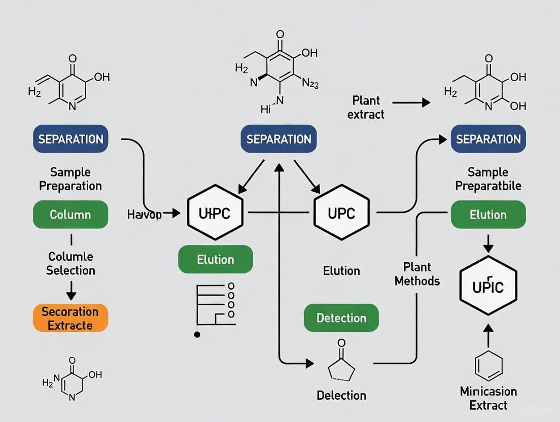
Workflow: From Plant Material to Analyzed Extract
The following diagram visualizes the complete integrated workflow for analyzing bioactive compounds in plants, from sample preparation to data acquisition.
This technical support center provides troubleshooting guides and FAQs to address common challenges in UHPLC analysis of complex plant extracts, supporting research on method optimization.
Troubleshooting Guides
Pressure Abnormalities
| Symptom | Potential Cause | Solution |
|---|---|---|
| No Pressure / Pressure Too Low | Formation of negative pressure in eluent reservoir; Leakage at piston seal or pump valves; Air in pump head [7]. | Avoid negative pressure using equalizing valves; Check tightness, replace seals or valves; Purge pump with water or isopropyl alcohol [7]. |
| Increase in Pressure / Pressure Too High | Blocked injector or capillaries; Blocked guard column or column inlet frit; Contamination of stationary phase [7]. | Clean injector and capillaries; Backflush column (if allowed) or replace; Wash column with strong solvent, follow manufacturer's regeneration procedure [7] [1]. |
Peak Deformation
| Symptom | Potential Cause | Solution |
|---|---|---|
| Peak Tailing | Interactions of basic analytes with silanol groups; Dead volume; Blocked frit; Wrong mobile phase pH [7] [1]. | Use high-purity silica or shield phases; Add competing base; Reduce extra-column volume; Adjust pH; Replace column [7] [1]. |
| Peak Fronting | Column overloading; Viscosity of sample or mobile phase too high; Contamination of stationary phase [7]. | Decrease injection volume or sample concentration; Increase temperature or change mobile phase; Wash column with strong solvent [7]. |
| Split Peaks | Blocked guard column or column inlet frit; Inappropriate injection solvent; Dead volume; Co-elution [7]. | Backflush or replace column; Use weaker injection solvent; Re-pack or replace column; Optimize sample preparation and method parameters [7]. |
| Peak Broadening | Injection volume too large; Detector response time too long; Contamination of stationary phase [7] [1]. | Inject smaller volume; Ensure detector flow cell volume ≤1/10 of smallest peak volume; Increase column temperature or wash column [7] [1]. |
Retention Time and Resolution Issues
| Symptom | Potential Cause | Solution |
|---|---|---|
| Retention Time Shifts | Eluent composition change; Reduced flow rate; Temperature fluctuation; Insufficient equilibration; Leakages [7]. | Check solvent mixing; Cover storage bottles; Control flow rate; Ensure constant temperature; Equilibrate with 10 column volumes; Check for leaks [7]. |
| Loss of Resolution | Contamination of mobile phase; Blocked pre-column; Ageing of stationary phase [7]. | Prepare fresh mobile phase; Replace pre-column; Flush column regularly and work within specifications [7]. |
Baseline and Signal Problems
| Symptom | Potential Cause | Solution |
|---|---|---|
| Baseline Noise | Air bubbles in mobile phase or column; Detector lamp defect; Contaminated detection cell; Air in pumps [7]. | Degas mobile phase; Store columns tightly sealed; Exchange lamp; Clean detection cell; Purge air from pumps [7]. |
| Negative Peaks | Absorption/fluorescence of analyte lower than mobile phase; Wrong polarization of analog output; Inappropriate reference wavelength (DAD) [1]. | Change detection wavelength; Use mobile phase with less background; Check cable polarity; Adjust reference wavelength settings [1]. |
Frequently Asked Questions (FAQs)
Method Development
How can I comprehensively characterize a botanical extract with limited constituent standards? A multi-detector approach is highly effective. As demonstrated in an ashwagandha root extract study, coupling UHPLC with Photodiode Array (PDA), Charged Aerosol Detection (CAD), and High-Resolution Mass Spectrometry (HRMS) enables detailed chemical profiling. This platform compensates for individual detector biases and allows for both semi-quantification and identification of a wide range of constituents, even without authentic standards for every compound [8].
What column chemistry is recommended for the analysis of complex natural products? The optimal column depends on your analytes. For cannabinoids, CORTECS Shield RP18, CORTECS C18, and ACQUITY UPLC HSS C18 columns have been successfully used [9]. For alkaloids in poppy extracts, a HILIC (Hydrophilic Interaction Liquid Chromatography) stationary phase is effective for separating polar compounds like benzylisoquinoline alkaloids [10].
System Operation
My method transfer from HPLC to UHPLC is causing peak shape issues. What should I check? Ensure all system components are optimized for UHPLC pressures and volumes. Use connecting capillaries with a small internal diameter (e.g., 0.13 mm) and low-volume flow cells. The extra-column volume should not exceed 1/10 of the volume of your narrowest peak to prevent peak broadening [1].
How can I prevent retention time fluctuations in my methods? Maintain consistent operating conditions. Use a column oven for stable temperature, ensure thorough mobile phase degassing, prepare fresh buffers daily to prevent microbial growth, and allow sufficient column equilibration (typically 10 column volumes) between runs, especially after gradient methods [7].
Data Quality
I suspect my peaks are co-eluting. How can I confirm this? Utilize a Diode Array Detector (DAD) to check peak purity by comparing UV spectra across the peak. For definitive confirmation, HRMS can detect trace ions from potential co-eluters that might not be visible in the chromatogram [11].
How can I improve the sensitivity and reliability of my quantification when standards are unavailable? Charged Aerosol Detection (CAD) is a valuable tool for semi-universal quantification. Since CAD response is less dependent on chemical structure than UV, it can provide more uniform quantification for analytes lacking standards. Ensure mobile phase compatibility and be aware that it can broaden peaks slightly more than UV due to the nebulization process [8] [1].
Experimental Protocols for Key Applications
Protocol 1: Comprehensive Profiling of Ashwagandha Root Extract
This protocol uses a multi-detector UHPLC platform for detailed characterization of complex botanical extracts [8].
1. Sample Preparation
- Weigh ashwagandha root extract and prepare a 20 mg/mL solution in 50:50 methanol-water.
- Vortex mix for 60 seconds, sonicate for 5 minutes, and vortex mix again for 60 seconds.
- Centrifuge the sample for 10 minutes and transfer the supernatant to an autosampler vial [8].
2. Instrumentation and Conditions
- System: UHPLC system coupled with PDA, CAD, and HRMS (e.g., Orbitrap) detectors.
- Column: Hypersil Gold aQ (2.1 × 150 mm, 1.9 µm).
- Mass Spectrometry: HRMS set to collect m/z 125–2000 at a resolution of 120,000. Use data-dependent acquisition with both CID and HCD fragmentation [8].
3. Standard Preparation
- Prepare individual stock solutions at 1.0 mg/mL in 50:50 methanol-water.
- Create combined standard mixes (e.g., 100 µg/mL each component) and perform serial dilutions to 20, 4, and 0.8 µg/mL for calibration [8].
Protocol 2: Analysis of Alkaloids in Poppy Tissues using HILIC-UHPLC-MS/MS
This high-throughput method is optimized for small tissue quantities and multi-tissue comparisons [10].
1. Tissue Extraction
- Use minimal tissue input (e.g., 5 mg of seeds, leaves, or capsules).
- Extract using a streamlined protocol designed to reduce matrix effects without extensive clean-up steps [10].
2. UHPLC-MS/MS Conditions
- Chromatography: HILIC (Hydrophilic Interaction Liquid Chromatography) separation.
- Detection: Tandem Mass Spectrometry (MS/MS) with Multiple Reaction Monitoring (MRM).
- Key Advantage: The method achieves sub-ng detection limits and is applicable across diverse cultivars and tissues [10].
3. Data Analysis
- Apply multivariate chemometric analysis to link metabolite profiles with cultivar identity.
- Use the framework for predictive chemotyping and targeted breeding [10].
The Scientist's Toolkit: Essential Research Reagents & Materials
| Item | Function | Application Example |
|---|---|---|
| Hypersil Gold aQ Column | UHPLC column with polar endcapping; stable in 100% aqueous mobile phases. | General profiling of medium-polar to polar compounds in plant extracts like ashwagandha [8]. |
| HILIC Stationary Phase | Separates polar compounds based on hydrophilic interactions and partitioning. | Analysis of highly polar alkaloids (e.g., in poppy extracts) and sugars [10]. |
| CORTECS C18 or Shield RP18 Columns | Robust UHPLC columns with solid-core particles (CORTECS) or polar-embedded groups (Shield). | Analysis of complex natural products like cannabinoids; Shield phases reduce tailing for basic compounds [9]. |
| Charged Aerosol Detector (CAD) | Semi-universal mass detector; response independent of chromophores. | Quantification of compounds in botanical extracts lacking UV chromophores or authentic standards [8]. |
| High-Resolution Mass Spectrometer (Orbitrap) | Provides accurate mass measurements for elemental composition and structural elucidation. | Identification of unknown constituents in complex plant matrices [8] [11]. |
Method Development and Troubleshooting Workflow
The following diagram outlines a systematic workflow for developing and troubleshooting UHPLC methods for complex plant extracts.
The Impact of Extract Composition on UHPLC Performance and Resolution
Troubleshooting Guide: Common UHPLC Issues with Plant Extracts
Complex plant extracts can present significant challenges during UHPLC analysis. The table below outlines common symptoms, their likely causes, and recommended solutions.
| Problem Symptom | Likely Cause | Recommended Solution | Preventive Measures |
|---|---|---|---|
| Rapid pressure increase or column overpressure [12] | Blockage of column frits by particulate matter (e.g., plant waxes, cellular debris) from unfiltered extracts. | Try backflushing the column (if manufacturer instructions allow) or replace the column[failed verification]. Install a guard column to protect the analytical column [12]. | Filter all samples prior to injection. Centrifuge samples as a high-throughput alternative to filtration [12]. |
| Poor peak shape (tailing or broadening) for basic compounds [13] | Detrimental interaction of analytes with metallic surfaces (e.g., stainless steel) in the LC hardware or column. | Use columns with inert (biocompatible) hardware. These columns have a passivated metal-free barrier that minimizes analyte adsorption and improves peak shape [13]. | Select an analytical column with a stationary phase designed for basic compounds, such as one with a positively charged surface [13]. |
| Insufficient separation of complex mixtures [14] [15] | The chromatographic conditions (mobile phase, column) lack the resolving power for the wide polarity range of compounds in the extract. | Switch from isocratic to gradient elution to better separate compounds with different polarities [15]. Consider comprehensive two-dimensional LC (LC×LC) for ultimate resolution [14]. | Optimize the mobile phase gradient. Use a column with alternative selectivity (e.g., phenyl-hexyl, biphenyl) for improved separation of specific isomers [13]. |
| Low analyte recovery/response for metal-sensitive compounds [13] | Analyte adsorption or complexation with active metal sites in the flow path. | Use inert guard cartridges and columns to enhance chromatographic response and recovery for metal-sensitive analytes [13]. | Ensure the entire system flow path, including the column, is rated as inert or bio-inert. |
Frequently Asked Questions (FAQs)
Q1: What is the most critical step in preparing a plant extract for UHPLC analysis to avoid system problems? Sample cleanup is paramount. Even with a simple extraction, filtration or centrifugation is essential to remove particulate matter that can clog the very small frits and tubing in a UHPLC system, preventing catastrophic pressure increases and column failure [12].
Q2: My method works for some plant samples but not others. Why would resolution suddenly degrade? The chemical composition of plant extracts can vary dramatically based on genetics, growing conditions, and plant part used [4]. A method optimized for one profile may be overwhelmed by a different concentration of co-extractives (like lipids or waxes) or new interfering compounds in another sample, leading to peak co-elution. A robust method development strategy that tests different columns and mobile phases is key [15].
Q3: How can I improve the separation of very polar compounds in my plant extract that don't retain on a standard C18 column? Reversed-phase (C18) columns are poor for highly polar compounds. Consider using Hydrophilic Interaction Liquid Chromatography (HILIC) columns [14] [15]. HILIC operates on a different separation mechanism and is highly effective for retaining and separating polar metabolites found in plants [13].
Q4: My peaks for certain compounds are tailing badly. Could the extract itself be causing this? Yes. While the extract matrix can contribute, severe tailing for basic compounds (like some alkaloids) is often due to interactions with metallic surfaces in the HPLC system. Investing in a column with inert or bio-inert hardware can significantly improve peak shape and analyte recovery for these sensitive compounds [13].
Detailed Experimental Protocols
High-Throughput Extraction and Analysis of Spinach Flavonoids
This validated protocol demonstrates an efficient approach for profiling flavonoids in plant tissue [4].
- Sample Homogenization: Fresh or frozen spinach tissue is homogenized in a 1:1 ratio with MilliQ water using a high-speed polytron homogenizer (e.g., 20,000 RPM for ~1 minute) [4].
- Extraction: The homogenate is mixed with a solvent like methanol or a hydro-alcoholic mixture (e.g., 70% ethanol/30% water). The mixture is stirred or shaken thoroughly [4] [16].
- Post-Extraction Processing: The extract is centrifuged to pellet solid debris. The supernatant is filtered or directly diluted in the initial UHPLC mobile phase (e.g., 1:1 MeOH:H₂O + 0.1% formic acid) prior to injection [4].
- UHPLC-MS/MS Conditions:
- Column: Reversed-phase C18 column [4].
- Mobile Phase: Binary gradient using water and methanol (or acetonitrile), both modified with 0.1% formic acid [4] [16].
- Gradient: Starts with a higher proportion of aqueous phase (e.g., 90%), ramping to a high proportion of organic phase (e.g., 95%) over a short runtime (e.g., 11.5 minutes) [4].
- Detection: Tandem mass spectrometry (MS/MS) with electrospray ionization (ESI) in negative or positive mode [4].
A Workflow for Systematic Troubleshooting
This diagram outlines a logical, step-by-step process to diagnose and resolve UHPLC issues related to extract composition.
Research Reagent Solutions Toolkit
The following table lists key materials and reagents essential for successful UHPLC analysis of complex plant extracts.
| Item Name | Function / Application | Key Considerations |
|---|---|---|
| C18 Reversed-Phase Column [15] [4] | The workhorse for separating a wide range of mid- to non-polar plant compounds (flavonoids, terpenoids). | Available in various particle sizes (e.g., 1.7 μm for UHPLC). Phenyl-hexyl and biphenyl phases offer alternative selectivity for isomers [13]. |
| HILIC Column [15] [14] | Separates highly polar and hydrophilic compounds that are poorly retained on C18 phases. | Useful for plant metabolites like sugars, amino acids, and some glycosylated compounds. Operates with a high-organic mobile phase [15]. |
| Inert/Bioinert Column [13] | Minimizes interaction of metal-sensitive analytes (e.g., phosphorylated compounds, some polyphenols) with hardware surfaces. | Critical for improving peak shape and recovery for challenging compounds. Often features a metal-free flow path or passivated surfaces [13]. |
| Guard Column [13] [12] | Protects the expensive analytical column from particulate matter and highly retained contaminants in crude extracts. | Extends analytical column lifetime. Should be packed with the same phase as the analytical column or a similar one [13]. |
| Acid Additives (Formic/Acetic) [4] [16] | Modifies the mobile phase pH to suppress ionization of acidic analytes, improving retention and peak shape in reversed-phase LC. | Commonly used at 0.1% concentration. Enhances ionization in ESI-MS detection [4]. |
| Buffers (Ammonium Formate/Acetate) | Provides better control of mobile phase pH compared to volatile acids alone, improving reproducibility for ionizable compounds. | Essential for HILIC and some difficult reverse-phase separations. MS-compatible [16]. |
| Solid Phase Extraction (SPE) Cartridges | Pre-concentrates target analytes and removes a significant portion of the interfering matrix before UHPLC analysis. | Can be selective (e.g., C18, SPE) or non-selective. Greatly reduces background noise and column contamination [4]. |
Troubleshooting Guides and FAQs for UHPLC Analysis of Plant Metabolites
Frequently Asked Questions (FAQs)
Q1: My peaks for compounds in a complex plant extract are broad and tailing. What could be the cause and how can I fix this?
Broad or tailing peaks often indicate issues with column chemistry or secondary interactions.
- Cause for Basic Compounds: Silanol interactions are a common cause for tailing peaks of basic compounds like alkaloids [1].
- Solution:
- Column Selection: Use high-purity silica (Type B) or specialized phases such as polar-embedded groups, charged surface hybrid (CSH), or phenyl-hexyl columns to improve peak shape for basic compounds [1] [17].
- Mobile Phase Additives: Add a competing base like triethylamine (TEA) or use buffers with sufficient ionic strength to mask silanol sites. Note that high ionic strength buffers may not be compatible with LC/MS [1].
Q2: I am getting low recovery or poor peak response for my analytes. What should I check?
Low response can stem from several parts of the workflow.
- Solution:
- Detector Settings: Ensure the detection wavelength (for DAD/PDA) or MS parameters are optimized for your specific compounds. For fluorescence detection (FLD), scan for the best excitation and emission wavelengths [1].
- Sample Solvent: The solvent used to dissolve the sample should not be stronger than the starting mobile phase, as this can cause peak broadening and splitting. Dilute samples in the starting mobile phase when possible [1].
- Extraction Efficiency: Review your extraction protocol. For instance, recovery of alkaloids can be optimized using mixed-mode solid-phase extraction (SPE) with cation-exchange properties, as demonstrated for indole alkaloids where recovery rates for different compounds varied from 51% to 88% [17].
Q3: How can I reduce my analysis time without compromising separation quality?
Faster separations are achievable by optimizing chromatographic parameters.
- Solution:
- Column Technology: Use shorter columns packed with smaller particles (e.g., sub-2 µm) common in UHPLC [18] [19].
- Gradient Optimization: Steepen the elution gradient. A method for onion flavonols was optimized using a Box-Behnken design, reducing runtime to 2.7 minutes [18].
- Flow Rate: Increasing the flow rate within the system's pressure limits can shorten run times [18].
Troubleshooting Common UHPLC Issues
The table below summarizes specific symptoms, their likely causes, and solutions based on the cited case studies and technical guides.
| Symptom | Possible Cause | Solution |
|---|---|---|
| Broad Peaks [1] | - Detector cell volume too large- Extra-column volume too large- Column degradation | - Use a flow cell volume ≤ 1/10 of the smallest peak volume- Use short, narrow-bore capillaries (e.g., 0.13 mm i.d.)- Replace column |
| Peak Tailing [1] [17] | - Silanol interactions (for basic compounds)- Column void- Inadequate buffer capacity | - Use a charged surface hybrid (CSH) or phenyl-hexyl column [17]- Replace column- Increase buffer concentration |
| Peak Fronting [1] | - Column overload- Blocked frit or channels in column | - Reduce sample amount- Replace pre-column frit or analytical column |
| Irreproducible Retention Times [1] | - Inconsistent column temperature- Insufficient buffer capacity- Contaminated column | - Use an eluent pre-heater- Increase buffer concentration- Flush column with strong eluent |
| Noisy Baseline / Low Sensitivity [1] | - Contaminated mobile phase or nebulizer (CAD)- Insufficient degassing (FLD)- Inappropriate detection settings | - Use high-purity solvents, clean detector nebulizer- Check degasser operation- Optimize wavelength (DAD) or gain (FLD) |
| Poor Peak Area Precision [1] | - Air in autosampler syringe or fluidics- Sample degradation- Leaking injector seal | - Purge autosampler, ensure sufficient sample volume- Use thermostatted autosampler- Check and replace injector seals |
Experimental Protocols from Case Studies
1. Protocol: High-Throughput Extraction and UHPLC-MS/MS Analysis of Spinach Flavonoids [4] [20]
This protocol enables the quantification of 39 flavonoid species in 11.5 minutes.
- Sample Preparation:
- Homogenize fresh or frozen spinach in a 1:1 ratio with MilliQ water.
- Aliquot and store homogenate at -80°C until extraction.
- Extraction:
- A high-throughput method allows processing of 48 samples in 60 minutes.
- Recovery rates are validated to be between 100.5 – 107.8%.
- UHPLC Conditions:
- Analytical Column: Not specified in detail, but a reverse-phase column is used.
- Mobile Phase: Components include water and methanol, both LC-MS grade with 0.1% formic acid.
- Gradient: Elution program is optimized for a total run time of 11.5 minutes.
- Detection: Tandem Mass Spectrometry (MS/MS).
- Quantification:
- Use an internal standard (e.g., taxifolin).
- For flavonoids without authentic standards, quantification is performed relative to a standard like quercetin-3-glucoside.
2. Protocol: UHPLC-PDA Analysis of Major Flavonols in Onion (Allium cepa) [18]
This method uses multiresponse optimization for rapid separation in under 2.7 minutes.
- Extraction:
- Methanolic extraction of onion bulbs.
- UHPLC Conditions:
- Analytical Column: Reverse-phase column.
- Mobile Phase: A) Acidic water; B) Methanol.
- Optimized Gradient: 9.9% B at start to 53.2% B at end.
- Flow Rate: 0.6 mL/min.
- Column Temperature: 55 °C.
- Detection: Photo-Diode Array (PDA).
- Validation: The method is validated for precision, linearity, and robustness.
3. Protocol: UHPLC-DAD Analysis of Protoberberine Alkaloids [19]
This method focuses on the rapid quantification of nine isoquinoline alkaloids from Berberis aristata.
- Extraction:
- Ultrasonic-assisted solid-liquid extraction using methanol.
- UHPLC Conditions:
- Column Technology: Core-shell particle technology column.
- Mobile Phase: A) Water + 0.1% Formic Acid; B) Acetonitrile + 0.1% Formic Acid.
- Gradient: A rapid gradient is used, compatible with the core-shell column.
- Detection: Diode Array Detector (DAD).
- Method Performance: The method demonstrates excellent resolution and a short analysis time.
Quantitative Data from Plant Metabolite Studies
The following tables summarize key quantitative findings from the research, providing benchmark values for method development and comparison.
Table 1: Bioactive Compound Concentrations in Plant Materials
| Plant Material | Bioactive Class | Target Compound(s) | Concentration | Reference |
|---|---|---|---|---|
| Spinach | Flavonoids | Total Flavonoids | 75.1 – 187.26 mg/100 g Fresh Weight | [4] [20] |
| Mangosteen Pericarp | Xanthones | α-Mangostin | Major compound (69.01% of total xanthones) | [3] |
| Mangosteen Pericarp | Xanthones | γ-Mangostin | 17.86% of total xanthones | [3] |
Table 2: Optimized Extraction Conditions and Recovery Rates
| Plant Material / Analyte | Extraction Method | Key Optimized Parameters | Recovery / Yield | Reference |
|---|---|---|---|---|
| Spinach Flavonoids | High-throughput | 48 samples/60 minutes | 100.5 – 107.8% | [4] [20] |
| Mangosteen Xanthones | Microwave-Assisted (MAE) | 2.24 min, 25 mL/g, 71% EtOH | TPC: 320.31 mg GAE/g | [3] |
| Monoterpene Indole Alkaloids | Solid-Phase Extraction (SPE) | Cation-exchange (Plexa PCX) sorbent | 51.13% (Harmaline) – 87.86% (Harmine) | [17] |
Workflow Diagram: UHPLC Method Development for Plant Extracts
The diagram below outlines a logical workflow for developing and troubleshooting a UHPLC method for complex plant extracts, based on the principles demonstrated in the case studies.
The Scientist's Toolkit: Essential Research Reagents and Materials
This table lists key reagents, solvents, and materials used in the featured UHPLC analyses of plant metabolites.
Table 3: Essential Reagents and Materials for Plant Metabolite UHPLC Analysis
| Item | Function / Application | Example from Case Studies |
|---|---|---|
| LC-MS Grade Solvents (Methanol, Acetonitrile, Water) | Mobile phase preparation; ensures low UV background and minimal MS interference. | Used in all cited UHPLC methods for flavonoid and alkaloid analysis [4] [17] [19]. |
| Acid Additives (Formic Acid, Acetic Acid) | Modifies mobile phase pH to suppress analyte ionization and improve peak shape in reversed-phase chromatography. | 0.1% Formic acid common in MS methods [4] [19]; 10 mM Ammonium Acetate for alkaloids [17]. |
| Buffers (Ammonium Acetate, Formate) | Provides ionic strength and controls pH for consistent retention times, especially critical for basic compounds. | 10 mM Ammonium Acetate used for alkaloid separation [17]. |
| Specialized UHPLC Columns (C18, Phenyl-Hexyl, CSH) | Stationary phase for compound separation. Choice depends on analyte chemistry (e.g., CSH for basic compounds). | Phenyl-Hexyl column provided superior resolution for indole alkaloids [17]; Core-shell C18 for protoberberine alkaloids [19]. |
| Solid-Phase Extraction (SPE) | Sample clean-up and pre-concentration; removes interfering matrix components. | Cation-exchange SPE (Bond Elut Plexa PCX) for purifying alkaloids [17]. |
| Authentic Standards | Compound identification and calibration curve generation for quantification. | Taxifolin, quercetin-3-glucoside for flavonoids [4]; α-mangostin for xanthones [3]; pure alkaloids [17] [19]. |
Advanced UHPLC Techniques and High-Throughput Workflows for Plant Analysis
FAQs: Addressing Common Challenges in Multi-Detector UHPLC
Q1: What are the primary advantages of using a multi-detector platform (PDA-CAD-HRMS) for analyzing complex plant extracts?
Integrating PDA, CAD, and HRMS detectors provides a complementary analytical approach that overcomes the limitations of using any single detector. Photodiode Array (PDA) detection identifies constituents with UV-chromophores, a standard in botanical analysis. Charged Aerosol Detection (CAD) provides semi-universal quantification, offering an unbiased response for compounds lacking a chromophore, such as sugars or certain lipids, ensuring they are not missed in the analysis. High-Resolution Mass Spectrometry (HRMS) delivers accurate mass data for confident constituent identification. This combination ensures a comprehensive chemical profile, supporting material authentication and robust safety assessments by providing both identification and semi-quantification of a wide array of constituents [8] [21].
Q2: How can I troubleshoot high backpressure in my UHPLC system when analyzing botanical extracts?
High system pressure is a common issue, often caused by clogged columns or frits due to sample contaminants or salt precipitation.
- Solution: Gradually flush the column with pure water at an elevated temperature (40–50°C), followed by methanol or another strong organic solvent. If possible, backflush the column. Using guard columns and inline filters, and ensuring all samples and solvents are filtered before injection, can prevent this issue [22] [23].
Q3: What steps should I take if I observe baseline noise or drift during a run?
Baseline disturbances can arise from several sources, including air bubbles, contaminated solvents/mobile phases, a contaminated detector flow cell, or a failing detector lamp.
- Solution: Thoroughly degas all mobile phases. Purge the system to remove air bubbles. Use high-purity solvents and clean the detector flow cell with a strong organic solvent. If the problem persists, the detector lamp may need replacement [22] [23].
Q4: Why are my peaks tailing or broadening, and how can I improve peak shape?
Peak tailing or broadening can result from column degradation, inappropriate stationary phase selection, sample-solvent incompatibility with the mobile phase, or secondary interactions with active sites on the column.
- Solution: Ensure the sample is dissolved in a solvent compatible with the initial mobile phase. Use a guard column. Flush the column to remove contamination. Consider modifying the mobile phase composition (e.g., pH, buffer concentration) or switching to a different column chemistry to mitigate active sites [22] [23].
Q5: How can I address shifting retention times in my UHPLC analysis?
Retention time instability is frequently caused by variations in mobile phase composition or preparation, poor column equilibration, especially in gradient methods, or inconsistent pump flow rates.
- Solution: Prepare mobile phases consistently and accurately. Allow sufficient time for the column to equilibrate with the starting mobile phase condition before starting a sequence. Regularly service and calibrate the pump to ensure stable flow rates [23].
Troubleshooting Guide: Common UHPLC Issues and Solutions
The following table summarizes frequent instrument-related problems, their likely causes, and corrective actions.
| Problem Symptom | Potential Causes | Recommended Solutions |
|---|---|---|
| High System Pressure [22] [23] | Clogged column or frit, mobile phase salt precipitation, blocked inline filter. | Flush column with warm water followed by organic solvent; backflush column; replace guard column or inline filter. |
| Baseline Noise & Drift [22] [23] | Air bubbles, contaminated mobile phase/solvents, contaminated detector flow cell, failing UV lamp. | Degas mobile phases; purge system; use high-purity solvents; clean flow cell; replace UV lamp. |
| Peak Tailing/Broadening [22] [23] | Column degradation, sample solvent stronger than mobile phase, active sites on column, extra-column volume. | Dilute sample in mobile phase; use guard column; replace column; modify mobile phase pH/buffer; use appropriate column chemistry. |
| Retention Time Shifts [23] | Inconsistent mobile phase composition, poor column equilibration, pump flow rate fluctuations, temperature variations. | Prepare fresh, consistent mobile phase; increase equilibration time; service pump; use a thermostatted column oven. |
| Low Signal Intensity [22] | Poor sample extraction/preparation, detector settings (e.g., time constant), air bubbles in system. | Optimize sample preparation; check and adjust detector settings; degas mobile phases and purge system. |
Experimental Protocol: Characterization of a Botanical Extract
The following methodology, adapted from published work on ashwagandha and grape seed extracts, details a comprehensive approach for profiling complex plant materials [8] [21].
Instrumentation and Materials
- UHPLC System: Ultra-high-performance liquid chromatography system capable of withstanding high backpressures (e.g., >15,000 psi).
- Detectors: A system configured with PDA (e.g., 190–800 nm), CAD, and HRMS (e.g., Orbitrap) detectors in series. An inverse gradient make-up flow can be implemented post-column to maintain consistent mobile phase composition for optimal CAD performance [8].
- Column: Reversed-phase column, such as a Hypersil Gold aQ (2.1 × 150 mm, 1.9 µm) or equivalent [8].
- Mobile Phase: Typically, a gradient of water and acetonitrile, both often modified with 0.1% formic acid or ammonium formate to aid ionization for MS.
- Standards: Chemical standards relevant to the botanical under investigation (e.g., withanolides for ashwagandha, proanthocyanidins for grape seed) for method validation and calibration [8] [21].
Sample Preparation
- Weigh out the botanical extract (e.g., 20 mg).
- Dissolve in a suitable solvent (e.g., 50:50 methanol–water) to a final concentration of 20 mg/mL.
- Vortex mix the sample for 60 seconds.
- Sonicate for 5 minutes.
- Vortex mix again for 60 seconds.
- Centrifuge for 10 minutes to pellet insoluble particulates.
- Transfer the supernatant to an autosampler vial for analysis [8].
Data Acquisition and Analysis
- PDA: Collect full UV-Vis spectra for each peak to aid in compound classification and purity assessment.
- CAD: Use for semi-quantification of all non-volatile constituents. The uniform response factor allows for estimating concentrations of unknowns against available standards [21].
- HRMS: Acquire data in both positive and negative ionization modes with a resolution of 120,000 or higher. Use data-dependent acquisition (DDA) to fragment precursor ions for structural elucidation [8].
- Data Integration: Process data from all detectors to create a consolidated list of constituents, including retention time, UV spectrum, accurate mass, fragment ions, and semi-quantitative abundance.
Workflow Visualization: Multi-Detector Analysis & Troubleshooting
UHPLC Multi-Detector Data Flow
Systematic UHPLC Troubleshooting Logic
The Scientist's Toolkit: Essential Research Reagents & Materials
The following table lists key materials required for setting up and performing a comprehensive multi-detector analysis of botanical extracts.
| Item | Function / Application | Example from Literature |
|---|---|---|
| UHPLC-grade Solvents (Methanol, Acetonitrile, Water) | High-purity mobile phase components to minimize baseline noise and contamination [8] [21]. | Honeywell (Morris Plains, NJ, USA) [8] [21]. |
| Volatile Mobile Phase Additives (Formic Acid, Ammonium Formate) | Modify mobile phase pH to improve chromatographic separation and enhance ionization efficiency in HRMS [8] [21]. | Aldrich Chemicals [21]. |
| Authenticated Botanical Reference Material | Serves as a benchmark to confirm the identity and authenticity of the test material, helping to detect adulteration [21]. | Voucher specimens deposited with Botanical Liaisons, LLC [21]. |
| Chemical Standards | Used for method development, calibration, and confirmation of constituent identity based on retention time and mass spectra. | Withanolide A, withanoside IV, and other withanolides for ashwagandha analysis [8]. Proanthocyanidin standards for grape seed extract analysis [21]. |
| Reversed-Phase UHPLC Column (e.g., C18, 1.7-1.9µm) | The core component for separating complex mixtures; sub-2-µm particles provide high resolution and efficiency. | Hypersil Gold aQ (2.1 × 150 mm, 1.9 µm) [8]. ACQUITY C18 or Shield RP18 (1.7-µm dp) [21]. |
Troubleshooting Guides
Pressure Abnormalities
| Symptom | Possible Cause | Solution |
|---|---|---|
| No Pressure / Pressure Too Low | - Leakage at pump seals or valves [7]- Air in pump heads or valves [7] | - Check and replace seals or valves [7]- Purge pumps with water or isopropanol [7] |
| Increase in Pressure / Pressure Too High | - Blocked injector or capillary tubing [7]- Blocked column inlet frit [1] [7] | - Clean or replace blocked components [7]- Backflush the column (if permitted) or replace the column/guard cartridge [1] [7] |
| Pressure Fluctuation | - Air or particulates in pump heads [7]- Leaking pump seals or valves [7] | - Purge pump; degas mobile phase [7]- Inspect and replace defective seals or valves [7] |
Peak Shape Issues
| Symptom | Possible Cause | Solution |
|---|---|---|
| Peak Tailing | - Silanol interaction with basic analytes [1] [7]- Extra-column volume too large [1] | - Use high-purity silica or shielded phases; add competing base to mobile phase [1]- Use shorter, narrower internal diameter capillaries [1] |
| Peak Fronting | - Column overload [1] [7]- Channels in the column packing [1] | - Reduce injection volume or sample concentration [1] [7]- Replace the column [1] |
| Split Peaks | - Blocked column frit [1] [7]- Inappropriate sample solvent [7] | - Backflush column or replace frit/column [1] [7]- Ensure sample is dissolved in a solvent weaker than the mobile phase [7] |
| Peak Broadening | - Detector flow cell volume too large [1]- Detector response time too slow [1] | - Use a micro-flow cell for UHPLC/microbore columns [1]- Set detector response time to <1/4 of the narrowest peak width [1] |
Retention Time and Baseline Problems
| Symptom | Possible Cause | Solution |
|---|---|---|
| Retention Time Shifts | - Mobile phase composition change [7]- Column temperature fluctuation [1] [7]- Insufficient column equilibration [7] | - Prepare fresh mobile phase; ensure proper solvent mixing [7]- Use a column oven for temperature stability [1] [7]- Equilibrate with 10-15 column volumes between runs [7] |
| Baseline Noise or Drift | - Contaminated mobile phase or eluent reservoir [1] [7]- Air bubbles in detector cell [7]- Strong UV-absorbing solvent in gradient [7] | - Use fresh, high-quality solvents; clean eluent system [1] [7]- Degas mobile phase thoroughly [7]- Use UV-transparent solvents for gradient elution [7] |
Frequently Asked Questions (FAQs)
Q1: How can I prevent my UHPLC column from degrading quickly when running complex plant extracts at high throughput?
A: To maximize column lifetime:
- Use a Guard Column: Always use a guard cartridge to trap particulates and highly retained compounds [7].
- Sample Cleanup: Employ solid-phase extraction (SPE) to remove proteins, polyphenols, and other interfering matrix components before UHPLC analysis [24] [25].
- Proper Flushing: Regularly flush the column with strong solvents according to the manufacturer's specifications to remove accumulated contaminants [1] [7].
- Pressure Management: Operate the column at 70-80% of its maximum pressure limit and avoid sudden pressure shocks [1].
Q2: I keep getting extra peaks (ghost peaks) in my chromatograms. What is the source and how can I eliminate them?
A: Extra peaks typically stem from contamination or carryover [26].
- Source: Common sources are the sample itself, the mobile phase, a contaminated autosampler needle, or carryover from a previous injection [1] [26].
- Solution: Run a blank injection. If peaks appear, systematically eliminate sources: prepare fresh mobile phase and blanks, perform intensive autosampler needle washing, and if needed, replace or clean the injection valve rotor seal [1].
Q3: My method requires high throughput, but I am losing resolution. What strategies can I use to speed up the analysis without compromising separation quality?
A: Several UHPLC principles can be leveraged:
- Elevated Temperature: Using temperatures of 90°C can increase peak capacity by 20-30% or decrease analysis time by 2-3-fold for identical peak capacity compared to 30°C [27].
- Sub-2µm Particles: Utilize columns packed with small particles for higher efficiency [27].
- Gradient Optimization: Steepening the gradient slope is a direct way to reduce run time, though this must be balanced against resolution needs [27] [27].
- Method Scouting: Use a method development approach that systematically tests different column chemistries and gradient conditions to find the optimal balance [26].
Q4: What is the best way to improve the stability of my analytes, especially phytohormones, during a high-throughput workflow?
A: Analyte stability begins with sample handling:
- Cold Extraction: Perform the initial extraction in a cold, acidified methanol-water buffer to inhibit enzymatic activity [24].
- Low Temperature Storage: Keep samples in a thermostatted autosampler at 4-10°C [1].
- SPE Purification: Use mixed-mode solid-phase extraction (e.g., Oasis MCX) to purify and concentrate unstable, low-abundance analytes like phytohormones away from degrading enzymes and matrix interferences [24].
High-Throughput Workflow for Plant Metabolite Analysis
The following diagram illustrates the integrated workflow for the high-throughput extraction, purification, and analysis of complex plant extracts.
Research Reagent Solutions
This table details key materials and reagents essential for implementing the described high-throughput workflow.
| Item | Function in the Workflow |
|---|---|
| Acidified Methanol-Water Buffer | Primary extraction solvent; methanol denatures proteins while acidification stabilizes acid-labile metabolites and prevents enzymatic degradation [24]. |
| Polyamide SPE Cartridges | Pre-fractionation step to selectively remove polyphenols (tannins), which are known to cause false positives in bioassays and interfere with analysis [25]. |
| Mixed-Mode SPE Cartridges (e.g., Oasis MCX) | Purification and grouping of analytes; separates cationic compounds (e.g., cytokinins), anionic compounds (e.g., auxins, gibberellins, JA, SA), and neutral compounds for cleaner analysis [24]. |
| UHPLC-grade Solvents (ACN, MeOH, Water) | Essential for generating low-background noise, maintaining system pressure, and ensuring reproducible chromatographic performance [27] [1]. |
| Sub-2µm Particle UHPLC Columns | Provides high-resolution separation necessary for resolving hundreds of metabolites in complex plant extracts within short run times [27]. |
| Formic Acid (LC-MS Grade) | A common mobile phase additive that promotes protonation of analytes, improving ionization efficiency and detection sensitivity in mass spectrometry [27]. |
Developing a robust Ultra-High Performance Liquid Chromatography (UHPLC) method for complex plant extracts requires a systematic approach to separate, identify, and quantify numerous compounds with diverse chemical properties. This process demands careful consideration of three fundamental pillars: column selection, mobile phase optimization, and elution gradient design. The intricate nature of plant matrices—containing acids, bases, neutrals, polar, and non-polar compounds—presents unique challenges that necessitate methodical troubleshooting and optimization. Within the broader context of optimizing UHPLC separation for complex plant extracts research, this guide provides targeted solutions to specific methodological problems, enabling researchers to achieve high-resolution separations with maximum efficiency and reproducibility. The following sections address common obstacles through detailed FAQs, structured protocols, and practical troubleshooting guides designed for scientists and drug development professionals.
Core Concepts and Column Selection
The Critical Role of Stationary Phase Chemistry
Why does column selection fundamentally impact my separation of plant metabolites?
A "C18" column is not universally equivalent to another "C18" column. The specific bonded phase chemistry, including factors like % carbon loading and the type of end-capping, drastically alters the selectivity and retention characteristics of a column [28]. In plant extract analysis, where compounds exhibit a wide range of polarities and structures, this selectivity is paramount for resolving closely eluting peaks. Proof of this concept is demonstrated in separations where identical test mixtures, mobile phases, and column dimensions yield vastly different chromatographic profiles solely due to differences in the packed bed's bonded phase chemistry [28].
Choosing Between Particle Types: FPP vs. SPP
Should I use fully porous (FPP) or superficially porous particles (SPP) for my method?
The choice between particle types involves a trade-off between efficiency, pressure, and practical handling. Superficially Porous Particles (SPP), such as 2.7 µm SPP, offer a compelling balance for many applications [29] [28]. They provide higher efficiency and resolution often approaching that of sub-2 µm Fully Porous Particles (FPP) used in UHPLC, but without generating the same high backpressures [29]. This allows for UHPLC-like performance on standard HPLC systems. Furthermore, SPP columns are noted for being robust and forgiving, as they are less prone to clogging compared to columns packed with sub-2µm materials, a significant advantage when dealing with complex plant sample matrices [28].
Essential Column Selection Guide
Table: Guidelines for Selecting Analytical Columns for Plant Extract Analysis
| Column Characteristic | Options | Application Considerations |
|---|---|---|
| Particle Type | Fully Porous (FPP, e.g., 1.9 µm) | Highest efficiency; requires UHPLC systems capable of high pressure [28]. |
| Superficially Porous (SPP, e.g., 2.7 µm) | High efficiency with lower backpressure; suitable for HPLC systems; clog-resistant [29] [28]. | |
| Column Chemistry | C18 (standard) | Good general retention for non-polar to medium-polarity compounds. |
| Biphenyl / Phenyl-Hexyl | Provides π-π interactions for separating planar molecules, aromatics, and compounds with double bonds [29] [28]. | |
| ARC-18 / Aqueous Stable C18 | Superior for 100% aqueous conditions; rugged at low pH (1.0–8.0); better retention for charged bases [29]. | |
| Column Dimensions | 50 x 2.1 mm (sub-2µm) | Optimal for fast UHPLC; run times of 3–5 min; higher backpressure [28]. |
| 100 x 3.0 mm (2.7µm SPP) | High efficiency on HPLC systems; lower backpressure; good compromise between speed and resolution [28]. | |
| pH Range | 1.0–8.0 (e.g., ARC-18) | Essential for low-pH applications to prevent phase degradation [29]. |
Figure 1: A logical workflow for selecting the appropriate UHPLC column based on the analytical goals and system capabilities.
Mobile Phase Optimization and Elution Strategies
Isocratic vs. Gradient Elution
When should I use a gradient instead of an isocratic method?
The decision hinges on the complexity and polarity range of your plant extract.
- Isocratic Elution employs a single, constant solvent composition throughout the separation. It is suitable for simple mixtures where the components have similar polarities. A drawback is that later-eluting peaks become progressively broader [28].
- Gradient Elution is the preferred technique for complex plant extracts. It involves a programmed change in solvent composition (e.g., increasing organic solvent percentage over time). This sharpens peaks throughout the chromatogram, shortens run times, and can achieve separations impossible under isocratic conditions [28]. After the run, a high-strength flush cleans the column of strongly retained compounds.
The Sample Solvent Strength Problem
Why do my peaks look distorted at the start of the chromatogram?
A primary cause is injecting your sample in a solvent that is stronger than the starting mobile phase [29] [1]. For a reversed-phase gradient starting with a high percentage of water, a sample dissolved in 100% acetonitrile is a very strong solvent. This causes analytes to travel too quickly through the initial part of the column instead of focusing at the head, leading to peak distortion, broadening, poor sensitivity, and shortened retention times [29].
Solution: Ensure the sample matrix is no stronger than the mobile phase condition at the time of injection. Ideally, dissolve or dilute samples in the starting mobile phase composition or a weaker solvent [29] [28]. Conversely, injecting in a weaker solvent (like 100% water) can sometimes be used intentionally for on-column compression to focus a large volume injection into a tight band [29].
Systematic Gradient Optimization
How do I develop a gradient method from scratch?
Begin with a screening gradient to scout the elution window for your entire plant extract. Based on the initial outcome, you can systematically optimize the gradient profile.
Figure 2: A decision tree for optimizing a gradient profile based on the initial chromatographic output.
Initial Screening Gradient Protocol:
- Column: Use a 100 x 3 mm column packed with 2.7 µm SPP particles (e.g., C18 or Biphenyl) [28].
- Mobile Phase: (A) Water (with modifier, e.g., 0.1% Formic Acid); (B) Acetonitrile (with modifier, e.g., 0.1% Formic Acid).
- Gradient Program: 5–100% B over 10–15 minutes.
- Flow Rate: 0.4–0.5 mL/min.
- Detection: UV-Vis DAD or MS.
- Interpret & Optimize: Follow the logic in Figure 2 to adjust the gradient's initial %B and slope until optimal separation is achieved within a desirable run time [28].
Troubleshooting Common Method Development Issues
Pressure and Baseline Anomalies
Table: Troubleshooting Guide for Pressure and Baseline Issues
| Symptom | Potential Cause | Solution |
|---|---|---|
| High Pressure | Clogged column frit from sample debris or salt precipitation [22]. | Flush column sequentially with warm water (40–50°C), methanol, and strong solvent [22]. Use guard columns [29]. |
| Pressure Fluctuation | Air bubbles in pump or check valves; insufficient mobile phase degassing [22]. | Purge pump. Degas mobile phases thoroughly (preferably with online degasser). Clean or replace check valves [22]. |
| Baseline Noise/Drift | Contaminated solvents/mobile phase; detector lamp issue; temperature instability [22]. | Use high-purity solvents. Clean detector flow cell. Replace old lamp. Maintain stable lab temperature [22]. |
| Negative Peaks | Sample solvent or analyte has lower UV absorption than the mobile phase [1]. | Change detection wavelength. Use mobile phase with less background absorption. Dissolve sample in mobile phase [1]. |
Peak Shape and Retention Problems
Table: Troubleshooting Guide for Peak Shape and Retention Issues
| Symptom | Potential Cause | Solution |
|---|---|---|
| Peak Tailing | (Basic compounds) Silanol interaction with silica [1] [7]. | Use high-purity silica (Type B) or shielded phases (e.g., ARC-18) [29] [1]. Reduce mobile phase pH to 2–3 [7]. |
| Peak Fronting | Column overload; sample solvent too strong; channeling in column [1] [7]. | Reduce injection volume/sample concentration [7]. Ensure sample is in correct solvent [29]. Replace column [1]. |
| Retention Time Shifts | Mobile phase composition change (evaporation, poor prep); insufficient column equilibration; leakage [22] [7]. | Prepare mobile phase consistently. Equilibrate with ~10 column volumes between gradient runs [29] [28]. Check for system leaks [22]. |
| Peak Splitting | Inappropriate injection solvent; blocked inlet frit; column void [1] [7]. | Inject in a weaker solvent [7]. Backflush column (if allowed) or replace frit/column [1] [7]. |
Advanced UHPLC Considerations
Managing System Volumes
Why does my gradient method behave differently when transferred to another UHPLC system?
The culprit is often a difference in gradient delay volume (or dwell volume)—the volume from the point of solvent mixing to the head of the column [28]. This volume is instrument-specific. On a system with a large delay volume, it takes longer for the programmed gradient to reach the column, causing a consistent shift in retention times and potentially compromising resolution, especially in fast, steep gradients.
Solution: Determine the delay volume for each instrument. Many data systems allow you to program an injection delay, so the sample is injected at the moment the gradient reaches the column head, compensating for the dwell volume difference [28].
Minimizing Band Broadening
How can I maximize resolution and sensitivity in my UHPLC method?
To achieve sharp, narrow peaks, it is critical to minimize extra-column volume (ECV)—the volume in the system that is outside the column itself (injector, tubing, detector flow cell) [28]. As column dimensions shrink to gain efficiency, the impact of ECV on peak broadening becomes more severe.
Guidelines for Minimizing ECV:
- Use narrow-bore tubing: Plumb systems with 0.005" ID tubing or smaller for all connections [28] [1].
- Keep tubing short: Use the shortest possible lengths of tubing between the injector, column, and detector.
- Match detector flow cell: Ensure the detector flow cell volume is appropriately small for the column format (should not exceed 1/10 of the smallest peak volume) [1].
Frequently Asked Questions (FAQs)
1. How do I calculate the appropriate injection volume when switching to a smaller ID column? Optimal injection volume is directly related to the column's cross-sectional area. When changing column diameter while keeping the length and phase the same, multiply your original volume by the ratio of the squares of the new and old column radii [29]. Formula: New Volume = Original Volume × (r_new² / r_old²) Example: Switching from a 4.6 mm ID to a 3.0 mm ID column: New Volume = 20 µL × (1.5² / 2.3²) ≈ 8.5 µL [29].
2. What is the recommended equilibration time between gradient runs? A general rule is to flush the column with the equivalent of 7-10 column volumes of the starting mobile phase before the next injection [29] [28]. This ensures the column is fully re-equilibrated to the initial conditions, which is critical for retention time reproducibility.
3. Can I use 100% aqueous mobile phase with any C18 column? No. Some traditional C18 phases can suffer from "phase collapse" or "dewetting" in highly aqueous conditions (>95% water), leading to loss of retention and reproducibility. For such applications, use specially designed aqueous-stable C18 columns (e.g., Restek Pinnacle DB Aqueous or Ultra Aqueous C18) [29].
4. My biphenyl column shows bleed in UV but not in MS. Is this normal? Yes. A small amount of phase bleed is inherent to all columns and may be visible under sensitive UV detection, especially with gradient elution. This is often negligible and may be reduced after conditioning, by using a shallower gradient, or by incorporating a gradient flush between runs [29].
The Scientist's Toolkit: Essential Research Reagents and Materials
Table: Key Reagents and Materials for UHPLC Method Development
| Item | Function & Importance | Recommendations |
|---|---|---|
| SPP Biphenyl Column | Provides orthogonal selectivity to C18 via π-π interactions; ideal for separating aromatic compounds in plant extracts [29] [28]. | 100 x 3.0 mm, 2.7 µm for high efficiency on HPLC/UHPLC systems [28]. |
| Aqueous-Stable C18 Column | Essential for methods requiring high aqueous content (>95%); prevents phase collapse [29]. | e.g., Restek Pinnacle DB Aqueous or similar. |
| HPLC-Grade Solvents | High-purity solvents minimize baseline noise and UV absorption background, crucial for sensitivity [22] [28]. | Acetonitrile and Methanol (UV-cutoff specified). Water from a purified system. |
| Mobile Phase Modifiers | Volatile acids/buffers (e.g., Formic Acid, Ammonium Acetate) control pH and ionization for improved peak shape and MS compatibility. | Use at 0.1% formic acid for acidity; 5-20 mM for buffers. |
| Guard Column | Protects the expensive analytical column from particulates and irreversible contaminants from crude extracts [29] [22]. | Choose a guard cartridge with the same phase chemistry as the analytical column. |
| 0.2 µm Syringe Filters | Removes particulate matter from samples prior to injection, preventing column frit blockage [28]. | Nylon or PTFE, compatible with sample solvent. |
| Narrow-Bore Connection Tubing | Minimizes extra-column volume to preserve the high efficiency gained from UHPLC columns [28] [1]. | 0.005" ID tubing for UHPLC systems. |
Troubleshooting Guides
Pressure Abnormalities
Problem: Unusual system pressure (too high, too low, or fluctuating).
| Symptom | Possible Cause | Recommended Action |
|---|---|---|
| High Pressure | Clogged column or frit, salt precipitation, sample contamination, blocked inlet frits, inappropriate flow rate [22]. | Gradually flush column with pure water at 40–50°C, followed by methanol or other organic solvents; backflush if applicable; reduce flow rate temporarily [22]. |
| Low Pressure | Leaks in tubing, fittings, or pump seals; excessively low flow rate [22]. | Inspect and tighten fittings (avoid overtightening); replace damaged seals or sleeves; increase flow rate to recommended levels [22]. |
| Pressure Fluctuations | Air bubbles in the system due to insufficient mobile phase degassing; malfunctioning pump or check valves [22]. | Thoroughly degas mobile phases (preferably with online degassing); purge air from the pump; clean or replace check valves [22]. Perform a system pressure test to check for leaks [30]. |
Peak Shape Problems
Problem: Peak tailing, fronting, or broadening, which can compromise resolution, precision, and accuracy [31].
| Symptom | Possible Cause | Recommended Action |
|---|---|---|
| Tailing of One/Few Peaks | Chemical effects: column degradation, active sites on stationary phase, mobile phase pH error, insufficient buffer concentration [31]. | Check mobile phase preparation (pH, new batch). Remove guard column; if problem persists, replace with a new column. For suspected buffer issues, double the buffer concentration [31]. |
| Tailing of All Peaks | Problem at the column inlet (e.g., void formation at the column head) [31]. | Replace the column. Improve sample cleanup or use a guard column to delay future column deterioration [31]. |
| Peak Fronting | Column overload (sample mass too high) or physical damage to the column (e.g., column collapse from operation outside pH/temp limits) [31] [32]. | For overload: reduce the amount of sample injected [32]. For column damage: replace column and ensure method operates within column's specified limits [31]. |
| Right-Triangle Shaped Peaks & Reduced Retention | Column Overload, often for ionizable analytes. The pore structure takes on a charge that repels similarly charged sample molecules, leading to ion exclusion [32]. | Confirm by reducing the sample mass on the column; retention time should increase and peak shape improve [32]. |
Retention Time Shifts
Problem: Inconsistent retention times between runs, affecting peak identification and quantification [30].
| Symptom | Possible Cause | Recommended Action |
|---|---|---|
| Decreasing Retention Time | - Mobile phase stronger than intended (evaporation, wrong composition) [30].- Increasing column temperature [30].- Column overload [30].- Increasing flow rate [30]. | - Prepare fresh mobile phase; cover reservoirs to prevent evaporation [30].- Use a column thermostat [30].- Reduce sample mass or use a larger column [30].- Confirm pump flow rate accuracy [30]. |
| Increasing Retention Time | - Mobile phase weaker than intended [30].- Decreasing column temperature [30].- Decreasing flow rate [30]. | - Prepare fresh mobile phase; cover reservoirs [30].- Use a column thermostat [30].- Confirm pump flow rate accuracy [30]. |
| Fluctuating Retention Time | - Insufficient mobile phase mixing or degassing [30].- Insufficient buffer capacity [31] [30].- Insufficient column equilibration [30].- Unstable flow rate or temperature [30]. | - Ensure mobile phase is well-mixed and degassed [30].- Use buffer concentrations preferably above 20 mM [30].- Pass 10-15 column volumes of mobile phase for equilibration (50+ for ion-pairing) [30].- Perform system pressure test; use a column thermostat [30]. |
Ghost Peaks
Problem: Unexplained, unexpected peaks in chromatograms that do not originate from the sample [33].
| Possible Cause | Recommended Action |
|---|---|
| Contamination from the system, sample, or mobile phase [33]. | Perform a blank injection. If ghost peaks appear, they are system-derived. Clean the injector, column, and other components [33]. |
| Mobile phase impurities or degradation [33]. | Use fresh, high-purity solvents. Ensure complete degassing to prevent bubbles [33]. |
| Carryover from previous injections [33]. | Implement a rigorous needle wash procedure and ensure the injection loop is properly cleaned between samples. |
The following table summarizes key validation parameters from a representative study developing a UHPLC-ESI-MS/MS method for phytochemical analysis in medicinal plants, illustrating typical performance targets [34].
| Parameter | Result / Range | Description / Context |
|---|---|---|
| Linearity (R²) | > 0.998 | For all 35 compounds (2 organic acids, 33 phenolic compounds) across their calibration ranges [34]. |
| LOD | 0.10 - 5.00 ng/mL | Limit of Detection, demonstrating high sensitivity [34]. |
| LOQ | 0.50 - 10.00 ng/mL | Limit of Quantification [34]. |
| Precision (RSD%) | < 4.95% | Relative Standard Deviation for both intra-day and inter-day precision, indicating high reproducibility [34]. |
| Accuracy (%) | 90.0 - 107.0 | Percent recovery, showing excellent agreement between measured and true values [34]. |
Workflow and Troubleshooting Logic
Frequently Asked Questions (FAQs)
Q1: What are the first steps I should take when I notice a sudden change in system pressure or peak shape? First, check the most recent change made to the system. Prepare a fresh batch of mobile phase, ensuring it is correctly mixed and degassed. Then, run a system suitability test or a blank injection to compare against a known good baseline. If the problem persists, inspect the column for damage and check for leaks in the system [22] [31].
Q2: How can I distinguish between column overload and detector overload? Column overload typically results in a right-triangle peak shape with a steep front and a trailing edge, accompanied by a decrease in retention time as the mass on column increases. Detector overload, on the other hand, produces flat-topped peaks where the detector response saturates, but retention time remains constant. The test for column overload is to reduce the injection volume or sample concentration; if the retention time increases and the peak shape becomes more Gaussian, you were overloading the column [32].
Q3: Why do my retention times keep drifting, and how can I stabilize them? Gradual retention time drift is often caused by changes in the mobile phase (e.g., evaporation of volatile solvents), column aging, or temperature fluctuations in the lab. To stabilize retention times: always cover mobile phase reservoirs, prepare fresh mobile phase frequently, use a column thermostat to maintain a constant temperature, and ensure the column is fully equilibrated before starting a sequence, especially in gradient methods [30].
Q4: I see peaks in my blank runs. Where are these "ghost peaks" coming from? Ghost peaks in blank injections typically originate from the system itself. Common sources include: contaminated mobile phase solvents, contaminants leaching from system components (e.g., tubing, seals), carryover from a previous sample in the injector, or a contaminated column. To eliminate them, use high-purity solvents, implement a rigorous cleaning and flushing protocol for the injector, and regularly maintain the system. Running a strong wash blank can help identify and flush out the source [33].
Q5: My method was working fine, but now one specific peak is tailing badly. What should I do? Tailing of one or a few peaks is usually a chemical issue. First, check the mobile phase pH and buffer concentration, as errors here can selectively impact ionizable compounds. If those are correct, the column may have developed active sites, often due to contamination or aging. Try flushing the column with a strong solvent according to the manufacturer's instructions. If flushing doesn't work, replacing the column (or just the guard column if one is used) will likely resolve the issue [31].
The Scientist's Toolkit: Essential Research Reagents & Materials
| Item | Function & Application in Natural Product Analysis |
|---|---|
| Solid-Phase Extraction (SPE) Cartridges (e.g., Oasis MCX, RP) | For sample cleanup and enrichment of target metabolites. Mixed-mode cation-exchange cartridges are particularly useful for separating different classes of phytohormones (e.g., cationic cytokinins from anionic auxins and ABA) from a complex plant extract matrix, reducing ion suppression and concentrating low-abundance analytes [24]. |
| High-Purity Solvents & Buffers | Essential for mobile phase preparation. High-purity solvents (LC-MS grade) minimize baseline noise and ghost peaks. Buffers (e.g., ammonium acetate/formate) control mobile phase pH, which is critical for reproducible retention of ionizable compounds. A buffer concentration of 5-10 mM is often a starting point for reversed-phase, but may need to be higher (≥20 mM) for ion-exchange or HILIC to ensure sufficient capacity [31] [30]. |
| UHPLC Columns (C18, HILIC) | The core separation component. C18 columns are the workhorse for reversed-phase separation of most semi-polar metabolites. HILIC (Hydrophilic Interaction Liquid Chromatography) columns are complementary, used to retain and separate highly polar compounds like sugars and amino acids that elute too quickly in reversed-phase mode [24]. |
| Analytical Standards | Certified reference materials are non-negotiable for method development and validation. They are used to confirm the identity of peaks (via retention time matching), create calibration curves for quantification, and determine recovery rates during sample preparation optimization [34]. |
| Guard Column | A small cartridge placed before the main analytical column. It protects the much more expensive analytical column by trapping particulate matter and chemical contaminants from crude plant extracts, significantly extending the analytical column's lifetime [22] [31]. |
Solving Common UHPLC Challenges and Leveraging AI for Peak Performance
Systematic Optimization Using Design of Experiments (DoE) and Response Surface Methodology
In the analysis of complex plant extracts using Ultra-High Performance Liquid Chromatography (UHPLC), achieving optimal separation of numerous bioactive compounds is a significant challenge. Traditional "one-factor-at-a-time" (OFAT) approaches to method development are inefficient, time-consuming, and often fail to identify optimal conditions due to their inability to account for interactive effects between multiple method parameters. Design of Experiments (DoE) is a systematic, statistical approach that overcomes these limitations by simultaneously varying multiple factors in a controlled manner. This allows for the efficient exploration of a large experimental space with a minimal number of runs.
Response Surface Methodology (RSM) is a powerful subset of DoE used for modeling and optimizing processes where the response of interest is influenced by several variables. The primary goal of RSM is to find the levels of the input factors (e.g., mobile phase composition, temperature) that optimize a response (e.g., chromatographic resolution, peak capacity) and to characterize the relationship between these factors and the response. RSM is particularly valuable for UHPLC method development as it can build a predictive mathematical model, typically a second-order polynomial, which accurately describes how critical method parameters influence the separation quality. This model can then be used to identify a robust "design space" where the method performs optimally, a key principle in Analytical Quality by Design (AQbD) [35] [36].
For research on complex plant extracts, which often contain compounds with a wide range of polarities and chemical properties, RSM provides a structured path to maximize the resolution of critical peak pairs, minimize analysis time, and enhance the detectability of key analytes, all while ensuring the method's robustness for quality control applications [37] [38].
Key Concepts and Terminologies
Factors (or Independent Variables): These are the input variables of the process that can be controlled and set by the experimenter. In UHPLC method development, typical factors include:
- % Organic Solvent (e.g., Acetonitrile or Methanol): Primarily controls analyte retention.
- pH of the Aqueous Mobile Phase: Critical for the separation of ionizable compounds.
- Column Temperature (°C): Affects retention, efficiency, and backpressure.
- Gradient Time (min): Determines the steepness of the elution profile.
- Flow Rate (mL/min): Impacts backpressure, analysis time, and to a lesser extent, selectivity.
Responses (or Dependent Variables): These are the measured outputs that define the quality and performance of the chromatographic method. Common responses include:
- Resolution (Rs): The degree of separation between two adjacent peaks. This is often the primary critical quality attribute.
- Retention Time (tR) of the Last Peak: A measure of the total analysis time.
- Peak Tailing Factor (Tf): A measure of peak shape.
- Number of Theoretical Plates (N): A measure of column efficiency.
Experimental Designs: These are predefined matrices that specify the exact set of factor-level combinations to be run.
- Central Composite Design (CCD): The most widely used design for RSM. It consists of a two-level factorial or fractional factorial design, augmented with center points and axial (star) points. This arrangement allows for the estimation of curvature and the fitting of a full quadratic model. The distance of the axial points from the center (α) can be chosen to make the design rotatable, meaning the prediction variance is consistent at all points equidistant from the center [35] [36].
- Box-Behnken Design (BBD): An alternative spherical, rotatable design that consists of a three-level incomplete factorial design. A key advantage of BBD is that it does not include points at the extremes of the factor ranges (e.g., all factors at their high or low levels simultaneously), which can be beneficial for avoiding experimental conditions that are impractical or outside instrument limits [39] [36].
Step-by-Step Experimental Protocol for RSM
Step 1: Define the Objective and Select Responses
Clearly state the goal of the method development. For example: "To optimize a UHPLC method for the separation of 15 major cannabinoids (e.g., CBD, THC) in a hemp extract with a minimum resolution of 2.0 between all critical peak pairs and a total run time of less than 10 minutes." Select measurable responses that reflect this objective, such as the resolution of the least-separated peak pair and the retention time of the last eluting compound [37].
Step 2: Identify and Screen Critical Factors
Use prior knowledge, scientific literature, and preliminary screening designs (e.g., Plackett-Burman designs) to identify the factors that have the most significant impact on the selected responses. For a reversed-phase UHPLC method for plant extracts, the pH of the mobile phase, the gradient profile, and the column temperature are often critical factors [37] [38].
Step 3: Select an Experimental Design
Choose an appropriate RSM design based on the number of critical factors. For 2-3 factors, a BBD is highly efficient. For 3-5 factors, a CCD is often preferred. Use statistical software (e.g., Minitab, Design-Expert, JMP) to generate the experimental run table. The software will randomize the run order to minimize the effect of uncontrolled variables.
Step 4: Execute Experiments and Record Data
Perform the UHPLC analyses according to the randomized run table. Ensure all instrument parameters are stable and that the system is properly calibrated. For each experimental run, record the values for all predefined responses from the resulting chromatogram.
Step 5: Model Data and Perform Statistical Analysis
Input the experimental response data into the statistical software. Perform multiple regression analysis to fit a quadratic model for each response. The generic form of a quadratic model for two factors (A and B) is:
Y = β₀ + β₁A + β₂B + β₁₁A² + β₂₂B² + β₁₂AB
Where Y is the predicted response, β₀ is the constant, β₁ and β₂ are linear coefficients, β₁₁ and β₂₂ are quadratic coefficients, and β₁₂ is the interaction coefficient.
The model's significance is evaluated using Analysis of Variance (ANOVA). Key outputs from the ANOVA include:
- p-value: Indicates the statistical significance of each model term. A p-value < 0.05 is generally considered significant.
- Lack-of-Fit Test: Checks whether the selected model adequately fits the data. A non-significant lack-of-fit (p-value > 0.05) is desirable.
- Coefficient of Determination (R²): Represents the proportion of variance in the response that is explained by the model. Values closer to 1.0 indicate a better fit.
- Adjusted R² and Predicted R²: These are more reliable indicators of model quality, especially with multiple factors. They should be in reasonable agreement with each other [35] [36].
Step 6: Data Visualization and Optimization
Use the software to generate contour plots and 3D response surface plots. These graphs visually represent the relationship between factors and the response, making it easy to identify optimal regions. Finally, use the software's numerical optimization feature to find a factor-level combination that provides the desired compromise between all responses. The software will generate one or more optimal solutions with a composite desirability function (D), which ranges from 0 to 1 (1 being the most desirable) [35].
Step 7: Validation of the Optimized Method
Confirm the predictive capability of the model by performing at least three replicate experiments at the suggested optimal conditions. Compare the experimental results with the model's predictions. The method should then be validated according to ICH guidelines for its intended use (e.g., specificity, accuracy, precision, linearity, and robustness).
Troubleshooting Guides and FAQs
FAQ 1: How do I choose between a Central Composite Design (CCD) and a Box-Behnken Design (BBD)?
Answer: The choice depends on your experimental constraints and goals.
- Choose a CCD if you are interested in predicting behavior at the extreme corners of the experimental space, as it includes factorial points at these extremes. CCD is also more efficient when you need to run a sequential experiment, starting with a factorial design and later adding axial points.
- Choose a BBD if you want to avoid extreme factor combinations for safety, practical, or cost reasons. BBD is also slightly more efficient than CCD for a three-factor system, requiring fewer runs (e.g., 15 runs for 3 factors vs. 17 for a CCD). However, BBD cannot detect extreme behavior at the corners of the factor space [39] [36].
FAQ 2: My model shows a high R² but a low predicted R². What does this mean and how can I fix it?
Answer: A high R² with a low predicted R² indicates that your model may be overfitted. It fits your current data well but will perform poorly in predicting new observations. This can happen if your model includes too many non-significant terms or if there is a significant lack-of-fit.
- Solution: Use the "Adjusted R²" and "Predicted R²" to guide you. Simplify your model by removing non-significant terms (p-value > 0.05) using a backward elimination procedure. Ensure you have sufficient replication to properly estimate pure error for the lack-of-fit test [36].
FAQ 3: During method validation, the optimal conditions from the RSM model do not perform as expected. What could be the cause?
Answer: Several factors could be responsible:
- Insufficient Model Resolution: The experimental design or model might have been too simple to capture the complex relationships in your system. Consider if a higher-order model or a different design is needed.
- Uncontrolled Noise: Uncontrolled variables (e.g., fluctuations in room temperature, mobile phase preparation, or column aging) introduced noise that distorted the model. Improve laboratory controls and consider using a guard column to protect the analytical column.
- Factor Range Too Narrow: The ranges selected for your factors might have been too narrow, making the model highly sensitive to small, uncontrollable variations. Slightly widening the factor ranges during the DoE phase can help build a more robust model [1] [40].
FAQ 4: How can I handle multiple, often conflicting, responses during optimization?
Answer: This is a common scenario, for example, when trying to maximize resolution while minimizing run time.
- Solution: Use the numerical optimization and desirability function in your statistical software. You will assign individual desirability functions for each response (e.g., a value of 1 for resolution above 2.0, and a value of 1 for a run time below 10 minutes). The software then searches for the factor settings that maximize the overall composite desirability (D), which is a geometric mean of the individual desirabilities. This provides a mathematically sound compromise between competing goals [35] [36].
Essential Research Reagent Solutions and Materials
The following table details key materials and reagents essential for conducting RSM-based UHPLC optimization for plant extract analysis.
Table 1: Essential Research Reagents and Materials for UHPLC Method Development
| Item | Function & Importance | Example Specifications |
|---|---|---|
| UHPLC Column | The heart of the separation; its chemistry (C18, phenyl, etc.), particle size (<2 µm), and dimensions dictate efficiency, resolution, and backpressure. | e.g., 100 mm x 2.1 mm, 1.7-µm C18; Acquity UPLC BEH Shield RP18 [38] |
| HPLC-Grade Solvents | High-purity solvents (water, acetonitrile, methanol) are critical for low UV background noise and to prevent column contamination. | 99.9% purity, far-UV grade, in amber glass bottles to prevent degradation [37] |
| Buffer Salts & Additives | Control mobile phase pH and ionic strength, which is crucial for the separation of ionizable compounds. Common buffers include phosphate, formate, and ammonium acetate. | e.g., Ammonium formate (99.999% purity for MS compatibility), Formic acid (99%+ for LC-MS) [38] |
| Analytical Standards | Pure compounds used to identify peaks in the complex plant extract and to construct calibration curves for quantification. | e.g., Certified reference standards for key bioactive compounds like Cannabidiol (CBD), Ursolic Acid [37] [39] |
| Guard Column | A short cartridge placed before the analytical column to trap particulates and chemical contaminants, significantly extending the life of the more expensive analytical column. | Must be packed with the same stationary phase as the analytical column [40] |
Workflow and Relationship Diagrams
RSM Optimization Workflow
Factor-Response Relationship
Quantitative Data from Case Studies
The application of RSM in complex separations is well-documented in recent literature. The table below summarizes quantitative outcomes from two relevant studies, demonstrating the efficacy of this approach.
Table 2: Summary of RSM Optimization Outcomes from Peer-Reviewed Studies
| Study & Application | RSM Design & Factors | Optimal Conditions | Response at Optimum |
|---|---|---|---|
| Ultrasound-Assisted Extraction \nand Analysis of Hemp Phytochemicals [37] | Box-Behnken Design (BBD)• Temp. (10-30°C)• Time (45-65 min)• Liquid-Solid Ratio (5-15 mL/g) | • Temp: 18°C• Time: 45 min• L/S Ratio: 10 mL/g | • CBD Yield: 222.45 mg/gDW• THC Content: 25.74 mg/gDW• Total Phenolic Content: 11.13 mg GAE/gDW |
| Microwave-Assisted Extraction of \nUrsolic Acid from Apple Pomace [39] | Box-Behnken Design (BBD)• Time (90-150 s)• Sample-Solvent Ratio (1:20-1:40)• Ethanol Conc. (75-85%) | • Time: 118.25 s• Ratio: 1:30.86• Ethanol: 82.23% | • Predicted UA Yield: 89.92%• Validated UA Yield: 88.87% |
The optimization of Ultra-High Performance Liquid Chromatography (UHPLC) for complex plant extracts presents unique challenges when extended to some of the most demanding analytes in modern science: per- and polyfluoroalkyl substances (PFAS), messenger RNA (mRNA)-based therapeutics, and 'sticky' biopharmaceutical compounds. These analytes push the boundaries of conventional chromatographic techniques due to their diverse chemical properties, complex matrices, and stringent regulatory requirements. PFAS compounds range from highly polar ultrashort-chain varieties to persistent long-chain forms. mRNA therapeutics are large, negatively charged, and highly sensitive to degradation. 'Sticky' biopharmaceuticals often exhibit nonspecific binding and aggregation. This technical support center provides targeted troubleshooting guides and FAQs to help researchers overcome these specific analytical hurdles, with methodologies framed within the context of optimizing separations for complex plant extract research.
Troubleshooting Guide: PFAS Analysis
Key Challenges and Solutions
Challenge: Poor Retention of Ultra-Short-Chain PFAS Ultra-short-chain PFAS like trifluoroacetic acid (TFA) show minimal retention on standard reversed-phase columns due to their high polarity, often eluting near the void volume where matrix interferences concentrate [41] [42].
- Solution: Employ specialized chromatographic approaches.
- Porous-shell C18 columns: Novel packing techniques can provide sufficient retention for highly polar compounds like TFA, enabling simultaneous analysis of both ultra-short and long-chain contaminants [42].
- Hydrophilic Interaction Liquid Chromatography (HILIC): Effective for polar PFAS separation [42].
- Ion-Exchange Chromatography: Suitable for ionic PFAS species [42].
- Trap column techniques: Pre-concentrate analytes and overcome sensitivity challenges caused by the ubiquitous presence of TFA in laboratory environments [42].
Challenge: Matrix Effects in Complex Samples Complex food and environmental matrices can cause significant interference, leading to ion suppression or enhancement in LC-MS applications [41] [43].
- Solution: Implement advanced sample preparation and detection.
- Solid-phase extraction (SPE): Using anion exchange cartridges improves recovery and reduces matrix effects [42].
- QuEChERS and DLLME: Effectively clean up samples [41].
- Tandem Mass Spectrometry (MS/MS): Provides the selectivity and sensitivity needed for detection at parts-per-trillion levels, using multiple reaction monitoring to minimize false positives [41].
Challenge: Achieving Regulatory Compliance Regulatory standards for PFAS, particularly in drinking water, have become increasingly stringent, with the EPA setting Maximum Contaminant Levels as low as 4 parts-per-trillion for PFOA and PFOS [41].
- Solution: Follow validated regulatory methods.
PFAS Analytical Parameters Table
Table 1: Method parameters for challenging PFAS separations
| PFAS Category | Recommended Column | Key Mobile Phase | Detection | Sample Preparation | Critical Note |
|---|---|---|---|---|---|
| Ultra-short-chain (e.g., TFA) | Poroshell 120 EC-C18 [42] or HILIC [42] | Acidic water/methanol or acetonitrile with ammonium additives [42] | LC-MS/MS (MRM) [42] | Direct injection with dilution or anion-exchange SPE [42] | Highly ubiquitous contaminant in labs; use trap columns to prevent false positives [42] |
| Long-chain & mixtures | C18 based columns (e.g., core-shell technology) [41] | Methanol/water with ammonium salts [41] | LC-MS/MS or HRMS [41] | SPE, QuEChERS, or DLLME [41] | Follow EPA Method 1633 for regulatory compliance [41] |
Experimental Protocol: Determination of Trifluoroacetic Acid (TFA) in Water
Objective: To determine trace levels of ultra-short-chain PFAS (TFA) in water samples using a robust, sensitive UHPLC-MS/MS method [42].
Materials and Reagents:
- Trifluoroacetic acid (99% purity)
- LC-MS grade methanol and acetonitrile
- Formic acid for LC-MS
- Deionized water (from Milli-Q system)
- Poroshell 120 EC-C18 column (3.0 × 50 mm, 2.7 μm)
Sample Preparation:
- Collect water samples in clean glass containers.
- Dilute samples 1:1 with LC-MS grade methanol.
- Centrifuge at 10,000 × g for 5 minutes.
- Transfer supernatant to LC vials for analysis.
Chromatographic Conditions:
- Column: Poroshell 120 EC-C18 (3.0 × 50 mm, 2.7 μm)
- Mobile Phase: A: 0.1% formic acid in water; B: 0.1% formic acid in acetonitrile
- Gradient: 5-95% B over 5 minutes
- Flow Rate: 0.4 mL/min
- Column Temperature: 30°C
- Injection Volume: 5 μL
MS/MS Parameters:
- Ionization Mode: Electrospray ionization (ESI) negative
- MRM Transitions: 112.9 → 69.0 (quantifier); 112.9 → 45.0 (qualifier)
- Source Temperature: 150°C
- Desolvation Temperature: 500°C
Validation Parameters:
- Follow SANTE/11312/2021 (V2) guidelines [42]
- Establish linearity (r ≥ 0.999)
- Precision (RSD < 15%)
- Accuracy (70-120% recovery)
- Limit of quantification (LOQ) appropriate for monitoring needs
Troubleshooting Guide: mRNA Therapeutic Characterization
Key Challenges and Solutions
Challenge: Assessing mRNA Integrity and Purity mRNA is large (300-1500 kDa), highly sensitive to nucleases, and prone to degradation and structural heterogeneity. Integrity is crucial as it directly impacts protein translation efficiency and therapeutic efficacy [44].
- Solution: Implement orthogonal analytical techniques.
- Capillary Gel Electrophoresis (CGE): Provides high-resolution assessment of mRNA size distribution, identifying truncated species and confirming the presence of full-length mRNA [44].
- Ion-Pair Reversed-Phase Liquid Chromatography (IP-RP LC): Separates mRNA from impurities based on hydrophobic interactions, particularly effective for identifying dsRNA impurities [44].
- Size Exclusion Chromatography (SEC): Identifies aggregates based on size separation [44].
Challenge: Characterizing Critical Quality Attributes Therapeutic mRNA must have a 5' cap for efficient translation and a 3' poly(A) tail for stability. Inefficient capping or incorrect tail length significantly reduces protein expression [44].
- Solution: Utilize advanced chromatographic techniques.
Challenge: Detecting Immunogenic Impurities Double-stranded RNA (dsRNA) impurities can elicit strong immune responses, compromising safety and efficacy [44].
- Solution: Employ specific detection methods.
mRNA Analytical Techniques Table
Table 2: Analytical techniques for mRNA quality attribute assessment
| Quality Attribute | Recommended Technique | Key Parameters | Purpose | Acceptance Criteria |
|---|---|---|---|---|
| Integrity/Purity | Capillary Gel Electrophoresis (CGE) [44] | Full-length mRNA percentage, size distribution | Confirm mRNA is intact and free of truncated forms | >70% full-length mRNA (commercial batches) [44] |
| Impurities (dsRNA) | ELISA [44] or IP-RP LC [44] | dsRNA concentration | Detect and quantify immunogenic impurities | Below level that triggers immune response |
| Capping Efficiency | Reversed-Phase HPLC with UV/MS [44] | Percentage of capped mRNA | Ensure efficient translation initiation | Typically >90% for therapeutic applications |
| Poly(A) Tail Length | Reversed-Phase HPLC with UV/MS [44] | Tail length distribution | Verify mRNA stability and translational efficiency | Consistent with design specifications |
| Sequence Verification | LC-MS/MS [44] or Direct RNA Sequencing [44] | Sequence confirmation, modification identification | Confirm correct genetic sequence | 100% match to target sequence |
Experimental Protocol: Analysis of mRNA Capping Efficiency by IP-RP LC
Objective: To determine the capping efficiency of in vitro transcribed (IVT) mRNA using ion-pair reversed-phase liquid chromatography [44].
Materials and Reagents:
- mRNA samples (1 mg/mL in nuclease-free water)
- IP-RP LC column (e.g., C18 or C8 with pore size > 300Å)
- HFIP (1,1,1,3,3,3-hexafluoro-2-propanol)
- Triethylamine (HPLC grade)
- Methanol (HPLC grade)
- Acetonitrile (HPLC grade)
Sample Preparation:
- Dilute mRNA sample to 0.1 mg/mL in nuclease-free water.
- Heat denature at 70°C for 2 minutes, then immediately place on ice.
- Centrifuge briefly before injection.
Chromatographic Conditions:
- Column: IP-RP column (e.g., 2.1 × 50 mm, 2.7 μm)
- Mobile Phase: A: 0.1 M TEAA/HFIP in water; B: Methanol or acetonitrile
- Gradient: 20-60% B over 20 minutes
- Flow Rate: 0.2 mL/min
- Column Temperature: 60°C
- Detection: UV at 260 nm
Data Analysis:
- Identify capped and uncapped mRNA peaks based on retention time using standards.
- Calculate capping efficiency as (Area of capped peak / Total area of mRNA peaks) × 100.
- Report percentage of capped mRNA.
Troubleshooting Guide: 'Sticky' Biopharmaceuticals
Key Challenges and Solutions
Challenge: Nonspecific Binding and Adsorption 'Sticky' compounds, including many biopharmaceuticals, tend to adsorb to surfaces in the chromatographic system, leading to peak tailing, carryover, and reduced recovery [43].
- Solution: Modify system components and mobile phases.
- Silanophilic Activity Minimization: Use ultra-inert HPLC systems with specially deactivated surfaces [43].
- Mobile Phase Additives: Incorporate additives such as dimethyl sulfoxide (1-5%) or chaotropic salts to compete for binding sites [43].
- System Passivation: Treat stainless steel components with nitric acid or phosphate solutions to create a protective oxide layer [43].
Challenge: Poor Peak Shape for Basic Compounds Ionizable compounds, particularly bases, can exhibit severe peak tailing due to interactions with residual silanol groups on stationary phases [43].
- Solution: Optimize stationary phase and mobile phase conditions.
- Specialty Stationary Phases: Use columns designed for basic compounds, such as charged surface hybrid (CSH) technology or specially end-capped phases [43].
- Mobile Phase pH Control: Operate at low pH (2-3.5) to suppress silanol ionization and protonate basic compounds [43].
- Competitive Amines: Add low concentrations (e.g., 0.1-0.5%) of triethylamine or other amines to block active sites [43].
Challenge: Carryover Between Injections 'Sticky' compounds can adhere to the autosampler needle, injection valve, and column, leading to contamination of subsequent injections [22] [43].
- Solution: Implement rigorous washing protocols.
- Needle Wash Optimization: Use strong solvent mixtures (e.g., DMSO/ACN/water) in both internal and external wash cycles [43].
- Column Cleaning: Develop regular column cleaning protocols with strong solvents to remove adsorbed compounds [22].
- System Flushing: Include a flush step with strong solvent at the end of each sequence [22].
UHPLC Method Development Framework for Complex Separations
Systematic Approach
Modern UHPLC method development for problematic compounds has evolved from trial-and-error to systematic, science-based strategies incorporating quality-by-design principles [43]. The framework below outlines a comprehensive approach:
Figure 1: UHPLC method development workflow for complex separations
Advanced Column Technologies
The selection of appropriate stationary phase chemistry is critical for addressing challenging separations:
Table 3: Advanced column technologies for complex separations
| Column Type | Mechanism | Best For | Limitations |
|---|---|---|---|
| Core-Shell [41] | Solid core with porous shell; improved mass transfer | Fast analysis with high efficiency; PFAS mixtures [41] | Lower surface area compared to fully porous |
| Monolithic [41] | Single piece of porous material; high permeability | Very fast flow rates; crude extracts | Limited chemistry options |
| HILIC [42] | Partitioning and polar interactions | Polar compounds; ultra-short-chain PFAS [42] | Long equilibration times |
| Mixed-Mode [43] | Multiple interaction mechanisms (RP, IE, HILIC) | Complex mixtures with diverse properties | Complex method development |
| Chiral [43] | Enantioselective interactions | Stereoisomers; chiral compounds | Limited application scope |
FAQs: Addressing Common UHPLC Challenges
Q1: What is causing my highly polar PFAS compounds (like TFA) to elute in the void volume, and how can I improve retention?
A: Ultra-short-chain PFAS such as trifluoroacetic acid (TFA) are highly polar and show minimal retention on standard reversed-phase columns. To address this:
- Use alternative separation mechanisms: HILIC, ion-exchange, or porous-shell C18 columns have demonstrated success in retaining TFA [42].
- Modify mobile phase: Incorporate ion-pairing reagents to increase hydrophobic interactions.
- Consider trap column techniques: These can pre-concentrate analytes before separation, improving overall sensitivity and retention [42].
Q2: How can I minimize adsorption and carryover for 'sticky' biopharmaceutical compounds in my UHPLC system?
A: 'Sticky' compounds pose significant challenges due to nonspecific binding:
- System passivation: Treat stainless steel components with 20-35% nitric acid or phosphate solutions to create an inert oxide layer [43].
- Mobile phase additives: Include DMSO (1-5%) or chaotropic salts to compete for adsorption sites [43].
- Enhanced washing protocols: Implement needle wash sequences using strong solvents between injections.
- Specialized columns: Use ultra-inert HPLC systems with specially deactivated surfaces to minimize silanophilic interactions [43].
Q3: What analytical techniques are most effective for characterizing the quality and purity of mRNA therapeutics?
A: mRNA therapeutics require multiple orthogonal techniques:
- Integrity assessment: Capillary gel electrophoresis provides high-resolution analysis of full-length mRNA and degradation products [44].
- Impurity profiling: IP-RP LC effectively separates mRNA from dsRNA impurities, while ELISA specifically detects immunogenic dsRNA [44].
- Critical quality attributes: Reversed-phase HPLC with UV or MS detection assesses 5' capping efficiency and poly(A) tail length [44].
- Sequence confirmation: LC-MS/MS and direct RNA sequencing verify sequence accuracy and modifications [44].
Q4: How can I improve peak shape for basic compounds that show severe tailing?
A: Peak tailing for basic compounds typically results from interactions with residual silanols:
- Use columns designed for basic compounds: Charged surface hybrid (CSH) technology or specially end-capped phases significantly reduce silanol interactions [43].
- Optimize mobile phase pH: Operate at low pH (2-3.5) to suppress silanol ionization and protonate basic analytes [43].
- Add competitive amines: Low concentrations (0.1-0.5%) of triethylamine can block active silanol sites [43].
- Increase buffer concentration: Higher buffer strength (e.g., 50-100 mM) can more effectively mask silanol activity [43].
Q5: What approach should I take when developing a single UHPLC method for analytes with diverse chemical properties?
A: For complex mixtures with diverse properties:
- Screen multiple column chemistries: Begin with a column screening approach using C18, HILIC, ion-exchange, and mixed-mode phases [43].
- Employ quality-by-design principles: Systematically vary critical parameters (pH, organic modifier, gradient slope) to map the design space [43].
- Consider mixed-mode columns: These combine multiple separation mechanisms (reversed-phase, ion-exchange, HILIC) in a single column [43].
- Implement tandem column setups: Coupling different column chemistries can address retention challenges for diverse analytes.
Q6: How can I make my UHPLC methods more environmentally friendly while maintaining performance?
A: Green UHPLC principles can be implemented through several strategies:
- Reduce solvent consumption: Use smaller diameter columns (e.g., 2.1 mm vs. 4.6 mm), shorter columns, and optimized gradients [45].
- Eliminate evaporation steps: In methods like pharmaceutical analysis in water, omit energy-intensive evaporation after solid-phase extraction [45].
- Substitute hazardous solvents: Replace acetonitrile with less toxic alternatives like ethanol where possible [45].
- Increase throughput: Shorter analysis times and faster gradients reduce overall solvent consumption per sample [45].
The Scientist's Toolkit: Essential Research Reagents and Materials
Table 4: Key reagents and materials for complex UHPLC separations
| Item | Function | Application Notes |
|---|---|---|
| Core-Shell C18 Columns [41] | High-efficiency separation with minimal backpressure | Ideal for PFAS mixtures and fast analysis; provides improved separation efficiency [41] |
| Porous-Shell C18 Columns [42] | Retention of highly polar compounds | Effective for ultra-short-chain PFAS like TFA; novel packing technique enables polar compound retention [42] |
| Ion-Pairing Reagents [44] | Enable retention of ionic compounds | Critical for mRNA analysis (IP-RP LC); use volatile pairs (e.g., HFIP/TEA) for MS compatibility [44] |
| Solid-Phase Extraction Cartridges [42] | Sample cleanup and concentration | Anion exchange cartridges effective for PFAS; reduce matrix effects [42] |
| LC-MS Grade Solvents [45] | High-purity mobile phases | Essential for sensitive detection; minimize background interference [45] |
| Mass Spectrometry Reference Standards [41] | Compound identification and quantification | Required for targeted analysis (e.g., PFAS monitoring); use isotopically labeled internal standards [41] |
| Column Ovens [22] | Temperature control | Improve retention time reproducibility and peak shape; particularly important for ionizable compounds [22] |
Addressing complex separations for PFAS, mRNA therapeutics, and 'sticky' biopharmaceutical compounds requires a sophisticated understanding of both the analytical challenges and the advanced UHPLC tools available to overcome them. Success hinges on selecting appropriate stationary phase chemistries, optimizing mobile phases and detection parameters, implementing robust sample preparation techniques, and understanding the fundamental properties of each analyte class. By applying the troubleshooting guides, experimental protocols, and FAQs provided in this technical resource, researchers can develop more robust, sensitive, and reliable UHPLC methods that advance their work with these challenging compounds. The continuous evolution of column technologies, detection systems, and method development approaches promises even greater capabilities for these critical separations in the future.
Troubleshooting Guides
FAQ: Addressing Common UHPLC Instrumental Challenges
1. Why is my chromatogram showing peak tailing or broadening for my plant extract analysis?
Peak shape issues are common when analyzing complex plant extracts and can stem from several sources [1].
- Symptom: Peak tailing, especially for basic compounds.
- Cause & Solution: Silanol interactions in the column can cause tailing. Use high-purity silica (Type B) or polar-embedded phase columns. Adding a competing base like triethylamine to the mobile phase can also help [1].
- Symptom: General peak broadening.
- Cause & Solution: Excessive extra-column volume (ECV) is a common culprit, particularly with UHPLC systems. Ensure all connection capillaries have the correct inner diameter (e.g., 0.13 mm for UHPLC) and are as short as possible. The total ECV should not exceed one-tenth of the volume of your narrowest peak [1].
- Symptom: Peak fronting.
- Cause & Solution: This can indicate a blocked column frit or channels within the column. Replace the pre-column frit or the entire column. Column overloading or dissolving the sample in a solvent stronger than the mobile phase can also cause fronting [1].
2. How can I ensure consistent retention times and resolution when transferring my UHPLC method between different instruments?
Instrument-to-instrument variation is a significant source of error during method transfer [46].
- Cause: Differences in dwell volume (the volume between the point where the mobile phases are mixed and the column head) can cause significant retention time shifts in gradient methods, especially with low-volume UHPLC columns [46].
- Solution: Calculate the dwell volume difference between the original and new instrument. To compensate, you can add an initial isocratic hold to the method when moving to an instrument with a larger dwell volume, or add an injection delay when moving to one with a smaller dwell volume [46].
3. What should I do if I get irreproducible peak areas in my analysis?
Poor peak area precision can be traced to the autosampler or the sample itself [1].
- Diagnosis: Perform multiple injections of the same sample.
- If the sum of all peak areas varies, the issue is likely with the injector.
- If only the areas of some peaks vary, the sample itself may be degrading or unstable [1].
- Common Autosampler Causes & Solutions:
- Air in the fluidics: Flush the autosampler fluidics according to the manufacturer's instructions.
- Clogged or deformed needle: Replace the injector needle.
- Leaking injector seal: Check and replace the seal [1].
Quantitative Data for Common UHPLC Issues
The table below summarizes frequent issues, their probable causes, and solutions to aid in rapid troubleshooting [1].
| Symptom | Possible Cause | Recommended Solution |
|---|---|---|
| No Peaks | No injection, detector failure | Verify sample drawn into loop. Check detector output with a test substance [1]. |
| Peak Tailing | Silanol interactions, column void | Use high-purity silica columns; add competing base to mobile phase; replace column [1]. |
| Peak Fronting | Blocked frit, column overload, strong sample solvent | Replace frit/column; reduce sample amount; dissolve in starting mobile phase [1]. |
| Broad Peaks | Large detector cell volume, high extra-column volume, slow detector response time | Use a low-volume flow cell; shorten & narrow connection capillaries; adjust detector time constant [1]. |
| Irreproducible Retention Times | Insufficient buffer capacity, column degradation, temperature fluctuations | Increase buffer concentration; replace column; use eluent pre-heater [1]. |
| High Background Noise (CAD) | Mobile phase contamination, contaminated nebulizer | Use high-purity mobile phases; wash detector nebulizer chamber [1]. |
Experimental Protocols
Detailed Methodology: Comprehensive Characterization of a Complex Plant Extract
This protocol, adapted from a study on ashwagandha root extract, details a robust methodology for analyzing complex botanical samples using a multi-detector UHPLC platform [8].
1. Sample Preparation:
- Material: Ashwagandha root extract.
- Procedure: Prepare a 20 mg/mL solution in 50:50 methanol-water. Vortex mix for 60 seconds, sonicate for 5 minutes, and vortex mix again for 60 seconds. Centrifuge the sample for 10 minutes and transfer the supernatant to an autosampler vial [8].
2. Instrumentation and Analytical Conditions:
- System: UHPLC system coupled with Photodiode Array (PDA), Charged Aerosol Detection (CAD), and high-resolution mass spectrometry (HRMS) detectors [8].
- Column: Hypersil Gold aQ, 2.1 × 150 mm, 1.9 µm particle size [8].
- Mobile Phase: (Specific gradients should be optimized for your extract. The ashwagandha study used water and acetonitrile) [8].
- Mass Spectrometry: HRMS detection with electrospray ionization (ESI) in both positive and negative modes. Data acquired at a resolution of 120,000 (at m/z 200) over a mass range of m/z 125-2000 [8].
3. Data Analysis:
- Use the PDA and CAD detectors for semi-quantification of constituents.
- Use the HRMS data for accurate mass measurement and identification of compounds by comparing with databases and standard compounds where available [8].
This multi-detector approach compensates for individual detector biases and provides a comprehensive chemical fingerprint of the plant extract, which is essential for authentication and subsequent toxicological evaluations [8].
Workflow Diagram: Multi-Detector UHPLC Analysis
The following diagram illustrates the logical workflow and instrument configuration for the comprehensive characterization of complex plant extracts.
The Scientist's Toolkit: Essential Research Reagents & Materials
The table below lists key reagents, standards, and materials used in the development and validation of robust UHPLC methods for complex plant extract analysis [8] [47] [45].
| Item | Function & Application |
|---|---|
| HPLC-Grade Solvents (Methanol, Acetonitrile, Water) | Used for mobile phase preparation and sample reconstitution to ensure minimal background noise and interference [8] [47]. |
| Chemical Standards (e.g., Withanolides for Ashwagandha) | Authentic reference compounds are critical for method development, calibration, and positive identification of compounds in the extract [8]. |
| Stable Isotope-Labeled Internal Standards (e.g., Ciprofol-d6) | Added to samples to correct for analyte loss during preparation and matrix effects in mass spectrometry, improving accuracy and precision [47]. |
| Acid/Base Modifiers (Formic Acid, Ammonium Acetate) | Mobile phase additives that enhance ionization efficiency in MS detection and help control pH for optimal chromatographic separation [47] [45]. |
| C18 UHPLC Columns (e.g., 2.1 x 150 mm, 1.9 µm) | The workhorse column chemistry for reversed-phase separation of a wide range of medium- to non-polar compounds in plant extracts [8] [48]. |
Troubleshooting Pathway: Peak Shape Issues
This decision tree provides a logical, step-by-step guide to diagnosing and resolving common chromatographic peak shape problems.
Trends in Automation and Cloud Integration for Remote Monitoring and Data Sharing
Technical Support Center
Frequently Asked Questions (FAQs)
Q1: What are the most common causes of poor peak shape in my UHPLC analysis of plant extracts, and how can I fix them?
A: Poor peak shape, such as tailing or fronting, is a frequent issue with complex plant extracts. The most common causes and solutions are [1]:
- Silanols Interacting with Basic Compounds: Plant extracts often contain basic alkaloids that can interact with acidic silanol groups on the silica column. Solution: Use high-purity (Type B) silica columns, polar-embedded phase columns, or polymeric columns. Add a competing base like triethylamine (TEA) to the mobile phase [1].
- Column Degradation or Voiding: The complex matrix of plant extracts can degrade the column over time, or cause voids from pressure shocks. Solution: Replace the column. As a preventative measure, avoid pressure shocks and operate columns at less than 70-80% of their pressure specification [1].
- Blocked Frit or Particles on Column Head: Particulate matter from the extract can clog the column inlet. Solution: Replace the pre-column frit or guard column. Ensure your sample preparation includes proper filtration [1].
- Sample Solvent Too Strong: If the sample is dissolved in a solvent stronger than the mobile phase, peak distortion can occur. Solution: Always dissolve or dilute the sample in the starting mobile phase composition [1].
Q2: How can I address retention time shifts when running large batches of samples for metabolomic profiling?
A: Retention time shifts are a serious challenge in large-scale untargeted analyses and can be caused by pressure changes, column contamination, or sample composition variations [49]. To address this:
- Use Advanced Data Analysis Platforms: Employ software with a "coarse-to-refined peak alignment strategy." These platforms use landmarks, such as in-source fragment ions from endogenous metabolites, to perform an initial coarse time-shift correction across all samples, followed by a refined alignment of individual extracted ion chromatograms (EICs) [49].
- Leverage Internal Standards: While selecting appropriate internal standards for complex plant extracts is difficult, introducing a set of well-chosen standards into every sample can serve as known landmarks for accurate alignment [49].
Q3: My peak area precision is unacceptable. How do I determine if the problem is with my autosampler or my sample?
A: You can perform a simple diagnostic test [1]:
- Perform multiple injections of the same sample.
- If the sum of all peak areas varies significantly, the issue is likely with the injector (e.g., a leaking seal, bubble in the syringe, or a clogged/deformed needle).
- If only the areas of some specific peaks are varying, the issue is likely sample-related (e.g., compound degradation or insufficient degassing).
- Verify by injecting a known, stable mixture. If the peak areas for this mixture do not vary, it confirms the problem is with your original sample stability [1].
Q4: What cloud and automation trends can help my lab implement effective remote monitoring and data sharing?
A: Several key trends for 2025 directly support these goals [50] [51] [52]:
- Hybrid Cloud and Multi-Cloud Strategies: These are now the norm, allowing labs to blend on-premise IT for sensitive data with public cloud services for scalability and collaboration, avoiding vendor lock-in [50] [52].
- AI-Driven Cloud Services: Cloud providers are embedding AI and machine learning tools (e.g., Amazon SageMaker, Azure Machine Learning, Google Vertex AI) that can automate data analysis, provide predictive insights, and optimize instrument workflows [50] [51].
- Automation-as-a-Service (AaaS): IT operations are increasingly providing self-service automation platforms. This allows researchers to build and manage their own data pipeline orchestration and remote monitoring workflows without deep technical expertise, governed by centralized IT guardrails [52].
Troubleshooting Guide: Common UHPLC Issues
The table below summarizes specific symptoms, their causes, and solutions for UHPLC analysis.
| Symptom | Possible Cause | Solution |
|---|---|---|
| No Peaks / Low Response [1] | Detector output failure; inappropriate settings; sample too volatile (for CAD). | Check detector and data transfer; inject test substance without column; optimize UV/FLD wavelengths; check mobile phase background. |
| Broad Peaks [1] | Extra-column volume too large; detector cell volume too large; column degradation. | Use shorter, narrower capillaries; use a smaller volume flow cell; replace the column. |
| Tailing Peaks [1] | Silanol interaction; column void; chelation with trace metals. | Use high-purity silica columns; add competing base/chelating agent to mobile phase; replace column. |
| Fronting Peaks [1] | Column overload; blocked frit; channels in column. | Reduce amount of sample; replace pre-column frit; replace analytical column. |
| Negative Peaks / Baseline Dips [53] | Incorrect baseline identification by the data system. | Manually reintegrate by adjusting the baseline to the correct position, ensuring all audit trail requirements are met [53]. |
| Inaccurate Impurity Quantification [53] | Incorrect integration method for fused peaks (using perpendicular drop instead of skimming). | Apply the "10% Rule": if the minor peak is less than 10% of the height of the major peak, skim the smaller peak off the tail of the larger one [53]. |
| Poor Peak Area Precision [1] | Autosampler issues (air in vial, clogged needle) or sample degradation. | Check sample volume and needle setting; reduce draw speed; degas sample; use thermostatted autosampler; replace needle. |
Experimental Protocols for Method Optimization
Protocol 1: Systematic Troubleshooting of Peak Tailing in Plant Extracts
- Initial Diagnosis: Inject a test mixture containing a basic compound. Observe if tailing is specific to basic peaks, indicating silanol interactions [1].
- Column Swap: Replace the current column with a high-purity silica (Type B) or a polar-embedded phase column. Re-inject the test mixture and your sample. If tailing is reduced, the cause was column chemistry [1].
- Mobile Phase Modification: If tailing persists, add a competing base like 0.1% triethylamine (TEA) to your mobile phase. Re-inject. This suppresses silanol activity [1].
- Check for Column Damage: If no improvement, the column may be degraded. Flush the column with a strong eluent (e.g., high organic content). If performance does not recover, replace the column [1].
Protocol 2: Minimizing Extra-Column Volume for UHPLC
- Audit Connections: Identify all capillaries between the injector and detector. For UHPLC, the inner diameter should be 0.13 mm (0.005 in.) or less [1].
- Minimize Length: Use the shortest possible capillary lengths that allow for comfortable connection.
- Use Appropriate Fittings: Use fingertight fitting systems (e.g., Viper or nanoViper) which are designed to minimize dead volume [1].
- Verify Detector Cell: Ensure the detector flow cell volume is appropriate for the column dimensions and expected peak volumes. It should not exceed 1/10 of the smallest peak volume [1].
Workflow Visualization
The following diagram illustrates the logical workflow for diagnosing and resolving common UHPLC issues, integrating the information from the FAQs and troubleshooting guide above.
The Scientist's Toolkit: Research Reagent Solutions
The table below lists key materials and their functions for optimizing UHPLC separation of complex plant extracts.
| Item | Function / Application |
|---|---|
| High-Purity Silica (Type B) Columns | Minimizes tailing of basic compounds (e.g., alkaloids) by reducing acidic silanol activity [1]. |
| Polar-Embedded Phase Columns | Provides alternative selectivity and improved peak shape for a wide range of compounds in complex matrices [1]. |
| UHPLC Guard Columns | Protects the expensive analytical column from particulate matter and contamination from plant extracts, extending column life [1]. |
| Viper or nanoViper Fingertight Fittings | Minimizes extra-column volume in the UHPLC system, which is critical for maintaining peak efficiency and resolution [1]. |
| HPLC-Grade Solvents and Additives | Ensures low UV background, minimal impurities, and reproducible chromatographic performance [1]. |
| Competing Bases (e.g., Triethylamine - TEA) | Added to the mobile phase to suppress silanol interactions and reduce peak tailing for basic analytes [1]. |
| Internal Standards for Metabolomics | Used as landmarks in the chromatogram to correct for retention time shifts during data analysis of large sample batches [49]. |
Ensuring Method Reliability: Validation, Green Metrics, and Technique Comparison
In the analysis of complex plant extracts using Ultra-High-Performance Liquid Chromatography (UHPLC), the reliability of your results is paramount. Method validation provides the documented evidence that your analytical procedure is suitable for its intended purpose, ensuring data integrity and regulatory compliance [54]. For researchers and scientists in drug development, understanding and troubleshooting the core validation parameters—Precision, Accuracy, Sensitivity, and Linearity—is a critical step in optimizing separations and guaranteeing the quality of your research. This guide addresses specific, high-impact issues you might encounter during these experiments.
Troubleshooting Guides & FAQs
Precision
FAQ: What is the difference between repeatability and intermediate precision? Answer: Precision is the closeness of agreement between individual test results from repeated analyses of the same homogeneous sample [54]. It is typically broken down into three levels:
- Repeatability (Intra-assay precision): Results are generated under the same operating conditions over a short time interval. It reflects the method's basic reliability [54].
- Intermediate Precision: Results are generated within the same laboratory but with variations like different days, different analysts, or different equipment. This assesses the method's robustness to normal laboratory variations [54].
- Reproducibility: Results are generated from collaborative studies between different laboratories [54].
Troubleshooting: High variability in peak areas (%RSD) during replicate injections.
- Possible Cause & Solution:
- Autosampler Issue: The autosampler may be drawing air from the vial. Check the sample filling height and the needle sampling height. A clogged or deformed injector needle can also be the cause [1].
- Sample Degradation: If the sample is unstable, its concentration may change during the analysis. Use appropriate, thermostatted autosampler conditions to prevent this [1].
- System Problem: Variations in the sum of all peak areas often point to an injector problem. Perform multiple injections of a known stable mixture to diagnose the issue [1].
Accuracy
FAQ: How is accuracy evaluated for a drug product assay? Answer: Accuracy is the measure of exactness of an analytical method, or the closeness of agreement between an accepted reference value and the value found [54]. For a drug product, accuracy is evaluated by spiking known quantities of the analyte into a placebo or sample matrix that contains all expected components except the analyte. Data should be collected from a minimum of nine determinations over at least three concentration levels covering the specified range (e.g., 50%, 100%, 150%). The data is reported as the percent recovery of the known, added amount [54] [55].
Troubleshooting: Recovery percentages are consistently outside the acceptable range (e.g., 98-102%).
- Possible Cause & Solution:
- Improper Standard Preparation: Review the procedures for weighing and dilution. Ensure reference standards are pure and correctly stored.
- Matrix Interference: The sample matrix may be suppressing or enhancing the analyte's signal. Re-evaluate the method's Specificity to ensure the analyte peak is free from interference from other components in the plant extract [54] [56].
- Sample Preparation Loss: The extraction process may be incomplete, or the analyte may be adsorbing to container surfaces. Optimize the extraction technique (e.g., using ultrasound or ultra-turrax as explored in plant extract studies) and ensure compatibility of labware [57].
Sensitivity (LOD & LOQ)
FAQ: What is the relationship between LOD and LOQ? Answer: The Limit of Detection (LOD) is the lowest concentration of an analyte that can be detected, but not necessarily quantified, under the stated conditions of the method. The Limit of Quantitation (LOQ) is the lowest concentration that can be quantified with acceptable precision and accuracy [54]. The LOQ is always a higher concentration than the LOD.
Troubleshooting: The signal-to-noise ratio at the estimated LOD/LOQ is unacceptable.
- Possible Cause & Solution:
- High Background Noise: A noisy baseline can mask the analyte signal. Check mobile phase quality and ensure the system and detector are clean and stable. Contaminated eluents or a contaminated detector nebulizer can be a source of noise [1].
- Insufficient Detector Response: The detector settings or the analyte's properties may not be optimal for low-level detection. For UV detection, scan for the best absorption wavelength. For fluorescence detection, optimize excitation and emission wavelengths and the photomultiplier gain [1].
- Sample Volatility (for certain detectors): In detectors like Charged Aerosol Detection (CAD), high sample volatility can lead to poor response. Check the vapor pressure of your analytes [1].
Linearity
FAQ: What is a typical acceptance criterion for the correlation coefficient (R²) in an assay method? Answer: For HPLC or UHPLC assay methods, a correlation coefficient (R²) of ≥ 0.999 is generally expected to confirm linearity [55]. This verifies the method can produce results directly proportional to the analyte concentration within a specified range.
Troubleshooting: The calibration curve shows a poor fit (low R²) or significant outliers.
- Possible Cause & Solution:
- Improper Standard Preparation: Errors in serial dilution are a common cause. Carefully prepare fresh standard solutions at a minimum of five concentration levels [54] [55].
- Insufficient Detector Dynamic Range: At high concentrations, the detector response may saturate and no longer be linear. Ensure the highest concentration point is within the detector's linear range.
- Carryover or Contamination: A contaminated autosampler needle or injector can lead to erroneous peak areas. Flush the sampler and replace parts prone to contamination, like the needle seal [1].
The table below summarizes the core validation parameters, their definitions, and typical experimental protocols and acceptance criteria based on ICH guidelines [54] [55].
Table 1: Key Analytical Method Validation Parameters at a Glance
| Parameter | Definition | Typical Experimental Protocol | Common Acceptance Criteria |
|---|---|---|---|
| Precision | Closeness of agreement between individual test results from repeated analyses [54]. | Repeatability: ≥ 9 determinations over 3 levels (3 reps each) or 6 at 100% [54]. Intermediate Precision: 2 analysts/different days using different equipment [54]. | % RSD (Repeatability): NMT 1% for assay, may be higher for impurities [54] [55]. |
| Accuracy | Closeness of agreement between the accepted reference value and the value found [54]. | Analyze a minimum of 9 determinations over 3 concentration levels (e.g., 50%, 100%, 150%) by spiking analyte into matrix [54]. | % Recovery: 98–102% for assay of drug substance/product; 80–120% for impurities [55]. |
| Sensitivity | |||
| ∟ LOD | Lowest concentration that can be detected [54]. | Based on signal-to-noise ratio or LOD = 3.3(SD/S). SD=standard deviation of response, S=slope of the calibration curve [54] [55]. | S/N Ratio: Approximately 3:1 [54] [55]. |
| ∟ LOQ | Lowest concentration that can be quantified with acceptable precision and accuracy [54]. | Based on signal-to-noise ratio or LOQ = 10(SD/S) [54] [55]. | S/N Ratio: Approximately 10:1. At LOQ, precision (%RSD) and accuracy (%Recovery) must also be acceptable [54] [55]. |
| Linearity | Ability of the method to obtain results directly proportional to analyte concentration [54]. | A minimum of 5 concentration levels covering a specified range (e.g., 80-120% of target) [54] [55]. | Correlation Coefficient (R²): ≥ 0.999 for assay methods [55]. |
Experimental Workflow for UHPLC Method Validation
The following diagram illustrates the logical sequence and relationships between key activities when developing and validating a UHPLC method for complex plant extracts.
The Scientist's Toolkit: Essential Research Reagents & Materials
Table 2: Key Reagents and Materials for UHPLC Analysis of Plant Extracts
| Item | Function in the Analysis | Example from Research |
|---|---|---|
| UHPLC C-18 Column | The stationary phase for reverse-phase separation; its particle size (e.g., 1.7 µm) and chemistry are critical for achieving high-resolution peaks in UHPLC [58]. | Waters ACQUITY UPLC BEH C18 (100 mm × 2.1 mm; 1.7 µm) [58]. |
| HPLC-Grade Solvents | High-purity mobile phase components (e.g., acetonitrile, methanol) and diluents are essential to minimize baseline noise and ghost peaks [58] [1]. | Acetonitrile (HPLC-grade) and Milli-Q purified water [58] [57]. |
| Acid Modifiers | Added to the aqueous mobile phase to control pH and suppress ionization of acidic/basic analytes, improving peak shape and retention [58]. | 0.1% v/v Orthophosphoric acid (pH 2.1) [58]. |
| Syringe Filters | Used to remove particulate matter from samples before injection, protecting the UHPLC column and system from blockage [58]. | 0.22 µm Polyvinylidene fluoride (PVDF) membrane filter [58]. |
| Sampling Swabs | For cleaning validation studies to recover residues from manufacturing equipment surfaces, a critical step in pharmaceutical production [58]. | Texwipe’s Alpha TX 714A swabs [58]. |
High-Performance Liquid Chromatography (HPLC) and Ultra-High-Performance Liquid Chromatography (UHPLC) are foundational analytical techniques in modern laboratories for separating, identifying, and quantifying components in complex mixtures. The core principle involves forcing a liquid mobile phase under high pressure through a chromatographic column packed with a stationary phase. Sample components interact differently with the stationary phase, leading to their sequential elution and enabling detailed analysis [22] [59].
HPLC has been a workhorse for decades, utilizing stationary phase particles typically ranging from 3 to 5 micrometers and operating at moderate pressures, generally up to 6,000 psi [60] [61]. It is renowned for its robustness and reliability in various applications, from pharmaceutical quality control to environmental monitoring.
UHPLC represents a significant technological advancement, designed to provide superior performance. It employs columns packed with much smaller particles, often less than 2 micrometers, and operates at dramatically higher pressures, exceeding 15,000 psi [60] [61] [59]. This fundamental difference in particle size and pressure tolerance is the key driver behind UHPLC's enhanced resolution, speed, and sensitivity compared to traditional HPLC.
Performance Comparison: Resolution, Speed, and Solvent Consumption
The choice between HPLC and UHPLC has direct implications for analytical performance and laboratory efficiency. The following section provides a quantitative and qualitative comparison based on key operational parameters.
Table 1: Quantitative Performance Comparison of HPLC and UHPLC
| Performance Parameter | HPLC | UHPLC |
|---|---|---|
| Typical Operating Pressure | Up to 6,000 psi (≈400 bar) [60] [59] | Exceeds 15,000 psi (≈1,000 bar) [60] [61] [59] |
| Stationary Phase Particle Size | 3–5 µm [60] [61] [59] | < 2 µm (sub-2 µm) [60] [61] [59] |
| Analysis Speed | Slower; longer run times due to larger particles and lower pressure [60] [59] | Faster; run times often reduced by more than 50% due to smaller particles and higher pressure [60] [61] |
| Chromatographic Resolution | Standard resolution and separation efficiency [59] | Superior resolution for separating closely eluting compounds [60] [61] |
| Sensitivity | Moderate sensitivity [59] | Higher sensitivity due to sharper peaks and improved signal-to-noise ratio [60] [59] |
| Solvent Consumption | Higher solvent consumption per analysis [61] | Reduced solvent consumption (often 80-90% less) due to faster runs and smaller column geometries [61] |
| Sample Volume | Typically requires larger sample volumes [59] | Requires smaller sample volumes due to improved separation efficiency [59] |
| Column Lifetime | Longer column lifespan due to lower operating pressures [59] | Shorter column lifespan due to higher pressures and smaller particles, potentially more prone to clogging [61] [59] |
Implications for Complex Plant Extract Analysis
The superior performance of UHPLC is particularly advantageous for the analysis of complex plant extracts. These samples, such as herbal medicines, contain thousands of constituents with wide polarity ranges and many structural analogues [62]. The enhanced resolution of UHPLC is critical for separating these closely related compounds, while the increased speed allows for higher throughput in screening applications. Furthermore, the reduced solvent consumption makes UHPLC a more environmentally friendly and cost-effective option for methods requiring extensive analysis time or large sample batches [61].
Troubleshooting Guides and FAQs for UHPLC/HPLC Experiments
This section addresses specific, common issues that researchers may encounter during chromatographic experiments, particularly in the context of analyzing complex plant matrices.
Pressure-Related Issues
Problem: System pressure is too high or rapidly increasing.
- Causes & Solutions:
- Clogged Column or Frit: Contamination from plant extract matrices is a common cause. Solution: Flush the column according to the manufacturer's instructions with a strong solvent. Use a guard column to trap particulates [22] [7].
- Blocked Capillaries or Injector: Solution: Inspect and clean or replace blocked tubing and injector components [7].
- Salt Precipitation in Mobile Phase: Solution: Ensure mobile phase components are compatible; flush the system thoroughly with water if precipitation occurs [22].
Problem: System pressure is too low or there is no pressure.
- Causes & Solutions:
- Leakage in the System: A leak in tubing, fittings, or pump seals. Solution: Inspect all connections, tighten if necessary, and replace damaged seals or sleeves [22] [7].
- Air in the Pump: Solution: Purge the pump according to the system's manual to remove trapped air [7].
- Leakage at Piston Seal or Pump Valves: Solution: Check tightness and replace worn seals or valves [7].
Peak Shape and Resolution Problems
Problem: Peak tailing, especially for basic compounds.
- Causes & Solutions:
- Silanol Interactions: Basic analytes can interact with acidic silanol groups on the silica surface. Solution: Use a high-purity silica (Type B) column, a shielded phase (e.g., polar-embedded groups), or a polymeric column. Add a competing base like triethylamine to the mobile phase [1].
- Column Void or Degradation: Solution: Replace the column. If a void is suspected, try flushing the column in the reverse direction (if permitted) [1].
- Inappropriate pH or Buffer Capacity: Solution: Adjust the mobile phase pH to suppress ionization or increase buffer concentration to ensure sufficient capacity [7] [1].
Problem: Peak splitting.
- Causes & Solutions:
- Blocked Guard Column or Column Inlet Frit: Solution: Backflush the column (if allowed) or replace the guard column/analytical column [7].
- Inappropriate Injection Solvent: The sample solvent is stronger than the mobile phase. Solution: Dissolve or dilute the sample in a solvent that is weaker than or matches the initial mobile phase composition [7] [63].
- Dead Volume in Flow Path: Solution: Check and ensure all capillary connections are tight and properly configured to minimize dead volume [7].
Problem: Loss of resolution over time.
- Causes & Solutions:
- Column Aging or Contamination: The stationary phase is degraded or fouled by sample components. Solution: Follow a regular column cleaning and regeneration protocol. For complex plant extracts, enhance sample pre-treatment (e.g., solid-phase extraction) to remove contaminants [7] [62].
- Blocked Pre-column: Solution: Replace the guard column in a timely manner to prevent contamination of the main analytical column [7].
- Change in Mobile Phase Composition: Solution: Prepare fresh mobile phase consistently and ensure the composition is accurate [22].
Baseline and Retention Time Issues
Problem: Baseline noise or drift.
- Causes & Solutions:
- Contaminated Mobile Phase or Solvents: Solution: Use high-purity HPLC-grade solvents and ultrapure water. Prepare fresh mobile phase daily and ensure the solvent reservoir is clean [22] [63].
- Air Bubbles in Detector Flow Cell: Solution: Degas mobile phases thoroughly using an online degasser or sonication. Purge the detector flow cell according to the manufacturer's instructions [7].
- Detector Lamp Issues: Solution: If the UV lamp is old or failing, replace it. Clean the detector flow cell if contaminated [22] [7].
Problem: Retention time shifts or fluctuations.
- Causes & Solutions:
- Insufficient Equilibration: The column is not equilibrated to the initial mobile phase conditions. Solution: Equilibrate the column with at least 10-15 column volumes of the initial mobile phase, especially after a gradient run [7].
- Mobile Phase Composition Variation: Solution: Prepare mobile phases accurately and consistently. Cover reservoirs to prevent evaporation of volatile components [22] [7].
- Temperature Fluctuation: Solution: Use a column oven to maintain a constant temperature [7].
- Leakage or Pump Problem: A leak or faulty pump valve causes inconsistent flow. Solution: Check for leaks and service the pump, including valve inspection [7].
Experimental Protocol: Method Transfer from HPLC to UHPLC
Transferring an existing HPLC method to a UHPLC platform is a common strategy to enhance throughput and resolution. The following workflow outlines a systematic approach for this process.
Figure 1: Workflow for transferring a method from HPLC to UHPLC.
Detailed Methodologies:
- Column Selection: Choose a UHPLC column with the same stationary phase chemistry (e.g., C18) as the original HPLC method but with smaller particles (e.g., 1.7-1.8 µm). The column dimensions should be scaled down (e.g., from 150 mm to 100 mm or 50 mm length; from 4.6 mm to 2.1 mm internal diameter) [60] [61].
- Flow Rate Scaling: Calculate the new flow rate for UHPLC to maintain the same linear velocity. A general formula is:
- Flow Rate (UHPLC) = Flow Rate (HPLC) × [Column Diameter (UHPLC) / Column Diameter (HPLC)]² × [Column Length (UHPLC) / Column Length (HPLC)] This typically results in a significantly lower flow rate (e.g., from 1.0 mL/min on a 4.6x150mm column to ~0.2-0.3 mL/min on a 2.1x50mm column) [60].
- Gradient Time Scaling: Adjust the gradient time to maintain the same number of column volumes. The scaling factor is based on the column void volume:
- Gradient Time (UHPLC) = Gradient Time (HPLC) × [Column Volume (UHPLC) / Column Volume (HPLC)] × [Flow Rate (HPLC) / Flow Rate (UHPLC)] This adjustment ensures the same elution strength profile [60].
- Injection Volume Scaling: Reduce the sample injection volume to minimize potential column overloading and maintain efficiency. The scaling is proportional to the column volumes:
- Injection Volume (UHPLC) = Injection Volume (HPLC) × [Column Volume (UHPLC) / Column Volume (HPLC)] [59].
- System Dispersion (Dwell Volume): UHPLC systems have a much lower dwell volume (the volume between the pump mixer and the column head). This can cause the gradient to reach the column faster than in HPLC. It is critical to factor this in, as it can lead to significant retention time shifts. The method may require an isocratic hold at the beginning or an adjustment of the gradient start time to compensate [7].
- Initial Run and Fine-Tuning: After the initial scaled run, fine-tune parameters like gradient slope, temperature, and mobile phase composition to achieve optimal peak shape and resolution for the complex plant extract [61].
The Scientist's Toolkit: Essential Research Reagent Solutions
Successful chromatography, especially for complex samples like plant extracts, relies on high-quality materials and reagents. The following table details key consumables and their functions.
Table 2: Essential Research Reagents and Materials for UHPLC/HPLC of Plant Extracts
| Item | Function & Importance |
|---|---|
| UHPLC Columns (sub-2µm) | The core of the separation. Columns with high-purity silica (e.g., Type B) provide better peak shape for basic compounds. Specific chemistries (C18, C8, HILIC) are selected based on analyte polarity [60] [61]. |
| Guard Columns | Small cartridges placed before the analytical column to trap particulate matter and highly retained compounds from crude plant extracts, significantly extending the life of the more expensive analytical column [22] [7]. |
| HPLC/UHPLC Grade Solvents | High-purity solvents (acetonitrile, methanol, water) are essential to minimize baseline noise, ghost peaks, and contamination that can interfere with sensitive detection, especially in LC-MS [22] [63]. |
| High-Purity Water | The foundation of aqueous mobile phases. Must be 18.2 MΩ-cm resistivity and free of organics and bacteria to prevent baseline drift and system contamination [1]. |
| Volatile Buffers & Additives | For LC-MS applications, additives like ammonium formate and ammonium acetate are essential as they are volatile and do not cause ion suppression. Acids like formic acid are used for pH control [64]. |
| Syringe Filters (0.22 µm or 0.45 µm) | Crucial for sample preparation. All plant extracts and samples should be filtered before injection to remove particulates that could clog the UHPLC system or column [22]. |
| Vial Inserts | Used in sample vials to allow for proper injection from small sample volumes, maximizing sample recovery and minimizing waste, which is critical for limited sample amounts [63]. |
| Solid-Phase Extraction (SPE) Cartridges | Used for sample pre-treatment to clean up crude plant extracts, remove interfering matrices, and pre-concentrate analytes of interest, thereby protecting the chromatographic column and improving detection [62] [1]. |
The integration of Green Analytical Chemistry (GAC) principles into Ultra-High-Performance Liquid Chromatography (UHPLC) represents a paradigm shift in modern laboratories, aligning analytical excellence with environmental responsibility. For researchers working with complex plant extracts, this involves a deliberate focus on reducing hazardous solvent consumption, minimizing waste generation, and lowering energy demands, all while maintaining the high-resolution separations required for metabolomic studies and bioactive compound analysis. The fundamental challenge lies in balancing the separation performance, practical practicality, and environmental sustainability of analytical methods, often referred to as the three pillars of white analytical chemistry.
The drive toward sustainable UHPLC is not merely an ecological consideration but a comprehensive approach that encompasses economic and social dimensions. Sustainability in the laboratory context is often confused with circularity; however, they are not identical. Sustainability is a broader concept balancing economic, social, and environmental pillars, while circularity focuses more specifically on minimizing waste and keeping materials in use for as long as possible [65]. For the plant researcher, adopting green UHPLC practices means contributing to both concepts by developing methods that consume less solvent, generate less waste, and utilize safer chemicals throughout the analytical workflow, from sample preparation to final analysis.
Green Metrics for UHPLC Method Assessment
Understanding and Applying Greenness Assessment Tools
Objective evaluation of a method's environmental impact requires specialized metrics. Several greenness assessment tools have been developed to provide quantitative and qualitative scores for analytical methods, allowing researchers to benchmark and improve their UHPLC practices.
Table 1: Common Greenness Assessment Tools for UHPLC Methods
| Tool Name | Type of Output | Key Assessment Criteria | Reported Example Scores |
|---|---|---|---|
| AGREEprep | Score 0-1 (1 = greenest) | Sample preparation aspects, including energy consumption, waste, and toxicity [65]. | 67% of standard methods score <0.2 [65]. |
| AGREE | Score 0-1 (1 = greenest) | Comprehensive method evaluation across multiple GAC principles [58]. | 0.67 for a tiopronin UHPLC method [58]. |
| GAPI | Pictorial (color-coded) | Evaluates the entire method lifecycle from sample collection to final determination. | N/A |
| BAGI | Numerical Score | Evaluates the greenness of analytical methods for sample preparation [58]. | 85.0 for a tiopronin UHPLC method [58]. |
| RGB 12 | Numerical Score | Assesses methods based on 12 principles of GAC [58]. | 82.1 for a tiopronin UHPLC method [58]. |
| AMGS | Single Numerical Score | Combines waste volume, energy use, and solvent benignity into a single metric [66]. | N/A |
These tools help answer a critical question for method development: "How can the fifth principle of green chemistry be quantitatively assessed in LC method development?" The answer involves tracking specific metrics such as waste volume, instrument energy use, and the benignity of solvents used, which includes their toxicity to people and aquatic life, flash point, and biodegradability [66].
Assessing Solvent Benignity
The choice of mobile phase solvents is one of the most significant factors determining a UHPLC method's greenness. When evaluating solvent alternatives, consider these properties:
- Toxicity: To humans and aquatic life.
- Flash Point: Indicator of flammability risk.
- Biodegradability: Environmental persistence.
- Manufacturing Cost & Energy: Including embodied energy.
- Recyclability: Potential for reuse within the laboratory [66].
Sustainable UHPLC Method Development Strategies
UHPLC Hardware Advantages
The core design of UHPLC systems inherently supports greener analysis compared to traditional HPLC. The environmental gains primarily stem from the use of very small, well-packed particles (often sub-2 µm fully porous or superficially porous particles). This technology directly impacts the van Deemter equation, which describes the relationship between flow rate and chromatographic efficiency.
- Reduced Eddy Diffusion (A-term): More uniform flow paths from well-packed small particles.
- Improved Mass Transfer (C-term): Shorter diffusion distances due to smaller particles [66].
The practical result is a flatter van Deemter curve, allowing you to operate at higher flow rates without significant efficiency loss. This enables the use of shorter columns and shorter run times, which directly translates to less solvent consumption and lower waste generation per analysis [66]. One study demonstrated a complete separation of major flavonols in onions in less than 2.7 minutes, drastically reducing solvent usage compared to traditional methods requiring over 30 minutes [18].
Green Solvent Alternatives
A key strategy in greening UHPLC methods is the replacement of hazardous solvents with safer alternatives. Recent research has investigated carbonate esters as potential greener alternatives to acetonitrile.
Table 2: Carbonate Esters as Green Solvent Alternatives in UHPLC
| Solvent | Polarity/Dipole Moment | Key Properties & Considerations | Applicable Chromatographic Modes |
|---|---|---|---|
| Dimethyl Carbonate (DMC) | Lower polarity | Partially water-miscible; requires co-solvent (e.g., methanol) for single-phase mobile phases [66]. | RPLC, NPLC |
| Diethyl Carbonate (DEC) | Lower polarity | Partially water-miscible; requires co-solvent for single-phase mobile phases [66]. | RPLC, NPLC |
| Propylene Carbonate (PC) | Higher polarity (Dipole ~4.9 D) | Higher viscosity (~2.5 cP); can increase backpressure; stronger elution power can shorten runs [66]. | RPLC, HILIC, NPLC |
Important Implementation Note: Carbonate esters are not fully miscible with water in all proportions. Therefore, a small amount of a co-solvent (e.g., methanol or acetonitrile) is required to maintain a single-phase mobile phase throughout the chromatographic run. Ternary phase diagrams are essential tools for identifying suitable solvent compositions that avoid phase separation during method development [66].
Sample Preparation and Green Sample Preparation (GSP)
The sample preparation stage often accounts for a significant portion of an analysis's environmental footprint. Adopting Green Sample Preparation (GSP) principles can substantially reduce this impact. Key strategies include:
- Maximizing Sample Throughput: Treating several samples in parallel to reduce energy consumption per sample [65].
- Accelerating Sample Preparation: Using vortex mixing or assisted fields (e.g., ultrasound, microwaves) to enhance extraction efficiency and speed while consuming less energy than traditional heating methods like Soxhlet extraction [65].
- Automation: Automated systems save time, lower reagent consumption, reduce waste, and minimize operator exposure to hazardous chemicals [65].
- Step Integration and Miniaturization: Streamlining multi-step processes into a single, continuous workflow and reducing sample sizes cuts down on resource use and waste production [65].
A specific example from pharmaceutical analysis in plant extracts shows an innovative approach: omitting the energy- and solvent-intensive evaporation step after solid-phase extraction (SPE) significantly reduces the method's environmental impact while maintaining analytical performance [45].
Troubleshooting Guides and FAQs
Frequently Asked Questions on Sustainable UHPLC
Q1: What are the main trade-offs when implementing greener UHPLC methods? While UHPLC cuts solvent use and shortens run times, leading to higher throughput with less waste, there are trade-offs. Systems and columns can be more costly, and they operate at higher pressures, requiring more careful solvent filtration and degassing. It is crucial to use fully miscible mobile phases and avoid very viscous blends to prevent excessively high pressures that can damage the system [66].
Q2: How can I overcome the sensitivity limitations when using green solvents with high UV cut-off? Some green solvents, like carbonate esters, have a higher UV cut-off than acetonitrile, which can raise the baseline and limit low-wavelength detection. This can be addressed by:
- Choosing a slightly longer detection wavelength when possible.
- Checking each solvent's UV transparency before finalizing a method.
- Using instrument settings, such as a reference wavelength, to reduce noise [66].
Q3: What is the "rebound effect" in green analytical chemistry? The rebound effect occurs when environmental improvements are offset by unintended consequences. For example, a novel, low-cost microextraction method might seem green, but its affordability could lead laboratories to perform significantly more analyses, increasing the total volume of chemicals used and waste generated. Similarly, automation might lead to over-testing simply because the technology allows it. Mitigation requires optimizing testing protocols and fostering a mindful laboratory culture [65].
Q4: My new green method works perfectly in development but fails during routine use. What could be wrong? This common issue often stems from the higher sensitivity of UHPLC systems. Re-evaluate your sample preparation. With the superior efficiency and sensitivity of UHPLC, traditional sample prep may be introducing contaminants or causing interferences that were masked by less sensitive HPLC systems. Ensure your sampling and preparation protocols match the capabilities of your UHPLC system [26].
Troubleshooting Common UHPLC Problems in a Green Context
Problem 1: High Backpressure After Switching to a Greener Solvent Blend
- Potential Cause: Higher viscosity of alternative solvents (e.g., propylene carbonate has a viscosity of ~2.5 cP vs. 0.37 cP for acetonitrile) [66].
- Solution: Adjust the method by reducing the flow rate slightly or using a shorter column. Check ternary phase diagrams to ensure the mobile phase composition remains in a low-viscosity, single-phase region.
Problem 2: Peak Splitting or Tailing in a Method Where Solvent Volume Was Reduced
- Potential Cause: Column overload or mismatch between sample solvent and mobile phase, which becomes more critical in fast, low-volume methods.
- Solution: Ensure the sample is dissolved in a solvent compatible with the initial mobile phase composition. For a method where the injection solvent is stronger than the mobile phase, the analyte can precipitate at the column head, causing peak splitting. Using a diluent that matches the initial mobile phase composition can often resolve this [26].
Problem 3: Fluctuating Baselines in Fast, Solvent-Efficient Gradients
- Potential Cause: Inadequate mobile phase degassing or temperature equilibration. This is more pronounced in rapid methods with high sensitivity detection.
- Solution: Always degas mobile phases thoroughly. Use a column oven to maintain a stable temperature, as thermal fluctuations significantly impact the baseline in short run times [26].
Problem 4: Unacceptable Loss of Resolution When Shortening a Method for Sustainability
- Potential Cause: The trade-off between analysis time and resolution has been miscalculated.
- Solution: Systematically re-optimize critical method parameters. Using a Design of Experiments (DOE) approach, such as a Box-Behnken design, can efficiently identify optimal conditions that balance analysis speed with sufficient resolution [18].
The Scientist's Toolkit: Essential Reagents and Materials
Table 3: Research Reagent Solutions for Sustainable UHPLC of Plant Extracts
| Item / Reagent | Function/Application | Green/Sustainable Considerations |
|---|---|---|
| C18-AQ (Aqua) or Polar-Endcapped C18 Columns | Stationary phase for retaining polar phenolic compounds without need for ion-pairing reagents [67]. | Enables analysis of polar compounds using mobile phases with higher water content, reducing organic solvent use. |
| ReproSil-Pur C18-AQ Sorbent | For hand-made SPE cartridges for clean-up of plant extracts [67]. | Reduces cost and waste versus commercial cartridges; high retention for diverse polyphenols improves efficiency. |
| Carbonate Esters (e.g., DMC, PC) | Green solvent alternatives for mobile phase preparation [66]. | Lower toxicity and better biodegradability profile compared to acetonitrile. |
| Methanol (as co-solvent) | Co-solvent for carbonate ester/water mobile phases [66]. | Often has a greener profile than acetonitrile; essential for maintaining miscibility in green solvent blends. |
| Tetrabutylammonium perchlorate | Additive to modify stationary-phase solvation and selectivity in HILIC modes [66]. | Provides a powerful, orthogonal "knob" for tuning retention, reducing the need for solvent-intensive gradient re-development. |
Experimental Workflow and Decision Pathways
The following workflow diagram outlines a systematic approach for developing and troubleshooting sustainable UHPLC methods for plant extract analysis.
Systematic Green UHPLC Method Development
Adopting sustainable UHPLC practices for complex plant extract analysis is an achievable and necessary goal for modern laboratories. By leveraging the inherent efficiency of UHPLC technology, making informed choices about solvents and sample preparation, and using standardized green metrics for guidance, researchers can significantly reduce their environmental footprint. This technical support guide provides a foundation for developing, optimizing, and troubleshooting methods that are not only analytically superior but also environmentally responsible. The integration of these practices ensures that the pursuit of scientific knowledge aligns with the principles of sustainability, creating a positive impact both inside and outside the laboratory.
Benchmarking Different UHPLC Systems and Consumables for Botanical Applications
Troubleshooting Guide: Resolving Common UHPLC Issues in Botanical Analysis
Pressure Fluctuations and Abnormal Readings
Problem: The UHPLC system is experiencing pressure fluctuations or abnormal pressure readings during analysis of botanical extracts.
Causes and Solutions:
| Pressure Symptom | Potential Causes | Recommended Solutions |
|---|---|---|
| Constant High Pressure [63] [22] | Clogged column, frit, or tubing; Salt precipitation; Sample contamination. | Flush column with pure water at 40-50°C followed by methanol or other organic solvents [22]; Replace clogged frits or filters; Reduce flow rate temporarily [63]. |
| Constant Low Pressure [63] [22] | Leak in tubing, fittings, or pump seals; Air bubbles in system; Low flow rate. | Inspect and tighten connections (avoid overtightening); Replace damaged seals or gaskets [22]; Purge system to remove air bubbles [63]. |
| Pressure Fluctuations [22] | Air bubbles due to insufficient mobile phase degassing; Malfunctioning pump or check valves. | Degas mobile phases thoroughly (use online degasser if available) [22]; Clean or replace check valves; Prime pump to remove air [63]. |
Baseline Noise and Drift
Problem: The chromatographic baseline is noisy, unstable, or drifting, making it difficult to integrate peaks accurately.
Causes and Solutions:
- Mobile Phase Contamination: Use high-purity, LC-MS grade solvents. Filter mobile phases through a 0.45-micron membrane before use [63].
- Dirty Detector Flow Cell: Clean the detector flow cell regularly according to the manufacturer's instructions. A dirty flow cell is a common source of noise [63] [22].
- Electrical Interference: Ensure the system is properly grounded to minimize electrical interference [63].
- Detector Lamp Issues: If using a UV detector, replace the lamp if it is near the end of its life or failing [22].
- Temperature Instability: Maintain a stable laboratory temperature to prevent baseline drift [22].
Poor Peak Shape and Resolution
Problem: Peaks are tailing, splitting, or too broad, leading to poor resolution between compounds in a complex botanical extract.
Causes and Solutions:
| Peak Anomaly | Potential Causes | Recommended Solutions |
|---|---|---|
| Peak Tailing/Broadening [22] | Column degradation; Inappropriate stationary phase; Sample-solvent mismatch. | Use compatible injection solvents; Replace or clean the column [22]; Consider a column with alternative selectivity (e.g., PFP) [68]. |
| Peak Splitting [63] | Poor column installation; Incorrect injection volume; Solvent mismatch. | Reinstall column ensuring proper tightness; Match injection solvent strength to the starting mobile phase [63]. |
| Poor Resolution [22] | Unsuitable column chemistry; Overloaded sample; Suboptimal method parameters. | Optimize mobile phase gradient and flow rate [22]; Improve sample preparation/clean-up [67]; Use a column more suited to the analytes (e.g., C18-AQ for polar phenolics) [67]. |
Retention Time Shifts
Problem: The retention times of target analytes are not stable from one run to the next.
Causes and Solutions:
- Mobile Phase Inconsistency: Prepare mobile phases consistently and accurately. Ensure that the proportions of organic solvent and aqueous buffer are reproducible [22].
- Column Equilibration: Allow sufficient time for the column to equilibrate with the starting mobile phase before starting a sequence of runs [22].
- Column Aging: As columns age and accumulate contamination, retention times can drift. Replace the column if the shift is significant and cannot be corrected [22].
- Pump Inaccuracy: Service pumps regularly to ensure they are delivering a consistent and accurate flow rate [22].
Frequently Asked Questions (FAQs)
Q1: How can I improve the separation of complex polyphenol mixtures in botanical extracts? A1: Consider using a pentafluorophenyl (PFP) stationary phase. Studies have shown that PFP columns provide multiple retention mechanisms (ionic, hydrogen bonding, dipole-dipole, π–π interactions) beyond simple hydrophobic interactions, leading to novel selectivity and enhanced retention for a wide range of phenolic compounds compared to conventional C18 columns [68]. This can be particularly useful for resolving challenging isomers.
Q2: What is a robust sample preparation method for analyzing diverse phenolic compounds in plants? A2: Solid-phase extraction (SPE) using a C18-AQ sorbent (hydrophilically end-capped C18) is highly effective. This method has been validated for simultaneously extracting 16 phenolics with a wide range of polarities (logKow 0.7–8.9). The hand-made SPE cartridges allow for efficient clean-up and pre-concentration of analytes from complex plant matrices like tobacco, wheat, and soybean, with recoveries up to 99.8% [67].
Q3: My UHPLC-MS/MS analysis of spinach flavonoids shows ion suppression. How can I reduce this? A3: Comprehensive chromatographic separation is key to reducing ion suppression. A well-optimized UHPLC method that separates 39 flavonoid species in 11.5 minutes can effectively reduce matrix interferences. The pre-separation simplifies the matrix entering the MS, thereby minimizing the ion suppression effect. Using a stable internal standard like taxifolin also helps correct for any variability [4].
Q4: How often should I perform routine maintenance on my UHPLC system to prevent problems? A4: Proactive maintenance is crucial. Key actions include [63] [22]:
- Regularly replace inlet frits and purge valves.
- Routinely flush and clean the system, especially when switching solvents.
- Weekly inspection and cleaning of the injection loop and detector flow cell.
- Scheduled replacement of pump seals and check valves as per manufacturer guidelines or at the first sign of pressure issues.
- Always use guard columns to protect the expensive analytical column.
Q5: What are the system suitability tests I should run to ensure my UHPLC is performing correctly? A5: Before starting critical analyses, run a test mixture to check [69]:
- Theoretical plate count (N): A measure of column efficiency.
- Peak symmetry (tailing factor): Indicates good peak shape and proper system functioning.
- Retention time repeatability: Confirms the system is stable.
- Resolution (Rs) between critical pairs: Ensures the method can distinguish closely eluting compounds.
Experimental Workflow for Method Optimization
The following diagram outlines a systematic workflow for developing and validating a UHPLC method for complex botanical extracts.
Method Optimization Workflow
The Scientist's Toolkit: Essential Research Reagents and Materials
The table below lists key consumables and reagents critical for successful UHPLC analysis of botanical compounds.
| Item | Function & Importance | Application Example |
|---|---|---|
| C18-AQ SPE Sorbent [67] | Hydrophilically end-capped C18; enables retention of very polar phenolic acids during sample clean-up and pre-concentration. | Simultaneous extraction of 16 phenolics with wide polarity range from plant tissues [67]. |
| PFP Chromatographic Column [68] | Pentafluorophenyl phase; provides multiple retention mechanisms (π-π, H-bonding, dipole-dipole) for superior selectivity of complex polyphenol mixtures. | Separating a broad range of phenolic compounds from sainfoin extracts where C18 phases showed insufficient resolution [68]. |
| LC-MS Grade Solvents [4] | High-purity solvents (MeOH, AcN, Water, FA); minimizes background noise and contamination, crucial for sensitivity and accurate quantification. | UHPLC-MS/MS analysis of trace-level flavonoids in spinach to ensure low baseline noise and no interfering peaks [4]. |
| Authentic Standards & Internal Standards [4] [67] | Required for peak identification (retention time matching) and quantitative calibration; stable isotope IS corrects for matrix effects. | Quantifying 39 spinach flavonoids; using taxifolin as an internal standard to correct for analytical variability [4]. |
| Guard Column [70] | Protects the analytical column from particulate matter and highly retained compounds in crude extracts, extending column life. | Essential for any analysis of crude or partially purified botanical extracts to prevent column clogging and degradation [70]. |
| 0.20 μm Porosity Membranes [70] | For filtering samples prior to injection; prevents particulate matter from damaging the UHPLC system or blocking the column. | Standard procedure for preparing purified botanical extracts for injection to ensure system longevity [70]. |
Conclusion
Optimizing UHPLC for complex plant extracts is a multifaceted process that requires a deep understanding of phytochemistry, advanced instrumentation, and rigorous validation. The integration of high-throughput workflows, multi-detector platforms, and AI-driven optimization is revolutionizing how researchers achieve precise separations. Future directions point towards increased automation, the adoption of green chemistry principles to reduce solvent use, and the application of these robust methods to accelerate the discovery and standardization of plant-based therapeutics. These advancements will be crucial for ensuring the quality, safety, and efficacy of natural products in biomedical and clinical research, ultimately bridging the gap between traditional plant medicine and modern pharmaceutical development.
References
- 1. HPLC Troubleshooting | Thermo Fisher Scientific - US [https://www.thermofisher.com/us/en/home/industrial/chromatography/chromatography-learning-center/high-performance-liquid-chromatography-hplc-support/hplc-troubleshooting.html]
- 2. 30 HPLC Problems and Solutions You Should Know [https://uhplcs.com/30-hplc-problems-and-solutions-you-should-know/]
- 3. Optimization of the antioxidant-rich xanthone extract from ... [https://pmc.ncbi.nlm.nih.gov/articles/PMC6812211/]
- 4. A comprehensive UHPLC-MS/MS and extraction method ... [https://www.frontiersin.org/journals/plant-science/articles/10.3389/fpls.2025.1496200/full]
- 5. RP-UHPLC method development and validation for the ... [https://pubmed.ncbi.nlm.nih.gov/39889446/]
- 6. UHPLC: Advancements and Challenges [https://lifesciences.danaher.com/us/en/products/chromatography-columns/topics/ultra-high-performance-liquid-chromatography-developments.html]
- 7. (U)HPLC Troubleshooting | HPLC Problem Guide [https://ymc.eu/expertise-portal/troubleshooting.html]
- 8. A multi-detector analytical approach for characterizing ... [https://pmc.ncbi.nlm.nih.gov/articles/PMC12367915/]
- 9. What is a good UPLC or UHPLC column choice for analysis of ... [https://support.waters.com/KB_Chem/Columns/WKB235905_What_is_a_good_UPLC_or_UHPLC_column_choice_for_analysis_of_cannabinoids]
- 10. A high-throughput HILIC-UHPLC-MS/MS approach for ... [https://www.sciencedirect.com/science/article/abs/pii/S0308814625039019]
- 11. Combining UHPLC and high-resolution MS [https://www.sciencedirect.com/science/article/abs/pii/S0165993614001927]
- 12. UPLC Column problems [https://www.chromforum.org/viewtopic.php?t=5564]
- 13. Innovations in Liquid Chromatography: 2025 HPLC ... [https://www.chromatographyonline.com/view/innovations-in-liquid-chromatography-2025-hplc-column-and-accessories-review]
- 14. HPLC 2025 Preview: Boosting the Separation Power of LC ... [https://www.chromatographyonline.com/view/boosting-the-separation-power-of-lc-lc]
- 15. Optimizing UHPLC : Chromatographic Conditions for Plant ... Extract [https://www.greenskybio.com/plant_extract/optimizing-uhplc-chromatographic-conditions-for-plant-extract-profiling.html]
- 16. - Ultra - High with Photodiode... Performance Liquid Chromatography [https://www.imrpress.com/journal/FBL/28/5/10.31083/j.fbl2805086/htm]
- 17. A Fast and Reliable UHPLC–MS/MS-Based Method for ... [https://pmc.ncbi.nlm.nih.gov/articles/PMC7397342/]
- 18. Development of a Rapid UHPLC-PDA Method for the ... [https://pmc.ncbi.nlm.nih.gov/articles/PMC8066725/]
- 19. A UHPLC-DAD method for quantification of berberine and ... [https://www.sciencedirect.com/science/article/abs/pii/S0889157524011840]
- 20. A comprehensive UHPLC-MS/MS and extraction method ... [https://pmc.ncbi.nlm.nih.gov/articles/PMC12066573/]
- 21. Multi-Detector Characterization of Grape Seed Extract to ... [https://www.frontiersin.org/journals/chemistry/articles/10.3389/fchem.2018.00334/full]
- 22. Expert Guide to Troubleshooting Common HPLC Issues [https://aelabgroup.com/expert-guide-to-troubleshooting-common-hplc-issues/]
- 23. HPLC Troubleshooting Guide [https://scioninstruments.com/us/blog/hplc-troubleshooting-guide-2/]
- 24. High-throughput quantification of more than 100 primary- and ... [https://plantmethods.biomedcentral.com/articles/10.1186/s13007-016-0130-x]
- 25. An Automated High-Throughput System to Fractionate ... [https://pmc.ncbi.nlm.nih.gov/articles/PMC2866159/]
- 26. Troubleshooting [https://www.chromacademy.com/hplc/troubleshooting/]
- 27. Metabolite profiling of plant extracts by ultra-high-pressure ... [https://www.sciencedirect.com/science/article/abs/pii/S0021967309008243]
- 28. Optimizing HPLC/UHPLC Systems [https://www.ssi.shimadzu.com/service-support/faq/liquid-chromatography/knowledge-base/optimizing-your-hplc-uhplc-system/index.html]
- 29. Frequently Asked Questions FAQs HPLC and UHPLC [https://www.restek.com/articles/frequently-asked-questions-hplc-and-uhplc?srsltid=AfmBOoqIZkUFVt5AVKiXMOWsDKsgz88nuxcqGq6cSm20J8ulbeqEYD30]
- 30. Troubleshooting Peak Retention Time Shifts and Non- ... [https://community.agilent.com/knowledge/lc-portal/kmp/lc-articles/kp1561.troubleshooting-peak-retention-time-shifts-and-non-reproducibility-in-lc-systems]
- 31. Troubleshooting Basics, Part IV: Peak Shape Problems [https://www.chromatographyonline.com/view/troubleshooting-basics-part-iv-peak-shape-problems]
- 32. Overload in Liquid Chromatography [https://www.chromatographyonline.com/view/overload-liquid-chromatography-0]
- 33. How to Identify Ghost Peaks in U/HPLC [https://www.phenomenex.com/knowledge-center/hplc-knowledge-center/ghost-peaks-uhplc-hplc-causes-preventions?srsltid=AfmBOopLfWbJlckDOFyweS8s462-4FMfrA_sK5NLvkJYrQaRXDit-y9u]
- 34. Development and validation of a UHPLC-ESI-MS ... [https://www.sciencedirect.com/science/article/abs/pii/S0026265X24007951]
- 35. All Topic Response Surface Methodology [https://www.theopeneducator.com/doe/Response-Surface-Methodology/All-Topic-Response-Surface-Methodology]
- 36. Response Surface Methodology - an overview [https://www.sciencedirect.com/topics/earth-and-planetary-sciences/response-surface-methodology]
- 37. Optimization, Kinetics, and Thermodynamics of Ultrasound- ... [https://pmc.ncbi.nlm.nih.gov/articles/PMC12508922/]
- 38. Analysis of Complex Pharmaceuticals by Ultrahigh ... [https://www.chromatographyonline.com/view/analysis-complex-pharmaceuticals-ultrahigh-pressure-liquid-chromatography-case-studies-and-quality-c]
- 39. Optimizing microwave-assisted extraction of ursolic acid ... [https://www.frontiersin.org/journals/nutrition/articles/10.3389/fnut.2025.1604863/full]
- 40. Frequently Asked Questions FAQs HPLC and UHPLC [https://www.restek.com/articles/frequently-asked-questions-hplc-and-uhplc?srsltid=AfmBOorh9ec3j1IlUGLUjscm_8omaR2Sc2iQJtd5OEYDaU_NuAa5eoJ9]
- 41. Modern HPLC Applications in PFAS Food Analysis [https://www.sepscience.com/modern-hplc-applications-in-pfas-food-analysis-technological-progress-and-regulatory-implications-12168]
- 42. Overcoming challenges in the analysis of the ubiquitous ... [https://www.sciencedirect.com/science/article/abs/pii/S0304389425008921]
- 43. How to Develop Methods for HPLC Problematic Compounds [https://eureka.patsnap.com/report-how-to-develop-methods-for-hplc-problematic-compounds]
- 44. Current Analytical Strategies for mRNA-Based Therapeutics [https://pmc.ncbi.nlm.nih.gov/articles/PMC11990077/]
- 45. Development and validation of a green/blue UHPLC-MS ... [https://www.nature.com/articles/s41598-025-15614-4]
- 46. UHPLC Instrument Variations and Approaches to Ease ... [https://www.chromatographyonline.com/view/uhplc-instrument-variations-and-approaches-ease-method-transfer-process-2]
- 47. Development and Validation of a UHPLC-MS/MS Method for ... [https://pmc.ncbi.nlm.nih.gov/articles/PMC12255265/]
- 48. Comprehensive UHPLC-Q-TOF-IMS based non-targeted ... [https://www.sciencedirect.com/science/article/abs/pii/S0305197825001413]
- 49. A new platform for untargeted UHPLC-HRMS data analysis ... [https://www.sciencedirect.com/science/article/abs/pii/S0003267021012198]
- 50. Cloud Transformation: Benefits, Strategies and Trends 2025 [https://www.valantic.com/en/research/digital-2030-trend-report/cloud-transformation-benefits-strategies-and-trends-2025/]
- 51. Cloud Computing in 2025: Key Strategic Predictions for ... [https://www.itconvergence.com/blog/top-strategic-cloud-computing-predictions-for-2025-and-onwards/]
- 52. Automation Trends 2025: Global State of IT ... [https://www.stonebranch.com/blog/it-automation-trends]
- 53. Peak Integration Errors: Common Issues and How to Fix ... [https://www.sepscience.com/hplc-solutions-129-peak-integration-part-3common-integration-errors-6962]
- 54. Analytical Method Validation: Back to Basics, Part II [https://www.chromatographyonline.com/view/analytical-method-validation-back-basics-part-ii]
- 55. HPLC Method Validation: Key Parameters and Importance [https://assayprism.com/hplc-method-validation/]
- 56. The 6 Key Aspects of Analytical Method Validation [https://www.elementlabsolutions.com/uk/chromatography-blog/post/6-key-aspects-of-analytical-method-validation]
- 57. Extraction optimization and UHPLC method development ... [https://www.sciencedirect.com/science/article/abs/pii/S0039128X1830028X]
- 58. Green UHPLC approach for the quantitative determination of ... [https://pmc.ncbi.nlm.nih.gov/articles/PMC12553291/]
- 59. HPLC vs UHPLC: Key Differences & Applications [https://www.phenomenex.com/knowledge-center/hplc-knowledge-center/hplc-vs-uhplc?srsltid=AfmBOoqFbBG0fX4KqKnOBnvTDpaufDVpc0UKcKWEPSVu16wVmjIhdkSO]
- 60. Comparing HPLC vs. UHPLC [https://www.creative-proteomics.com/resource/comparing-hplc-vs-uhplc.htm]
- 61. Difference Between UHPLC and Traditional HPLC [https://lifesciences.danaher.com/us/en/products/chromatography-columns/topics/uhplc-vs-hplc-columns.html]
- 62. Chemical separation and characterization of complex ... [https://www.sciencedirect.com/science/article/abs/pii/S0165993619303000]
- 63. Troubleshooting Common HPLC Problems - Mastelf [https://www.mastelf.com/troubleshooting-common-hplc-problems-solutions-for-better-results/]
- 64. Application of HPLC and ESI-MS techniques in the ... [https://pmc.ncbi.nlm.nih.gov/articles/PMC5790745/]
- 65. HPLC 2025 Preview: The Road To Sustainable Analytical ... [https://www.chromatographyonline.com/view/hplc-2025-preview-the-road-to-sustainable-analytical-chemistry]
- 66. Green Solvents and UHPLC: Balancing Chromatographic ... [https://www.chromatographyonline.com/view/green-solvents-and-uhplc-balancing-chromatographic-performance-with-environmental-sustainability]
- 67. Optimization of Plant Extract Purification Procedure for ... [https://www.mdpi.com/2297-8739/8/2/13]
- 68. Optimization of a high-performance liquid chromatography ... [https://www.sciencedirect.com/science/article/abs/pii/S0021967310010472]
- 69. Making Life Easier when HPLC and UHPLC Troubleshooting [https://www.sepscience.com/ats-making-life-easier-when-hplc-and-uhplc-troubleshooting-6557]
- 70. HPLC: Operative Guide for Botanical Extracts Analysis [https://extractionmagazine.com/2024/08/19/hplc-operative-guide-for-botanical-extracts-analysis/]