Advanced LC-HR-ESI-MS for Plant Extract Profiling: A Comprehensive Guide for Natural Product Research and Drug Discovery
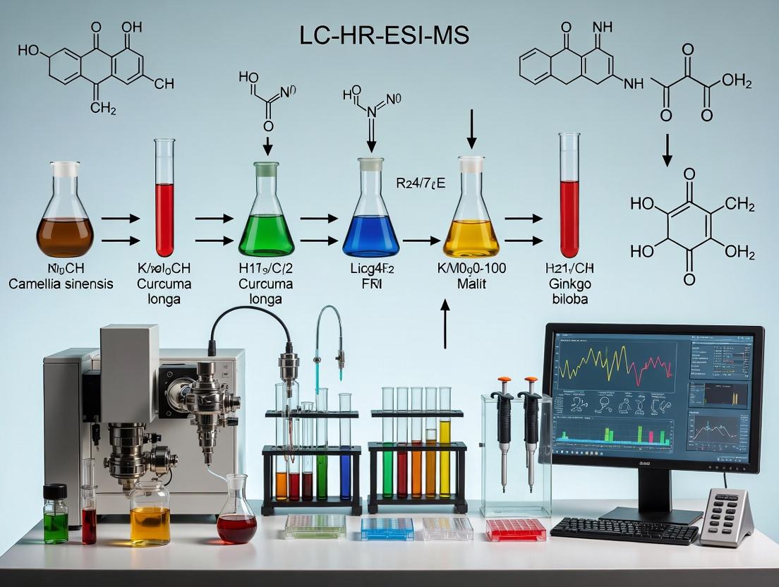
This article provides a detailed, expert-level guide to leveraging Liquid Chromatography-High Resolution Electrospray Ionization Mass Spectrometry (LC-HR-ESI-MS) for the comprehensive comparison and analysis of complex plant extracts.
Advanced LC-HR-ESI-MS for Plant Extract Profiling: A Comprehensive Guide for Natural Product Research and Drug Discovery
Abstract
This article provides a detailed, expert-level guide to leveraging Liquid Chromatography-High Resolution Electrospray Ionization Mass Spectrometry (LC-HR-ESI-MS) for the comprehensive comparison and analysis of complex plant extracts. Tailored for researchers, scientists, and drug development professionals, it explores the fundamental principles of the technique, details optimized methodologies for real-world application, offers practical troubleshooting and optimization strategies, and establishes robust frameworks for data validation and comparative analysis. The content synthesizes current best practices to enable accurate metabolite profiling, biomarker discovery, and the reliable assessment of extract quality and bioactivity for natural product-based research.
Understanding LC-HR-ESI-MS: The Essential Foundation for Plant Metabolomics
This application note explores the strategic decoupling of liquid chromatography (LC), high-resolution mass spectrometry (HRMS), and electrospray ionization (ESI) parameters to optimize the analysis of complex plant extracts. Framed within a thesis on comparative phytochemistry, we demonstrate how independent optimization of each "triad" component enhances metabolite coverage, reduces ion suppression, and improves reproducibility for robust comparative studies in natural product-based drug discovery.
In traditional LC-HR-ESI-MS workflows for plant extract analysis, parameters are often optimized as a monolithic block, leading to suboptimal conditions where compromises in chromatography or ionization limit detection. The "triad" approach advocates for the systematic, independent optimization of each segment:
- LC Fractionation: Aimed at reducing complexity per unit time delivered to the MS.
- ESI Ionization: Tuned for efficient and stable droplet formation and ion generation from diverse chemistries.
- HRMS Detection: Optimized for sensitivity, resolution, and mass accuracy across a broad m/z range.
This decoupling is critical for comparative research, where consistent, comprehensive metabolite profiling is paramount for identifying genuine biological variation over technical artifacts.
Application Notes: Optimized Parameters for Plant Extracts
Independent LC Method Development (Offline MS Detection)
Initial LC development should use standardized UV/VIS or CAD detection to establish baseline separation for major compound classes without the variability of ESI.
Table 1: Decoupled LC Method Development Protocol
| Parameter | Exploratory Range for Plant Extracts | Recommended Starting Point | Primary Optimization Goal |
|---|---|---|---|
| Column Chemistry | C18, PFP, HILIC, RP-Amide | C18 (100 x 2.1 mm, 1.7-1.9 µm) | Class-specific separation |
| Gradient | 5-95% B in 10-60 min | 5-95% Acetonitrile in 20 min | Peak capacity > 200 |
| Mobile Phase A | Water + 0.1% Formic Acid or 5 mM Ammonium Formate | Water + 0.1% Formic Acid | Protonation / Adduct control |
| Mobile Phase B | ACN or MeOH + same additive | ACN + 0.1% Formic Acid | Evaporation efficiency |
| Flow Rate | 0.2 - 0.4 mL/min | 0.3 mL/min | ESI compatibility |
| Injection Volume | 1-5 µL (of 1 mg/mL extract) | 2 µL | Column loading capacity |
ESI Source Tuning Using Standard Infusion
A mixture of standard compounds representing key phytochemical classes (e.g., alkaloid, flavonoid, terpenoid, phenolic acid) is infused directly to tune ESI parameters independently of LC flow.
Table 2: ESI Source Optimization via Standard Infusion
| Standard Mixture (1 µg/mL each) | Ionization Mode | Key Adducts Monitored | Tuning Objective |
|---|---|---|---|
| Quercetin, Berberine, Ursolic Acid, Chlorogenic Acid | Positive & Negative | [M+H]+, [M+Na]+, [M-H]-, [M+FA-H]- | Maximize S/N for all classes |
| ESI Parameter | Tested Range | Optimal Value (Q-TOF System) | Impact on Plant Metabolites |
| Capillary Voltage | 2.5 - 4.0 kV | +3.2 kV (Pos), -2.8 kV (Neg) | Impacts [M+H]+/[M+Na]+ ratio |
| Cone Voltage / Fragmentor | 20 - 120 V | 40 V (soft), 100 V (in-source CID) | Controls in-source fragmentation |
| Source Temperature | 100 - 150 °C | 120 °C | Aids desolvation of polar compounds |
| Desolvation Gas Temp | 200 - 500 °C | 350 °C | Critical for non-polar terpenoids |
| Nebulizer Gas Pressure | 20 - 60 psi | 40 psi | Stable spray for gradient elution |
HRMS Calibration and Data Acquisition
Post-ESI tuning, HRMS parameters are set for mass accuracy (< 2 ppm RMS) and resolving power (> 30,000 FWHM) using a separate calibration solution.
Table 3: HRMS Data Acquisition Settings
| Parameter | Setting for Comparative Profiling | Rationale |
|---|---|---|
| Mass Range | m/z 50 - 1200 | Covers primary & secondary metabolites |
| Scan Rate | 5 Hz | Sufficient points per chromatographic peak |
| Collision Energy | Low (6 eV) & Ramped (20-50 eV) in parallel | Simultaneous MS1 and All-Ions Fragmentation |
| Reference Mass | Lock-mass (e.g., Leu-Enkephalin) or continuous | Ensures < 2 ppm mass accuracy during runs |
| Data Format | Profile mode | Enables precise isotopic pattern analysis |
Integrated Protocol: Decoupled Method for Plant Extract Comparison
Protocol Title: Comprehensive LC-HR-ESI-MS Profiling of Plant Extracts via the Decoupled Triad Approach.
Step 1: Sample Preparation.
- Prepare dried plant material (e.g., 100 mg) from multiple biological replicates (n≥5) per species/condition.
- Extract using 1 mL of methanol:water (80:20, v/v) with 0.1% formic acid in an ultrasonic bath for 30 min.
- Centrifuge at 14,000 g for 10 min. Filter supernatant through a 0.22 µm PTFE membrane. Dilute to a final concentration of 1 mg/mL for LC-MS analysis.
Step 2: Decoupled LC Optimization (Offline).
- Using a standardized plant extract (e.g., green tea or Ginkgo biloba), run gradients from Table 1 on different columns with UV detection at 254 nm and 330 nm.
- Select the column and gradient yielding the highest peak count and best Gaussian shape for major peaks. Fix this method.
Step 3: Direct Infusion ESI Tuning.
- Continuously infuse the standard mixture from Table 2 at 10 µL/min via a syringe pump.
- Tune source parameters (Table 2) to maximize the stable ion current for all standard classes in both ionization modes. Fix these source settings.
Step 4: HRMS Calibration and Acquisition Template.
- With the ESI source optimized, infuse the manufacturer's calibration solution.
- Calibrate the mass axis to achieve RMS error < 2 ppm.
- Create an acquisition method using the calibrated mass axis, the settings from Table 3, and the fixed LC gradient timetable.
Step 5: Batch Acquisition for Comparative Study.
- Analyze all sample extracts in randomized order, bracketed by blank injections (80:20 MeOH:Water) and pooled quality control (QC) samples (a mix of all extracts).
- Acquire data in both positive and negative ESI modes as separate injections.
Step 6: Data Processing for Comparison.
- Use software (e.g., Compound Discoverer, MZmine, XCMS) to perform:
- Peak picking (S/N threshold > 5).
- Alignment (retention time tolerance < 0.1 min, mass tolerance < 5 ppm).
- Gap filling.
- Normalization (to total ion count or internal standard).
- Compound annotation using accurate mass (± 5 ppm), isotopic pattern, and MS/MS library matching (e.g., GNPS, NIST).
Visualization of Workflows & Relationships
Diagram 1: Decoupled Triad Optimization Workflow
Diagram 2: Comparative Analysis Experimental Flow
The Scientist's Toolkit: Key Research Reagent Solutions
Table 4: Essential Materials for the Decoupled Triad Protocol
| Item | Function / Role in Protocol | Example Product/Catalog |
|---|---|---|
| Mixed Phytochemical Standard | For decoupled ESI source tuning; validates ionization across compound classes. | Custom mix of Quercetin, Berberine, Ursolic Acid, Chlorogenic Acid. |
| LC-MS Grade Solvents | Minimizes background noise, ensures reproducible chromatography and ionization. | Acetonitrile, Methanol, Water with 0.1% Formic Acid. |
| Hybrid Stationary Phases | Provides orthogonal selectivity for complex plant mixtures during LC optimization. | C18, PFP, HILIC columns (e.g., 2.1 x 100 mm, 1.7 µm). |
| Mass Calibration Solution | Enables sub-2-ppm mass accuracy critical for molecular formula assignment. | ESI-L Low Concentration Tuning Mix (Agilent) or similar. |
| Internal Standard Mix | For data normalization and monitoring system stability during long batches. | Stable Isotope-Labeled Compounds (e.g., Caffeic Acid-d3, Apigenin-d6). |
| Solid Phase Extraction (SPE) Cartridges | For pre-fractionation or clean-up of crude extracts to reduce matrix effects. | Strata-X (Polymeric Reversed-Phase) 30 mg/1 mL tubes. |
| Retention Time Index Standards | Aids in alignment and compound identification across multiple batches. | Homologous series of alkyl benzoates or PFAs. |
Why High Resolution is Non-Negotiable for Plant Extract Analysis
Within the thesis context of developing a robust Liquid Chromatography-High Resolution-Electrospray Ionization-Mass Spectrometry (LC-HR-ESI-MS) method for plant extract comparison, the criticality of high-resolution mass spectrometry (HRMS) is paramount. Plant extracts represent one of the most chemically complex matrices, containing thousands of primary and secondary metabolites spanning a wide dynamic range. This application note details why high mass resolution and accuracy are indispensable for meaningful comparative phytochemical analysis, providing specific protocols and data to support this claim.
The Imperative for High Resolution: Key Data
Table 1: Comparison of MS Resolving Power Impact on Plant Extract Analysis
| Parameter | Low Resolution (Unit Mass, e.g., Quadrupole) | High Resolution (≥ 30,000 FWHM, e.g., Q-TOF, Orbitrap) | Implication for Plant Research |
|---|---|---|---|
| Mass Accuracy | 100-500 ppm | 1-5 ppm | Confident elemental composition assignment for unknowns. |
| Isobar Separation | Cannot separate isobars (e.g., C₆H₁₂O₆ vs C₁₂H₁₂). | Resolves nominal mass isobars (e.g., reserpine [m/z 609.2812] from an isobar at m/z 609.2124). | Prevents misidentification; essential for flavonoids, glycosides. |
| Dynamic Range in Complex Mix | Limited by chemical noise. | Enhanced due to extraction of exact ion chromatograms. | Detects low-abundance bioactive compounds amidst major constituents. |
| Metabolite Annotation Confidence | Low, relies on retention time and library match. | High, uses exact mass, isotope patterns, fragmentation. | Enables non-targeted discovery and reliable database queries (e.g., against GNPS, HMDB). |
| Differential Analysis | Prone to false positives/negatives from co-elution. | Accurate peak picking and alignment across samples. | Essential for finding statistically significant markers between plant varieties or treatments. |
Table 2: Representative HRMS Data for Discriminating Similar Flavonoids
| Compound | Molecular Formula | Theoretical [M-H]⁻ m/z | Measured [M-H]⁻ m/z (Orbitrap) | Mass Error (ppm) | Resolving Power Required* (FWHM) |
|---|---|---|---|---|---|
| Kaempferol-3-O-glucoside | C₂₁H₂₀O₁₁ | 447.0933 | 447.0928 | -1.1 | 18,500 |
| Luteolin-7-O-glucuronide | C₂₁H₁₈O₁₂ | 461.0725 | 461.0720 | -1.1 | 72,000 |
| Apigenin-8-C-glucoside (Vitexin) | C₂₁H₂₀O₁₀ | 431.0984 | 431.0979 | -1.2 | 25,000 |
| Apigenin-6-C-glucoside (Isovitexin) | C₂₁H₂₀O₁₀ | 431.0984 | 431.0979 | -1.2 | 167,000 |
*Minimum resolving power required to differentiate from closest common plant metabolite interference.
Detailed Experimental Protocols
Protocol 1: LC-HR-ESI-MS Method for Untargeted Plant Extract Profiling
1. Sample Preparation:
- Weigh 50.0 mg of lyophilized, homogenized plant material.
- Add 1.0 mL of 80% methanol/20% water (v/v) with 0.1% formic acid.
- Sonicate for 30 minutes at 4°C, then centrifuge at 14,000 x g for 15 minutes.
- Filter supernatant through a 0.22 µm PTFE syringe filter into an LC vial. Store at -20°C until analysis.
2. LC Conditions:
- Column: C18 reversed-phase (2.1 x 100 mm, 1.7 µm particle size).
- Mobile Phase A: Water with 0.1% formic acid.
- Mobile Phase B: Acetonitrile with 0.1% formic acid.
- Gradient: 5% B to 95% B over 25 min, hold 5 min, re-equilibrate.
- Flow Rate: 0.3 mL/min. Column Temp: 40°C. Injection Volume: 2 µL.
3. HR-ESI-MS Conditions (Orbitrap Exploris 120 example):
- Ionization Mode: ESI positive and negative, separate injections.
- Source Parameters: Capillary Temp 320°C, Sheath Gas 40, Aux Gas 10, Spray Voltage ±3.5 kV.
- Mass Analyzer: Full-scan from m/z 100-1500.
- Resolving Power: 60,000 FWHM (at m/z 200).
- Mass Accuracy: Calibrated daily with external calibration mix; lock mass (e.g., phthalates) or internal calibration recommended.
- Data-Dependent MS/MS: Top 5 most intense ions per cycle, stepped NCE 20, 40, 60.
Protocol 2: Data Processing for Comparative Analysis
1. Raw Data Conversion: Convert vendor files (.raw) to open format (.mzML) using MSConvert (ProteoWizard). 2. Feature Detection & Alignment: Use software (e.g., MZmine 3, XCMS Online) with HRMS-optimized parameters: * Noise Level: Adjusted to instrument baseline. * m/z tolerance: 5 ppm. * RT tolerance: 0.1 min. * Grouping: Use gap-filling to account for missing peaks. 3. Compound Annotation: * Query exact mass against databases (PlantCyc, COSMOS, NAP) with 5 ppm tolerance. * Interpret MS/MS spectra using CFM-ID, SIRIUS, or GNPS molecular networking. 4. Statistical Comparison: Export peak area table for multivariate analysis (PCA, OPLS-DA) in R or SIMCA to identify discriminating ions.
Visualizations
Diagram 1: HRMS Workflow for Plant Extract Comparison
Diagram 2: HRMS Resolves Isobars for Confident ID
The Scientist's Toolkit: Research Reagent Solutions
Table 3: Essential Materials for LC-HR-ESI-MS Plant Analysis
| Item | Function & Rationale |
|---|---|
| UHPLC-grade Solvents (Acetonitrile, Methanol, Water) | Minimizes background chemical noise and ion suppression, ensuring reproducible chromatography and ionization. |
| MS-grade Additives (Formic Acid, Ammonium Acetate) | Volatile buffers and pH modifiers that enhance ionization efficiency in ESI positive or negative mode without fouling the source. |
| Stable Isotope-labeled Internal Standards (e.g., ¹³C-quercetin) | Corrects for matrix effects and instrument variability, enabling semi-quantitative comparison across samples. |
| Instrument Calibration Solution | Daily verification of sub-ppm mass accuracy is non-negotiable for reliable molecular formula assignment. |
| Solid Phase Extraction (SPE) Cartridges (C18, HILIC) | For sample clean-up or fractionation to reduce complexity and concentrate low-abundance metabolites. |
| Reference Standard Compound Library | Essential for validating retention times and fragmentation patterns of key plant metabolite classes (alkaloids, phenolics, terpenes). |
| High-Purity Nitrogen/Argon Gas | Source and collision gases for ESI operation and HRMS/MS fragmentation. |
The comparative analysis of complex plant extracts using Liquid Chromatography-High Resolution Electrospray Ionization Mass Spectrometry (LC-HR-ESI-MS) demands rigorous assessment of instrument performance. Three key metrics—Resolution, Mass Accuracy, and Dynamic Range—directly determine the confidence of metabolite identification, the depth of coverage, and the ability to quantify compounds across vast concentration differences. This protocol outlines their definitions, calibration methodologies, and application in ensuring reproducible and meaningful data for phytochemical comparison and drug discovery workflows.
Definitions and Impact on Plant Extract Analysis
- Resolution (R): The ability of the mass analyzer to distinguish between two ions of similar mass-to-charge ratio (m/z). High resolution is critical for separating isobaric compounds (e.g., flavonoids with identical nominal mass) prevalent in plant matrices.
- Mass Accuracy: The difference between the measured m/z value and the true theoretical m/z of an ion, typically expressed in parts per million (ppm). High mass accuracy (< 5 ppm) is essential for reducing false positives in database searches for metabolite annotation.
- Dynamic Range: The ratio between the highest and lowest concentration of an analyte that can be detected with a specified linear response. It determines the ability to quantify both major abundant metabolites and trace-level bioactive compounds simultaneously.
Quantitative Benchmark Data for Common HR-MS Platforms
Table 1: Typical Performance Metrics of Common HR-MS Analyzers in Plant Metabolomics
| Mass Analyzer Type | Typical Resolution (FWHM at m/z 200) | Typical Mass Accuracy (ppm) | Linear Dynamic Range | Key Strengths for Plant Analysis |
|---|---|---|---|---|
| Time-of-Flight (TOF) | 20,000 - 60,000 | < 5 ppm | 10³ - 10⁴ | Fast acquisition, ideal for untargeted profiling. |
| Orbitrap | 15,000 - 500,000 | < 3 ppm (internal calibration) | 10³ - 10⁴ | Exceptional resolution and accuracy for complex mixtures. |
| Quadrupole-TOF (Q-TOF) | 20,000 - 50,000 | < 5 ppm (post-calibration) | 10³ - 10⁴ | Combines MS/MS capability with good resolution. |
| FT-ICR | > 1,000,000 | < 1 ppm | 10² - 10³ | Ultra-high resolution for definitive formula assignment. |
Table 2: Calibration Standard Compounds for Performance Verification
| Compound | Formula | Theoretical [M+H]+ (m/z) | Use Case |
|---|---|---|---|
| Caffeine | C₈H₁₀N₄O₂ | 195.08765 | Low-mass calibration, system suitability. |
| Reserpine | C₃₃H₄₀N₂O₉ | 609.28066 | Mid-mass calibration, ESI performance check. |
| Ultramark 1621 | Perfluorinated phosphazine | Multiple (e.g., 922.00980) | Broad-range mass calibration for TOF systems. |
| Leucine Enkephalin | C₂₈H₃₇N₅O₇ | 556.27644 | Lock mass for continuous internal calibration (Orbitrap, Q-TOF). |
Experimental Protocols for Performance Assessment
Protocol 4.1: Daily System Suitability and Mass Accuracy Test
Objective: Verify instrument performance meets pre-defined criteria before sample analysis. Materials: Caffeine standard (1 ppm in 50:50 Water:Acetonitrile + 0.1% Formic Acid), Lock mass solution (e.g., Leucine Enkephalin). Procedure:
- Tune and calibrate instrument according to manufacturer specifications.
- Infuse calibration standard via syringe pump or via LC flow injection at 10 µL/min.
- Acquire data in positive ion mode for 1 minute.
- Process the acquired spectrum: Identify the protonated molecule [M+H]⁺ of caffeine.
- Calculate Mass Accuracy: [(Measured m/z - 195.08765) / 195.08765] * 10⁶.
- Acceptance Criterion: Mass accuracy ≤ 5 ppm.
- For instruments with lock mass capability, enable continuous internal calibration during chromatographic runs.
Protocol 4.2: Resolution Measurement Workflow
Objective: Determine the resolving power at a specific m/z. Materials: Caffeine or reserpine standard. Procedure:
- Acquire a high-resolution spectrum of the standard as in Protocol 4.1.
- Select the monoisotopic peak of interest (e.g., m/z 195.08765 for caffeine).
- Measure the peak width at half maximum (FWHM) in m/z units.
- Calculate Resolution (R): R = m/z / Δm (FWHM).
- Example: For caffeine peak at m/z 195.08765 with FWHM of 0.005 m/z, R = 195.09 / 0.005 ≈ 39,018.
Protocol 4.3: Dynamic Range and Linearity Assessment
Objective: Establish the concentration range over which the instrument response is linear for quantitative analysis. Materials: Reserpine or a target analyte, prepared in a series of concentrations across 5-6 orders of magnitude (e.g., 0.1 pg/µL to 1000 pg/µL) in solvent matched to sample matrix. Procedure:
- Analyze concentration series in triplicate via LC-MS.
- Integrate the extracted ion chromatogram (EIC) peak area for each concentration.
- Plot peak area (log scale) against concentration (log scale).
- Fit a linear regression model to the data in the linear region.
- Define Dynamic Range: The range where the coefficient of determination (R²) ≥ 0.99 and signal-to-noise ratio (S/N) > 10 for the lower limit of quantitation (LLOQ).
Visualization of Concepts and Workflows
Key Metrics Drive Confident Metabolite ID
Performance QC in Plant Analysis Workflow
The Scientist's Toolkit: Research Reagent Solutions
Table 3: Essential Materials for LC-HR-ESI-MS Performance Validation
| Item | Function & Rationale |
|---|---|
| High-Purity Calibration Standards (e.g., Caffeine, Reserpine) | Provides known m/z values for verifying mass accuracy and resolution. Must be ≥ 98% purity to avoid interfering signals. |
| Perfluorinated Calibration Mix (e.g., Ultramark, PFTBA) | Supplies multiple, evenly spaced m/z signals across a wide range for comprehensive mass axis calibration in TOF and FT-ICR systems. |
| Lock Mass Solution | A reference compound infused during analysis for real-time, internal mass correction, dramatically improving mass accuracy (e.g., Leucine Enkephalin for ESI+). |
| Quality Control (QC) Pooled Sample | A homogeneous mixture of all plant extracts being studied. Injected repeatedly throughout the run to monitor system stability, retention time drift, and signal reproducibility. |
| LC-MS Grade Solvents (Water, Acetonitrile, Methanol) | Minimizes chemical noise and ion suppression, ensuring consistent electrospray formation and baseline signal. |
| Volatile Ion-Pairing Agents (Formic Acid, Ammonium Acetate) | Enhances protonation/deprotonation in ESI and improves chromatographic peak shape for acidic/basic metabolites without leaving residues. |
| Reference Plant Extract (e.g., Green Tea, Ginkgo) | A well-characterized, complex natural matrix used as a system suitability test to ensure overall method robustness for the intended sample type. |
Application Notes: Advancing Comparative Phytochemical Profiling
Within the scope of a thesis focused on developing an LC-HR-ESI-MS method for the comparative analysis of plant extracts, a primary challenge is the comprehensive separation and detection of a vast array of metabolites with divergent polarities, concentrations, and structural complexities. The following notes detail key considerations and recent data.
1. Scale of Metabolite Diversity: A single plant extract can contain thousands of unique metabolites, spanning from highly polar primary metabolites (e.g., sugars, amino acids) to non-polar secondary metabolites (e.g., terpenes, fatty acids). The dynamic range of concentrations can exceed 9 orders of magnitude, with crucial bioactive compounds often present in trace amounts.
Table 1: Representative Metabolite Classes and Associated Analytical Challenges
| Metabolite Class | Polarity Range | Typical Concentration Range | Key Detection Challenge |
|---|---|---|---|
| Organic Acids | High | Medium-High (μM-mM) | Matrix suppression, co-elution with sugars |
| Flavonoid Glycosides | Medium-High | Low-Medium (nM-μM) | Isomeric separation, in-source fragmentation |
| Alkaloids | Medium | Very Low-Low (pM-μM) | Ionization efficiency, background interference |
| Terpenoids (e.g., Taxanes) | Low | Very Low (pM-nM) | Low ionization, poor chromatographic retention on C18 |
| Chlorophylls/Carotenoids | Non-polar | High (in raw extract) | Column fouling, signal saturation |
2. The Critical Role of Chromatographic Separation: Reversed-phase (C18) chromatography remains the workhorse but is insufficient alone. Recent implementations employ serially coupled columns (e.g., HILIC + C18) or utilize mixed-mode stationary phases to increase metabolome coverage. Data from recent studies show a 40-60% increase in detected features when using orthogonal separation modes compared to C18 alone.
Table 2: Impact of Chromatographic Strategy on Feature Detection
| Chromatographic Strategy | Average Features Detected (per Salvia spp. extract) | Increase vs. Std. C18 | Remarks |
|---|---|---|---|
| Standard C18 (Acetonitrile/Water + 0.1% FA) | 1,250 ± 150 | Baseline | Misses most polar organics |
| HILIC (Acetonitrile/Ammonium Acetate buffer) | 900 ± 100 | -28% | Excellent for polar metabolites, poor for non-polar |
| Serial HILIC → C18 (2D-LC setup) | 2,100 ± 200 | +68% | Maximum coverage; requires complex method development |
| Mixed-Mode (C18/Anion Exchange) | 1,700 ± 180 | +36% | Good compromise for ionizable compounds |
3. High-Resolution Mass Spectrometry (HRMS) for Deconvolution: HR-ESI-MS in both positive and negative modes is mandatory. A resolving power (RP) > 60,000 FWHM (at m/z 200) is necessary to separate isobaric ions (e.g., quercetin-3-O-glucoside, m/z 463.0882 [M-H]⁻ vs. kaempferol-7-O-glucuronide, m/z 461.0726 [M-H]⁻). Data-dependent MS/MS acquisition (dd-MS²) with dynamic exclusion is standard for identification, but data-independent acquisition (DIA) methods like SWATH are gaining traction for more reproducible cross-sample comparisons.
Detailed Experimental Protocols
Protocol 1: Two-Phase Extraction for Broad Metabolite Coverage Objective: To comprehensively extract metabolites of wide-ranging polarity from 100 mg of dried, powdered plant material (e.g., Echinacea purpurea aerial parts). Materials: Cryogenic mill, lyophilizer, ultrasonicator, centrifugal vacuum concentrator. Reagents: LC-MS grade Methanol (MeOH), Acetonitrile (ACN), Water (H₂O), Dichloromethane (DCH), Formic Acid (FA). Procedure: 1. Preparation: Lyophilize fresh plant material for 48h. Powder using a cryo-mill. Weigh 100 mg ± 0.5 mg into a 15 mL polypropylene centrifuge tube. 2. Polar Phase Extraction: Add 5 mL of 80% aqueous MeOH (v/v, 0.1% FA). Sonicate in an ice-water bath for 15 min. Centrifuge at 10,000 x g, 4°C for 10 min. Transfer supernatant to a new tube. 3. Non-Polar Phase Extraction: Re-suspend the pellet in 5 mL of DCM:MeOH (2:1, v/v). Sonicate for 15 min (ice-bath). Centrifuge as before. Combine this supernatant with the first extract in a glass vial. 4. Post-Processing: Evaporate the combined extract to dryness under vacuum at 35°C. Reconstitute the residue in 1.5 mL of 50% ACN/H₂O (0.1% FA). Vortex for 2 min, sonicate for 5 min. Centrifuge at 14,000 x g for 15 min. Transfer the clarified supernatant to an LC-MS vial. Store at -80°C until analysis.
Protocol 2: LC-HR-ESI-MS Method for Comparative Profiling Objective: To separate and detect metabolites in plant extracts for untargeted comparative analysis. LC Conditions: Column: C18 column with polar embedded groups (e.g., 2.1 x 150 mm, 1.7 μm). Mobile Phase A: H₂O + 0.1% Formic Acid. Mobile Phase B: Acetonitrile + 0.1% Formic Acid. Gradient: 2% B (0-2 min), 2% to 98% B (2-45 min), 98% B (45-48 min), re-equilibration to 2% B (48-55 min). Flow Rate: 0.25 mL/min. Column Temp: 40°C. Injection Volume: 2 μL (partial loop). HRMS Conditions (Q-TOF or Orbitrap-based): Ionization: ESI, positive/negative switching. Capillary Voltage: ±3.0 kV. Nebulizer Gas: 35 psig. Drying Gas: 10 L/min, 325°C. Mass Range: m/z 70-1200. Acquisition Mode: Data-dependent (dd-MS²). Top 10 most intense precursors per cycle, exclude after 2 spectra for 30s. Dynamic precursor selection threshold: 1000 counts. Resolution: > 60,000 (for TOF: > 40,000 FWHM) in MS¹ mode; > 15,000 for MS². Collision Energies: Ramped (e.g., 20, 40, 60 eV for small molecules).
Visualization
Title: Workflow for Comparative Plant Metabolomics
Title: Key Biosynthetic Pathways in Plant Secondary Metabolism
The Scientist's Toolkit: Key Research Reagent Solutions
Table 3: Essential Materials for Plant Metabolite Separation & Detection
| Item | Function/Benefit | Example/Note |
|---|---|---|
| Mixed-Mode UPLC Columns (e.g., C18/phenyl with polar embedded groups) | Enhances retention of polar metabolites vs. standard C18, reducing "column dead time" losses. | Waters Cortecs T3, Phenomenex Kinetex F5. |
| HILIC Columns (e.g., Amide, Silica) | Separates highly polar metabolites (organic acids, sugars) that elute near void on RP columns. | Waters BEH Amide, Thermo Syncronis HILIC. |
| LC-MS Grade Solvents & Additives (Water, MeOH, ACN, FA, Ammonium Acetate) | Minimizes background ions, reduces ion suppression, and ensures column longevity and reproducibility. | Must be ≥ 99.9% purity, low particulate. |
| Stable Isotope Labeled Internal Standards (SIL-IS) | Corrects for matrix effects and ionization variability in semi-quantitative workflows. | e.g., C¹³-labeled amino acids, phenolic acids. |
| Solid Phase Extraction (SPE) Plates (Mixed-mode, C18) | Enables high-throughput cleanup and fractionation to reduce complexity and concentrate analytes. | Used prior to LC-MS for complex extracts. |
| Mass Spectral Databases & Software | Critical for annotation using accurate mass, RT, and MS/MS fragmentation patterns. | GNPS, METLIN, NIST, mzCloud, Compound Discoverer. |
| Quality Control (QC) Pool Sample | Created by combining aliquots of all study extracts; injected repeatedly to monitor system stability. | Essential for data normalization in untargeted studies. |
Application Notes
1. Chemotaxonomy and Phylogenetic Analysis: Liquid Chromatography-High Resolution-Electrospray Ionization-Mass Spectrometry (LC-HR-ESI-MS) enables the generation of comprehensive phytochemical profiles from plant extracts. By applying multivariate statistical analysis (e.g., PCA, OPLS-DA) to the high-resolution m/z and intensity data, researchers can cluster plant species or accessions based on their metabolite composition. This chemical fingerprinting provides a powerful, complementary approach to molecular phylogenetics for taxonomic classification and understanding evolutionary relationships. Key discriminating ions can be annotated to identify chemotaxonomic markers.
2. Standardization and Quality Control: For herbal drug development, batch-to-batch consistency is critical. LC-HR-ESIMS facilitates the multi-parametric standardization of complex plant extracts. It allows for the simultaneous quantitation (using external/internal standards) and qualification of multiple marker compounds—both known actives and characteristic metabolites—against a validated reference extract fingerprint. This ensures not only the content of specific markers but also the overall chemical profile, guarding against adulteration and ensuring pharmacological reproducibility.
3. Biomarker Hunting for Bioactivity: In the context of bioactivity-guided fractionation, LC-HR-ESI-MS is integral for dereplication (early identification of known compounds) and for correlating specific mass features with biological assay results. By analyzing a series of related plant extracts or fractions and their bioactivity scores, chemometric tools can pinpoint m/z features (potential novel biomarkers) whose abundance positively correlates with the measured biological effect. This guides the targeted isolation of novel bioactive lead compounds.
Table 1: Quantitative Metrics for LC-HR-ESI-MS in Core Applications
| Application | Key Measured Parameters | Typical Data Analysis Methods | Primary Output |
|---|---|---|---|
| Chemotaxonomy | m/z, RT, Intensity for 100s-1000s of features per sample. | PCA, HCA, OPLS-DA, ANOSIM. | Chemical phylogenies, identification of taxon-specific markers. |
| Standardization | Intensity/Area of 5-50 target ions; similarity indices (e.g., Pearson correlation). | Targeted quantification, fingerprint alignment, similarity analysis. | Certificate of Analysis with quantified markers & fingerprint match >90% to reference. |
| Biomarker Hunting | m/z, RT, Intensity correlated with bioassay IC50/% inhibition. | Correlation analysis (Pearson/Spearman), OPLS-DA, Volcano plots. | List of candidate biomarker ions with p-value & correlation coefficient (e.g., r > 0.8). |
Experimental Protocols
Protocol 1: LC-HR-ESI-MS Fingerprinting for Chemotaxonomic Comparison
Objective: To generate and compare chemical fingerprints of 20 different Salvia species extracts.
- Sample Prep: Weigh 50 mg of dried, powdered leaf material per species. Extract with 1 mL 80% methanol in water (v/v) via ultrasonication (30 min). Centrifuge (15,000 x g, 10 min), filter supernatant (0.22 µm PTFE).
- LC Conditions: Column: C18 (100 x 2.1 mm, 1.7 µm). Gradient: 5-95% B over 25 min (A: 0.1% Formic acid in H2O; B: Acetonitrile). Flow: 0.3 mL/min. Temperature: 40°C.
- HRMS Conditions: ESI source in positive & negative polarity modes. Full scan range: m/z 100-1500. Resolution: 70,000 (at m/z 200). Spray voltage: 3.5 kV. Sheath gas: 40 arb. Aux gas: 10 arb.
- Data Processing: Use software (e.g., Compound Discoverer, MZmine) for peak picking (S/N > 3), alignment (RT window 0.2 min, m/z tol. 5 ppm), and gap filling. Export a feature table (m/z, RT, intensity).
- Statistical Analysis: Import normalized intensity table into SIMCA/P or R. Perform Pareto-scaled PCA to visualize clustering. Use OPLS-DA to find discriminating features (VIP > 1.5). Tentatively annotate key markers via accurate mass (± 5 ppm) and MS/MS databases (e.g., GNPS).
Protocol 2: Multi-Marker Standardization of aEchinacea purpureaRoot Extract
Objective: To quantify three marker compounds and verify fingerprint consistency across 10 production batches.
- Standard & Sample Solutions: Prepare calibration curves (0.1-50 µg/mL) for cichoric acid, echinacoside, and alkamide dodeca-2E,4E,8Z,10E-tetraenoic acid isobutylamide. Prepare sample extracts as in Protocol 1 (10 mg/mL final).
- LC-HRMS Quantification: Use a targeted SIM/dd-MS2 method. For each marker, define an inclusion list with its exact m/z. LC conditions as in Protocol 1. Acquire full scans and targeted MS2 for confirmation.
- Data Analysis: Integrate extracted ion chromatograms (XIC) for each marker m/z (± 5 ppm). Plot calibration curves. Calculate concentration in each batch.
- Fingerprint Consistency: For each batch, generate a total ion chromatogram (TIC). Use professional software to calculate the similarity index (e.g., cosine correlation) of each batch TIC against the Master Reference Fingerprint (from a validated control batch). Accept batches with >92% similarity and marker content within 85-115% of specification.
Protocol 3: Biomarker Hunting for Antioxidant Activity inGinkgo bilobaLeaf Extracts
Objective: To identify LC-HRMS features correlating with DPPH radical scavenging activity across 15 different Ginkgo extracts.
- Bioactivity Profiling: Perform DPPH assay in triplicate for each extract. Express result as % scavenging at a standard concentration (e.g., 100 µg/mL). Calculate IC50 values.
- Chemical Profiling: Analyze all 15 extracts in randomized order using untargeted LC-HR-ESI-MS (as in Protocol 1).
- Correlation Analysis: Create a feature/ion intensity matrix. In R, perform Spearman rank correlation between the normalized intensity of each ion and the corresponding % DPPH scavenging. Apply false discovery rate (FDR) correction.
- Identification: Prioritize ions with a strong positive correlation (Spearman's ρ > 0.75, FDR-adjusted p < 0.01). Acquire MS/MS data for these ions, either in parallel (dd-MS2) or via a follow-up injection. Annotate using accurate mass MS/MS matching to public libraries (GNPS, MassBank) or isolated standard comparison.
Visualizations
Title: Core LC-HRMS Workflow for Plant Extract Research
Title: Biomarker Hunting via Bioassay-Correlation Workflow
The Scientist's Toolkit: Essential Research Reagents & Materials
| Item | Function in LC-HR-ESI-MS Plant Research |
|---|---|
| U/HPLC-Grade Solvents (Acetonitrile, Methanol, Water) | Essential for mobile phase and sample preparation to minimize background noise and system contamination. |
| Acid Modifiers (Formic Acid, Acetic Acid, 0.1%) | Improves chromatographic peak shape (especially for acids) and enhances positive ion mode ESI response. |
| Solid Phase Extraction (SPE) Cartridges (C18, Diol, Polyamide) | For sample clean-up, fractionation, or targeted enrichment of compound classes (e.g., phenolics, alkaloids). |
| Reference Standard Compounds | Critical for method validation, absolute quantification, and confirming metabolite identification via RT & MS/MS match. |
| Stable Isotope-Labeled Internal Standards (e.g., 13C-, 2H-labeled analogs) | Enables precise quantification by correcting for matrix effects and ionization variability in complex extracts. |
| Chemical Derivatization Reagents (e.g., MSTFA for silylation, Dansyl chloride) | Enhances detection, separation, or MS response of poorly ionizing metabolite classes (e.g., sugars, some alkaloids). |
| Quality Control Reference Material (e.g., Certified Plant Extract, Pooled QC Sample) | Injected repeatedly to monitor LC-HRMS system stability, reproducibility, and data quality throughout sequence runs. |
| MS Calibration Solution (e.g., Pierce LTQ Velos ESI Positive Ion Calibration Solution) | For regular external mass calibration of the HRMS instrument to ensure sustained sub-5 ppm mass accuracy. |
Building Your Method: A Step-by-Step LC-HR-ESI-MS Protocol for Plant Extracts
Application Notes & Protocols
Thesis Context: This document details the standardized sample preparation protocol for the LC-HR-ESI-MS-based metabolomic comparison of phytochemical profiles in medicinal plant extracts (Panax ginseng vs. Panax quinquefolius). Robust, reproducible preparation is paramount for generating high-quality, comparable data in chemotaxonomic and drug discovery research.
Experimental Protocols
Protocol 1.1: Sequential Solvent Extraction
Objective: To comprehensively extract phytochemicals of varying polarities.
- Homogenization: 100 mg of lyophilized, powdered plant root tissue is combined with 1 mL of extraction solvent and a 5 mm stainless steel bead in a 2 mL microcentrifuge tube.
- Cold Maceration: Samples are incubated at 4°C for 30 minutes to allow solvent penetration.
- Mechanical Disruption: Samples are homogenized using a high-speed bead mill homogenizer at 30 Hz for 3 minutes, then immediately placed on ice.
- Sequential Extraction:
- Step A (Polar Metabolites): Use 80% methanol/water (v/v) containing 0.1% formic acid. Sonicate in an ice-water bath for 15 minutes. Centrifuge at 15,000 × g, 4°C for 15 minutes. Transfer supernatant (Extract A) to a new vial.
- Step B (Mid-Polar to Non-Polar Metabolites): To the pellet from Step A, add 1 mL of 100% ethyl acetate. Vortex vigorously, sonicate for 15 minutes (ice-water bath). Centrifuge as above. Transfer supernatant (Extract B) to a new vial.
- Pooling & Concentration: Combine Extracts A and B. Evaporate to dryness under a gentle stream of nitrogen at 35°C.
- Reconstitution: Reconstitute the dried extract in 200 µL of initial LC mobile phase (e.g., 5% acetonitrile in water, 0.1% formic acid). Vortex for 1 min, sonicate for 5 min.
- Clarification: Centrifuge at 18,000 × g, 4°C for 10 minutes. Transfer the clarified supernatant to a low-volume LC vial with insert for analysis.
Protocol 1.2: Solid-Phase Extraction (SPE) Clean-up
Objective: To remove pigments, lipids, and other co-extracted interferents.
- Cartridge Conditioning: A mixed-mode cation-exchange SPE cartridge (e.g., Oasis MCX, 30 mg) is conditioned with 1 mL methanol, followed by 1 mL of 2% formic acid in water.
- Sample Loading: The entire reconstituted sample from Protocol 1.1 is loaded onto the cartridge.
- Wash: Interferents are removed with 1 mL of 2% formic acid in water, followed by 1 mL of methanol.
- Elution: Target analytes (including many alkaloids and semi-polar compounds) are eluted with 1 mL of 5% ammonium hydroxide in methanol.
- Post-Processing: The eluate is evaporated to dryness under nitrogen and reconstituted in 100 µL of LC mobile phase for analysis.
Protocol 1.3: Stability Assessment Protocol
Objective: To evaluate short-term (autosampler) and long-term (storage) stability.
- Preparation: Prepare a pooled Quality Control (QC) sample from all study extracts.
- Short-Term Stability: Inject the QC sample at time 0, then after 6, 12, and 24 hours in the autosampler (maintained at 10°C). Monitor peak area and retention time shift of 10 selected marker compounds.
- Long-Term Stability: Aliquot the QC sample and store at:
- -80°C (benchmark)
- -20°C
- 4°C Aliquots are analyzed at 1, 4, and 12 weeks. % Change relative to the -80°C benchmark is calculated.
- Freeze-Thaw Stability: Subject QC aliquots to three freeze-thaw cycles (-80°C to 25°C). Analyze after each cycle.
Data Presentation
Table 1: Comparison of Extraction Efficiency for Marker Compounds
| Compound Class (Example) | 80% MeOH Extraction Yield (µg/g) | Sequential (MeOH + EtOAc) Yield (µg/g) | % Increase |
|---|---|---|---|
| Ginsenoside Rb1 (Polar) | 452.3 ± 12.1 | 467.8 ± 9.5 | 3.4% |
| Ginsenoside Rg1 (Polar) | 321.7 ± 8.4 | 335.2 ± 10.2 | 4.2% |
| Polyacetylenes (Mid-polar) | 45.2 ± 5.1 | 89.6 ± 7.3 | 98.2% |
| β-Sitosterol (Non-polar) | 8.1 ± 1.2 | 22.4 ± 2.1 | 176.5% |
| Total Feature Count (LC-MS) | 1250 ± 45 | 1870 ± 62 | 49.6% |
Table 2: SPE Clean-up Recovery Rates (%) for Key Analytes
| Analytic | Without Clean-up (Area Count) | With MCX Clean-up (Area Count) | Matrix Effect Reduction (%) | Recovery (%) |
|---|---|---|---|---|
| Choline | 1,250,450 | 1,180,500 | 95% | 94.4 |
| Trigonelline | 890,200 | 845,690 | 97% | 95.0 |
| Caffeic Acid | 450,300 | 423,282 | 98% | 94.0 |
| (Internal Std.) | 1,000,000 | 955,000 | N/A | 95.5 |
Table 3: Autosampler (10°C) Stability of Selected Markers
| Compound | 0h (Peak Area) | 24h (Peak Area) | % Change | Retention Time Shift (min) |
|---|---|---|---|---|
| Adenosine | 1,504,300 | 1,488,257 | -1.07% | +0.02 |
| Ferulic Acid | 675,800 | 661,284 | -2.15% | +0.01 |
| Ginsenoside Rf | 2,125,600 | 2,114,172 | -0.54% | 0.00 |
Visualizations
Title: LC-MS Plant Analysis Workflow
Title: Extract Stability Stressors & Mitigation
The Scientist's Toolkit: Essential Research Reagents & Materials
| Item | Function & Rationale |
|---|---|
| Lyophilizer (Freeze Dryer) | Removes water from fresh plant tissue via sublimation, halting enzymatic activity and enabling stable, powdered starting material for reproducible extraction. |
| Cryogenic Mill / Bead Homogenizer | Provides efficient, rapid mechanical cell lysis in a cooled environment, ensuring complete release of intracellular metabolites while minimizing thermal degradation. |
| LC-MS Grade Solvents (MeOH, ACN, Water) | Ultra-high purity solvents are essential to minimize background chemical noise, ion suppression, and column contamination in sensitive HR-ESI-MS detection. |
| Formic Acid (Optima Grade) | Used as a mobile phase additive (0.1%) to promote protonation [M+H]+ of analytes in positive ESI mode, improving ionization efficiency and signal stability. |
| Mixed-Mode SPE Cartridges (e.g., Oasis MCX) | Provide selective clean-up by combining reversed-phase and ion-exchange mechanisms, effectively removing salts, acids, and neutral interferents while retaining target ions. |
| Silanized / Low-Bind Microcentrifuge Tubes & Vials | Reduce non-specific adsorption of low-abundance or hydrophobic compounds to plastic surfaces, maximizing recovery and data accuracy. |
| Nitrogen Evaporator | Enables gentle, concentrated removal of volatile organic solvents from extracts without excessive heat, preventing loss of thermolabile compounds. |
| Certified Reference Standards | Pure chemical standards for key plant metabolites (e.g., ginsenosides) are required for method validation, quantification, and confirming compound identities via accurate mass. |
| Internal Standard Mix (Stable Isotope Labeled) | Added at the very beginning of extraction, these correct for variability in sample preparation, ionization efficiency, and instrument performance throughout the run. |
Within the framework of developing a robust LC-HR-ESI-MS method for the comparative metabolomic analysis of complex plant extracts, chromatography optimization is paramount. Achieving comprehensive polarity coverage is essential to capture the diverse chemical space of primary and secondary metabolites. This application note details a systematic strategy for optimizing the chromatographic system—focusing on column chemistry, gradient design, and mobile phase modifiers—to maximize metabolite detection and resolution for accurate comparative research.
Column Selection Strategy
The stationary phase is the primary determinant of selectivity. For broad-polarity coverage, a multi-column screening approach is recommended. The following table summarizes the performance characteristics of modern column chemistries.
Table 1: Column Chemistries for Broad Polarity Coverage in Plant Metabolomics
| Column Chemistry | Phase Description | Polarity Coverage | Typical Applications in Plant Extracts | Key Interaction Mechanisms |
|---|---|---|---|---|
| C18 (Bridged Hybrid) | Octadecyl silica with hybrid organic/inorganic backbone | Moderate to Non-polar | Flavonoids, terpenoids, fatty acids | Hydrophobic (van der Waals) |
| HILIC (e.g., Amide, Zwitterionic) | Polar stationary phase | High to Polar | Sugars, amino acids, organic acids, glycosides | Hydrophilic partitioning, hydrogen bonding, electrostatic |
| Phenyl-Hexyl | Aromatic ring with hexyl spacer | Moderate | Isomeric separation of flavonoids, aromatic compounds | π-π interactions, hydrophobic |
| PFP (Pentafluorophenyl) | Fluorinated aromatic phase | Broad, alternative selectivity | Polar isomers, halogenated compounds, acidic/basic metabolites | Dipole-dipole, π-π, charge-transfer |
| C18 + Ion-Pairing | Standard C18 with ion-pair reagents | Extended to ionic species | Organic acids, phosphorylated compounds | Hydrophobic + ionic pairing |
Protocol 1.1: Rapid Column Screening
- Materials: UHPLC system, columns (e.g., C18, HILIC, PFP), standard mixture (log P range -4 to 10), plant extract (e.g., Arabidopsis thaliana leaf).
- Method: Use a generic, linear gradient (e.g., 5-95% organic in 15 min) with a standard mobile phase (0.1% Formic Acid in Water/Acetonitrile).
- Injection: 2 µL of standard mix and 5 µL of filtered plant extract (1 mg/mL).
- MS Detection: Full-scan HR-ESI-MS in positive and negative modes (m/z 50-1200).
- Analysis: Evaluate based on peak capacity, number of detected features, and spread of peaks across the chromatographic space. Select 2-3 complementary columns for orthogonal screening.
Gradient Optimization for Polarity Elution
The gradient profile must be tuned to the selected column to achieve uniform peak distribution.
Table 2: Optimized Gradient Profiles for Different Column Chemistries
| Column Type | Initial %B | Final %B | Gradient Time (min) | Flow Rate (mL/min) | Post-Time (min) | Notes |
|---|---|---|---|---|---|---|
| C18 (Standard) | 5% | 95% | 20 | 0.3 | 5 | Suitable for moderate non-polar metabolites. |
| C18 (Extended Polarity) | 1% | 99% | 25 | 0.3 | 7 | Better for very hydrophobic compounds (e.g., chlorophylls). |
| HILIC (Amide) | 95% | 50% | 15 | 0.4 | 8 | High starting organic. Equilibration critical. |
| Shallow Mixed-Mode | 5% | 50% | 40 | 0.25 | 5 | Used for extremely complex samples; increases peak capacity. |
Protocol 2.1: Scouting Gradient Formation
- Setup: Install selected C18 column. Prepare mobile phase A (Water + Modifier) and B (Acetonitrile + Modifier).
- Run a series of 5 gradients: 10, 20, 40, 60, and 90-minute gradients from 1% to 99% B.
- Analyze the total ion chromatogram (TIC) and base peak chromatogram (BPC). Plot the number of detected features vs. gradient time. Choose the time yielding >90% of maximum features.
- Refine the gradient shape by inserting isocratic holds (e.g., at 5% B for 2 min) or shallow segments (e.g., 30-50% B over 10 min) to resolve congested regions observed in the plant extract chromatogram.
Mobile Phase Modifier Optimization
Modifiers control ionization efficiency, peak shape, and selectivity, especially for ionizable analytes.
Table 3: Common Modifiers and Their Effects in LC-HR-ESI-MS
| Modifier | Typical Conc. | Effect on Positive ESI | Effect on Negative ESI | Primary Use Case |
|---|---|---|---|---|
| Formic Acid | 0.1% | Strong signal enhancement | Signal suppression | General metabolomics, positive mode favored. |
| Ammonium Formate | 5-10 mM | Moderate enhancement | Moderate enhancement | Better for both ion modes; volatile buffer. |
| Acetic Acid | 0.1-1% | Moderate enhancement | Less suppression than formic acid | Acidic compounds, some alkaloids. |
| Ammonium Hydroxide | 0.1% | Severe suppression | Strong signal enhancement | Basic compound analysis, negative mode. |
| Trifluoroacetic Acid (TFA) | 0.01-0.05% | Excellent peak shape (ion pairing) | Severe suppression + ion suppression | Peptides, but use with caution in MS. |
Protocol 3.1: Modifier Screening for Dual ESI Polarity Coverage
- Prepare mobile phases with four different modifiers:
- Condition A: 0.1% Formic Acid.
- Condition B: 10 mM Ammonium Formate, pH ~3.
- Condition C: 0.1% Acetic Acid.
- Condition D: 10 mM Ammonium Bicarbonate, pH ~8 (for negative mode emphasis).
- Perform the optimized gradient (from Protocol 2.1) for each condition in duplicate.
- Acquire data in both positive and negative HR-ESI-MS modes alternately.
- Process data using non-targeted feature finding (e.g., MZmine, XCMS). Metrics: Total feature count per mode, feature overlap between modes, and signal intensity for a set of internal standards spanning pKa values.
Visualization of the Method Development Workflow
Workflow for LC Method Optimization
The Scientist's Toolkit: Essential Research Reagent Solutions
Table 4: Key Reagents and Materials for LC-HR-ESI-MS Method Development
| Item | Function/Description | Example Product/Note |
|---|---|---|
| UHPLC-QTOF or Orbitrap MS | High-resolution mass spectrometer for accurate mass and sensitivity. | Necessary for untargeted metabolomics. |
| Analytical Column Set | Different chemistries for orthogonal separation. | e.g., C18, HILIC, PFP (2.1 x 100 mm, 1.7-1.9 µm). |
| LC-MS Grade Solvents | Minimizes background noise and ion suppression. | Water, Acetonitrile, Methanol. |
| Mobile Phase Additives | Analytical grade modifiers for pH and ionic strength control. | Formic Acid, Ammonium Acetate, Ammonium Hydroxide. |
| Metabolite Standard Mix | For system suitability, retention time calibration, and polarity coverage assessment. | Covering logP from -4 to 10 (e.g., uracil, caffeine, reserpine). |
| Syringe Filters | For sample cleanup prior to injection. | 0.22 µm, PTFE or Nylon. |
| Data Processing Software | For feature detection, alignment, and statistical analysis. | MZmine, MS-DIAL, Compound Discoverer. |
A systematic, iterative approach to column selection, gradient design, and modifier optimization is critical to develop an LC-HR-ESI-MS method with comprehensive polarity coverage for plant extract comparison. The protocols outlined herein enable researchers to construct a robust chromatographic method that, when integrated with HR-MS detection, forms the foundation for reliable, high-quality metabolomic data essential for drug discovery and phytochemical research.
Within the broader thesis framework, which aims to develop a robust LC-HR-ESI-MS method for the comparative metabolomic analysis of plant extracts, precise mass spectrometer parameter tuning is foundational. The reliability of comparative data—critical for identifying chemotaxonomic markers or novel bioactive compounds—is directly contingent upon optimal instrument configuration. This document details application notes and protocols for tuning three pivotal HR-MS parameter domains: Electrospray Ionization (ESI) source conditions, mass analyzer resolution settings, and mass scan ranges.
Key Parameter Domains: Protocols and Data
Electrospray Ionization (ESI) Source Condition Optimization
Objective: To maximize ion generation and transmission for a broad range of phytochemicals (e.g., alkaloids, flavonoids, terpenoids) while minimizing in-source fragmentation and adduct formation.
Experimental Protocol: Source Condition Tuning
- Standard Solution Preparation: Prepare a mixed standard solution (1 µg/mL in 50% MeOH with 0.1% formic acid) containing compounds representative of your plant extract's chemical space (e.g., quercetin, berberine, rutin, salicylic acid).
- LC Conditions: Use an isocratic flow (50:50 Water:Acetonitrile, 0.1% Formic Acid) at 0.3 mL/min into the MS.
- Initial Parameter Baseline: Set to manufacturer defaults.
- Systematic Optimization:
- Capillary Voltage: Vary from 2.5 kV to 4.0 kV in 0.25 kV steps. Monitor the total ion count (TIC) and signal-to-noise (S/N) for the [M+H]+ of each standard.
- Cone Voltage/Orifice Voltage: For the optimal capillary voltage, vary this parameter from 20 V to 80 V in 10 V steps. Monitor the parent ion intensity vs. in-source fragment formation.
- Desolvation Temperature: Vary from 250°C to 500°C in 50°C steps. Monitor sensitivity and stability of thermolabile compounds.
- Desolvation Gas Flow: Vary from 600 L/hr to 1000 L/hr.
- Source Temperature: Typically held between 100°C and 150°C.
- Data Evaluation: Plot response (peak area) vs. parameter value for each key analyte. The optimal condition is the value that maximizes S/N for the broadest set of standards without inducing excessive fragmentation.
Table 1: Optimized ESI Source Conditions for Plant Metabolite Analysis
| Parameter | Typical Range for Positive Mode | Optimized Setting (Example) | Primary Function & Impact |
|---|---|---|---|
| Capillary Voltage (kV) | 2.8 - 3.5 | 3.2 | Electrospray plume formation; too low reduces sensitivity, too high increases arcing. |
| Cone Voltage (V) | 30 - 60 | 40 | Ion guidance; higher values induce in-source fragmentation (CID). |
| Source Temperature (°C) | 120 - 150 | 130 | Aids droplet desolvation. |
| Desolvation Temperature (°C) | 350 - 450 | 400 | Complete solvent evaporation. Critical for LC flow rates >0.2 mL/min. |
| Desolvation Gas Flow (L/Hr) | 800 - 1000 | 900 | Removes solvent vapors; aids ion desolvation. |
| Cone Gas Flow (L/Hr) | 50 - 150 | 50 | Focuses the spray into the sampling cone. |
Mass Analyzer Resolution Settings
Objective: To select a resolution setting that provides sufficient accurate mass measurement for formula prediction while maintaining adequate scan speed and sensitivity for LC peak definition.
Experimental Protocol: Resolution vs. Sensitivity/Speed Trade-off
- Preparation: Infuse a standard with a known accurate mass (e.g., leucine enkephalin, [M+H]+ = 556.2766) at a constant concentration.
- Resolution Variation: Set the mass analyzer (e.g., Q-TOF, Orbitrap) to a series of defined resolution settings (e.g., 15k, 30k, 60k, 120k FWHM at m/z 200 for Orbitrap; 20k, 40k, 60k for TOF).
- Data Acquisition: Acquire profile data for 1 minute at each setting.
- Measurement:
- Record the peak width (FWHM in Da or m/z) at ~m/z 556.
- Record the absolute peak intensity (counts per second).
- Calculate the scan rate or transient length (ms/scan) for each setting.
- Analysis: Plot (a) Measured Resolution vs. Set Resolution, (b) Log(Peak Intensity) vs. Resolution, and (c) Scan Time vs. Resolution.
Table 2: Impact of Resolution Settings on Key Performance Metrics
| Resolution (FWHM @ m/z 200) | Approx. Scan Time | Relative Sensitivity | Mass Accuracy (ppm) | Recommended Use Case |
|---|---|---|---|---|
| 10,000 - 25,000 | Fast (<0.1s) | High | <5 ppm | High-speed profiling, UPLC peak definition (≥10 pts/peak). |
| 30,000 - 60,000 | Medium (0.1-0.5s) | Medium-High | <3 ppm | General untargeted metabolomics, accurate mass screening. |
| 70,000 - 120,000 | Slow (0.5-1.5s) | Medium-Low | <1-2 ppm | Isomeric separation, complex mixture analysis, isotope fine structure. |
| >120,000 | Very Slow (>1.5s) | Low | <1 ppm | Specialized research on isotope patterns or very high complexity samples. |
Mass Scan Range Selection
Objective: To define a scan range that captures all ions of interest while maximizing cycle time and sensitivity by avoiding wasted scans on empty regions.
Experimental Protocol: Defining the Analytical Scan Range
- Preliminary Full Scan: Perform an initial LC-HR-MS run on a representative plant extract with a broad scan range (e.g., m/z 50 - 1200 or 1500).
- TIC and BPC Examination: Visually inspect the total ion chromatogram and base peak chromatogram for overall quality.
- Mass Spectrum Examination:
- Extract and average mass spectra across all chromatographic peaks.
- Create a frequency histogram of detected ions (with intensity > 10^3 counts) across the m/z range.
- Range Determination:
- Lower Limit: Set to m/z 100 to avoid intense solvent cluster ions and background chemical noise (
- Upper Limit: Identify the m/z value above which no significant ions (>1% of base peak intensity) are detected. Add a 50-100 m/z margin. For most plant secondary metabolites, m/z 1200 is often sufficient.
- Lower Limit: Set to m/z 100 to avoid intense solvent cluster ions and background chemical noise (
- Validation: Re-run the sample with the narrowed scan range and compare the number of detected features and the average cycle time to the preliminary run.
Table 3: Recommended Scan Ranges for Plant Extract Analysis
| Extract Type / Analysis Goal | Recommended Scan Range (m/z) | Rationale |
|---|---|---|
| General Untargeted Profiling | 100 - 1200 | Captures vast majority of secondary metabolites (flavonoids, alkaloids, saponins). Excludes low-mass noise. |
| Polar Metabolomics (Primary Metabolites) | 50 - 1000 | Includes low molecular weight organic acids, sugars, amino acids. |
| Targeted Analysis of Large Molecules (Triterpenoids) | 200 - 1500 | Ensures capture of high mass ions from glycosylated compounds. |
| Fast Screening / High-Throughput | 100 - 900 | Narrower range reduces cycle time, increasing points per UPLC peak. |
Integrated Workflow Diagram
Diagram 1: HR-MS Parameter Tuning Workflow for Plant Extracts.
The Scientist's Toolkit: Key Research Reagents & Materials
| Item Name | Specification / Example | Function in HR-MS Tuning |
|---|---|---|
| Tuning & Calibration Solution | Sodium formate cluster ions or proprietary mix (e.g., API-TOF Tuning Mix). | Provides accurate m/z reference peaks for mass calibration and instrument performance validation. |
| System Suitability Standard Mix | Custom mix of phytochemical standards (e.g., reserpine, chlorogenic acid, rutin). | Assesses overall system performance (sensitivity, resolution, mass accuracy) under optimized conditions. |
| LC-MS Grade Solvents | Water, Methanol, Acetonitrile, with 0.1% Formic Acid or Ammonium Acetate. | Minimizes background noise and ion suppression; ensures stable ESI spray formation. |
| In-Source CID Calibrant | Caffeine or other compound with known fragmentation pattern. | Used to empirically optimize cone/orifice voltage by monitoring parent and fragment ion intensities. |
| Lock Mass Solution | Leucine Enkephalin or HP-0921, infused via reference sprayer or post-column. | Provides a real-time internal m/z correction during LC-MS runs, ensuring <2 ppm mass accuracy. |
| Data Processing Software | Vendor-specific (e.g., XCMS Online, Compound Discoverer, MZmine) and in-house databases. | For feature detection, alignment, and statistical comparison of plant extract HR-MS datasets. |
Data-Dependent and Data-Independent Acquisition Strategies (DDA vs. DIA)
Application Notes
In the context of an LC-HR-ESIMS thesis for plant extract comparison, the choice of acquisition strategy is fundamental. DDA and DIA offer complementary approaches for untargeted and comprehensive profiling of complex phytochemical mixtures.
DDA (Data-Dependent Acquisition): Ideal for discovery-phase identification of major and mid-abundance compounds. It selects the most intense precursor ions from an MS1 scan for subsequent fragmentation (MS2). This is highly effective for building spectral libraries from plant extracts but can suffer from stochastic sampling, limiting reproducibility and coverage of low-abundance ions.
DIA (Data-Independent Acquisition): Fragments all ions within sequential, pre-defined m/z isolation windows across the full mass range. This provides a complete record of all detectable analytes, ensuring high reproducibility and quantitative accuracy. It is superior for large-scale comparative studies of plant extracts where comprehensive coverage and consistent quantification across many samples are paramount. Analysis requires specialized software and often a project-specific spectral library.
Table 1: Core Characteristics of DDA and DIA in Plant Extract Analysis
| Feature | DDA (Data-Dependent Acquisition) | DIA (Data-Independent Acquisition) |
|---|---|---|
| Primary Goal | Novel compound identification, library generation. | Comprehensive, reproducible quantification across samples. |
| Precursor Selection | Intensity-based, stochastic. Top N most intense ions per cycle. | Systematic, sequential isolation of all ions in defined windows. |
| Coverage | Biased towards high-abundance ions; gaps in low-abundance data. | Comprehensive, uniform coverage of all ions within acquired range. |
| Reproducibility | Lower due to stochastic precursor selection. | Very high; acquisition is identical across all injections. |
| Quantitative Precision | Moderate; can be affected by dynamic exclusion. | Excellent due to consistent MS2 data for all analytes. |
| Data Complexity | Simpler; direct MS2-to-precursor linkage. | Complex; requires deconvolution software (e.g., DIA-NN, Skyline). |
| Ideal Use Case | Initial profiling of unknown extract, building a spectral library. | Large-scale cohort studies, precise comparison of treatment groups. |
Table 2: Performance Metrics in a Model Plant Extract Study
| Metric | DDA Result | DIA Result |
|---|---|---|
| Average Compounds Identified per Run | 250-400 (highly variable) | 450-600 (consistent) |
| CV (%) for Peak Areas (Major Compound) | 15-25% | 5-10% |
| CV (%) for Peak Areas (Low-Abundance Compound) | >30% (if triggered) | 8-12% |
| Required Spectral Library | Essential for identification. | Project-specific library or public repository. |
| Throughput (Post-Acq. Analysis) | Faster | Slower, computationally intensive. |
Experimental Protocols
Protocol 1: DDA Method for Initial Plant Extract Profiling and Library Generation
Objective: To create a comprehensive MS2 spectral library for a plant extract of interest.
Materials: See "The Scientist's Toolkit" below.
Procedure:
- Sample Preparation: Weigh 10 mg of dried, powdered plant material. Extract with 1 mL of 80% methanol/water (v/v) with 0.1% formic acid in a sonicator for 30 minutes. Centrifuge at 14,000 x g for 10 min. Filter supernatant through a 0.22 µm PVDF membrane. Dilute 1:10 with starting mobile phase for injection.
- LC Conditions:
- Column: C18 (2.1 x 100 mm, 1.7 µm).
- Flow Rate: 0.3 mL/min.
- Temperature: 40°C.
- Gradient: 5% B to 95% B over 25 min (A: Water + 0.1% FA; B: Acetonitrile + 0.1% FA). Hold 95% B for 3 min, re-equilibrate.
- HRMS DDA Parameters (Q-TOF system):
- Ionization: ESI, positive and negative modes, separate runs.
- Mass Range (MS1): 100-1500 m/z.
- Scan Rate: 12 spectra/sec.
- MS2 Acquisition:
- Isolation Window: 1.2 m/z.
- Collision Energies: Ramped (e.g., 20, 40, 60 eV).
- Select top 10 most intense ions per cycle.
- Dynamic Exclusion: 15 sec.
- Data Processing: Use vendor software (e.g., Compound Discoverer, MS-DIAL) to perform feature detection, alignment, and compound identification via database search (e.g., GNPS, mzCloud). Export consensus MS2 spectra to create a project-specific library (.MSP or .BLIB format).
Protocol 2: DIA Method for Comparative Quantitative Analysis of Plant Extracts
Objective: To quantify differences in phytochemical profiles across multiple plant extract samples (e.g., different cultivars, treatments).
Procedure:
- Sample Preparation & LC: Follow Protocol 1 steps 1 and 2. Use a randomized injection order with quality control (QC) pooled samples.
- HRMS DIA Parameter Optimization:
- Perform a preliminary DDA run to estimate the precursor m/z distribution.
- Define DIA windows: For a range of 100-1500 m/z, use 20-40 variable windows, narrower on dense regions (e.g., 100-300 m/z) and wider on sparse regions.
- DIA Acquisition (Q-Orbitrap system):
- Mass Range (MS1): 100-1500 m/z, Resolution: 60,000.
- MS2 DIA Scans: Resolution: 30,000.
- Isolation Scheme: 30 variable windows (e.g., 100-210, 210-310, ..., 1300-1500 m/z).
- Collision Energy: Stepped, normalized (e.g., 25, 35, 45%).
- Cycle Time: ~2-3 seconds.
- Data Processing & Quantification:
- Use specialized DIA software (DIA-NN, Skyline).
- Import the project-specific spectral library from Protocol 1.
- Set search parameters: precursor and fragment mass tolerance (e.g., 10 ppm), digestion enzyme set to "none."
- The software will extract fragment ion chromatograms from the DIA data, match to library spectra, and report integrated peak areas.
- Export a matrix of compound intensities across all samples for statistical analysis (e.g., PCA, ANOVA).
Visualizations
DDA Acquisition Logic Flow
DIA Sequential Window Acquisition
The Scientist's Toolkit
Table 3: Essential Research Reagent Solutions for LC-HR-ESI-MS Plant Analysis
| Item | Function in Protocol |
|---|---|
| Hybrid Quadrupole-Orbitrap or Q-TOF Mass Spectrometer | High-resolution accurate mass (HRAM) measurement for precursor and fragment ions. Essential for compound identification. |
| Reverse-Phase C18 UHPLC Column (1.7-1.9 µm particles) | Provides high-efficiency chromatographic separation of complex plant metabolite mixtures. |
| LC-MS Grade Solvents (Water, Acetonitrile, Methanol) | Minimize background noise and ion suppression; ensure reproducibility. |
| Mass Spectrometry-Compatible Acid Modifiers (Formic Acid, Acetic Acid) | Promotes protonation/deprotonation in ESI source, improving ionization efficiency and chromatographic peak shape. |
| Solid Phase Extraction (SPE) Cartridges (C18, HLB) | For sample clean-up to remove salts and pigments that cause ion suppression. |
| Chemical Reference Standards (e.g., polyphenols, alkaloids) | For verification of retention time and fragmentation patterns, and generating calibration curves. |
| Data Analysis Software (e.g., Compound Discoverer, MS-DIAL, DIA-NN, Skyline) | For processing complex DDA/DIA datasets, feature detection, identification, and quantification. |
| Spectral Library (e.g., GNPS, mzCloud, in-house) | Critical for annotating MS2 spectra in both DDA and DIA workflows. |
Within the broader thesis investigating an LC-HR-ESI-MS method for the comparative analysis of plant extracts, the transformation of raw instrumental data into interpretable chemical features is a critical step. This protocol details established workflows for both untargeted (discovery) and targeted (validation) analysis, enabling comprehensive metabolite profiling and precise quantification.
Foundational Data Processing Workflow
The initial data processing steps are common to both analytical approaches, converting raw chromatograms into a structured data matrix.
Protocol 2.1: Raw Data Conversion and Peak Picking
- Input: Raw data files (.raw, .d, .wiff formats).
- Conversion: Use conversion tools (e.g., MSConvert from ProteoWizard) to translate vendor-specific files into an open format (.mzML, .mzXML).
- Peak Detection: Process files using software (e.g., XCMS, MZmine 3).
- Centroiding: Apply to profile MS data.
- Noise Filtering: Set signal-to-noise threshold (S/N > 3-5).
- Peak Picking: Use the centWave algorithm (XCMS) or ADAP chromatogram builder (MZmine) with the following typical parameters for Q-TOF data:
Table 1: Typical Peak Picking Parameters for Plant Extract LC-HR-ESI-MS
| Parameter | Untargeted Analysis | Targeted Analysis | Function |
|---|---|---|---|
| Peak Width | 5-30 s | Defined by standard RT | Expected chromatographic peak width. |
| Mass Accuracy | < 5 ppm | < 5 ppm | Tolerance for m/z alignment. |
| SN Threshold | 3-6 | 10 | Minimum signal-to-noise for peak detection. |
| Integration | Automatic (sum, apex) | Manual review | Method for peak area quantification. |
| m/z Tolerance | 0.001-0.01 Da | 0.001 Da or 5 ppm | Tolerance for grouping adducts/isotopes. |
- Output: A peak list with m/z, retention time (RT), and intensity for each sample.
Diagram 1: Foundational Data Processing Workflow (78 chars)
Untargeted Analysis Workflow
This workflow aims to comprehensively detect all measurable analytes to identify differentially abundant features.
Protocol 3.1: Feature Alignment, Gap Filling, and Annotation
- Alignment: Correct RT drift across batches using the obiwarp or peak groups method (tolerance: 5-15 s adjusted).
- Correspondence: Group peaks from different samples representing the same feature (mz tolerance: 0.005-0.01 Da; RT tolerance: 10-30 s).
- Gap Filling: Re-integrate missing peaks in samples where they were not initially detected (fillChromPeaks function).
- Annotation (Putative):
- Isotope/Adduct Grouping: Use CAMERA or built-in algorithms to group features from the same metabolite.
- Database Search: Query processed m/z against public (e.g., GNPS, MassBank, PubChem) or in-house libraries. Use [M+H]⁺/[M-H]⁻ for ESI.
- MS/MS Fragmentation: If available, compare experimental MS2 spectra to spectral libraries (Cosine score > 0.7).
Table 2: Output Metrics from Untargeted Analysis of 10 Plant Extracts
| Processing Step | Typical Number of Features | Key Metric | Purpose | ||
|---|---|---|---|---|---|
| Initial Peak Picking | 5,000 - 15,000 per sample | Peak Area | Raw feature detection. | ||
| After Alignment & Filtering | 2,000 - 8,000 aligned features | CV < 30% in QCs | Remove irreproducible signals. | ||
| After Annotation (Putative) | 50 - 500 compounds | MS1 & MS/MS match score | Assign chemical identity. | ||
| Differential Features | 10 - 200 features | p-value < 0.05, FC > | 2 | Identify significant changes. |
Diagram 2: Untargeted Analysis for Discovery (91 chars)
Targeted Analysis Workflow
This workflow quantifies specific, pre-defined metabolites with high precision and accuracy.
Protocol 4.1: Targeted Feature Extraction and Quantification
- Compound List Definition: Create a target list with metabolite name, formula, exact mass, adduct form, and expected RT.
- Chromatogram Extraction: Extract Ion Chromatograms (XICs) for each target ion (m/z window: ±5-10 ppm).
- Peak Integration: Manually review or use automated integration with manual verification for each sample. Set consistent baseline limits.
- Calibration & Quantification:
- Use a dilution series of authentic standards (5-8 concentration levels).
- Generate a linear or quadratic calibration curve (R² > 0.99).
- Calculate concentration in unknown samples via regression.
- Quality Control: Include continuing calibration verifications (CCVs) and check standard recovery (80-120%).
Table 3: Calibration Data for Targeted Flavonoid Analysis (Hypothetical)
| Compound | Calibration Range (ng/mL) | Linear Equation | R² | LOD (ng/mL) | LOQ (ng/mL) |
|---|---|---|---|---|---|
| Quercetin | 1 - 500 | y = 12540x + 850 | 0.9987 | 0.3 | 1.0 |
| Kaempferol | 5 - 1000 | y = 8900x + 620 | 0.9991 | 1.5 | 5.0 |
| Apigenin | 2 - 750 | y = 11000x + 310 | 0.9989 | 0.6 | 2.0 |
Diagram 3: Targeted Analysis for Validation (86 chars)
The Scientist's Toolkit: Essential Research Reagent Solutions
Table 4: Key Materials for LC-HR-ESI-MS Plant Metabolomics Workflows
| Item | Function in Workflow | Example / Specification |
|---|---|---|
| LC-MS Grade Solvents | Mobile phase preparation; minimizes background ions and system contamination. | Acetonitrile, Methanol, Water (with 0.1% Formic Acid). |
| Authentic Chemical Standards | Targeted quantification: used to generate calibration curves and confirm identities. | Commercial phytochemical standards (e.g., polyphenols, alkaloids). Purity > 95%. |
| Stable Isotope-Labeled Internal Standards | Corrects for matrix effects and variability in sample preparation/injection. | ¹³C- or ²H-labeled analogs of target analytes (if available). |
| Quality Control (QC) Pool Sample | Monitors system stability; used for feature filtering (CV) in untargeted analysis. | Pooled aliquot of all study samples. |
| Procedure Blanks | Identifies background contamination originating from solvents, tubes, and preparation. | Sample prepared without plant material. |
| Retention Time Index Standards | Aids in alignment and putative identification by calibrating RT across runs. | Homologous series (e.g., alkyl carboxylic acids). |
| Database/Software Subscription | Critical for metabolite annotation via spectral and accurate mass matching. | GNPS, MassBank, PubChem, Compound Discoverer, MZmine. |
Solving Common LC-HR-ESI-MS Challenges in Plant Analysis: A Troubleshooting Manual
Diagnosing and Fixing Ion Suppression in Complex Matrices
Ion suppression is a critical matrix effect in liquid chromatography-high resolution-electrospray ionization-mass spectrometry (LC-HR-ESI-MS) that adversely impacts sensitivity, accuracy, and reproducibility. In plant extract comparison research, the complex and variable chemical background of extracts introduces significant challenges for reliable metabolite profiling and biomarker discovery. This article details diagnostic protocols and remediation strategies to ensure data integrity within a thesis focused on developing a robust LC-HR-ESI-MS method for comparative phytochemical analysis.
Diagnostic Protocols for Ion Suppression
Protocol 1: Post-Column Infusion Assay
Purpose: To visualize regions of chromatographic ion suppression/enhancement. Materials:
- LC-HR-ESI-MS system.
- Syringe pump.
- T-connector.
- Standard solution of a representative analyte (e.g., quercetin for polyphenol-rich extracts).
- Blank matrix extract (e.g., solvent-extracted plant matrix).
Method:
- Prepare a neat solution of the analyte (e.g., 1 µg/mL in mobile phase).
- Connect the syringe pump containing this solution via a T-connector between the HPLC column outlet and the ESI source.
- Inject the blank matrix extract onto the LC column and start the chromatographic gradient.
- Simultaneously, infuse the analyte at a constant rate (e.g., 10 µL/min).
- Acquire MS data in selected ion monitoring (SIM) mode for the analyte's [M+H]+ or [M-H]- ion.
- The resulting chromatogram shows the response of the continuously infused analyte. A dip in the signal indicates ion suppression caused by co-eluting matrix components.
Protocol 2: Standard Addition Method
Purpose: To quantify the absolute matrix effect (ME%) for specific target analytes. Materials:
- Pure standard compounds.
- Blank matrix extract.
- Solvent standards at equivalent concentrations.
Method:
- Prepare a series of matrix-matched standards by spiking known concentrations of the target analyte(s) into the blank matrix extract.
- Prepare an identical series of standards in pure solvent.
- Analyze all samples in triplicate using the LC-HR-ESI-MS method.
- Calculate the Matrix Effect (ME%) for each concentration:
- ME% = (Peak Area of matrix-matched standard / Peak Area of solvent standard) x 100%
- Interpret results: ME% = 100% (no effect); <100% (ion suppression); >100% (ion enhancement).
Quantitative Assessment of Ion Suppression
Table 1: Example Matrix Effect Data for Key Metabolites in a Ginkgo biloba Extract
| Metabolite Class | Compound | Retention Time (min) | ME% in Leaf Extract | ME% in Bark Extract | Severity |
|---|---|---|---|---|---|
| Flavonol Glycosides | Rutin | 12.4 | 65% | 45% | High |
| Terpene Lactones | Ginkgolide A | 18.7 | 88% | 92% | Low |
| Proanthocyanidins | Procyanidin B2 | 9.8 | 32% | 28% | Severe |
| Hydroxycinnamic Acids | Chlorogenic Acid | 5.2 | 110% | 95% | Mild Enhancement/Suppression |
Remediation Strategies and Experimental Protocols
Protocol 3: Enhanced Sample Cleanup via Solid-Phase Extraction (SPE)
Purpose: To remove interfering matrix components prior to LC-MS analysis. Materials:
- Mixed-mode cation/anion exchange SPE cartridges (e.g., Oasis MCX or MAX).
- Conditioning and elution solvents (MeOH, water, acidified/basified solutions).
- Vacuum manifold.
Method:
- Condition the SPE cartridge with 5 mL methanol followed by 5 mL acidified water (pH 2 for MCX) or basified water (pH 10 for MAX).
- Load the crude plant extract (acidified/basified as per cartridge selection).
- Wash with 5 mL acidified/basified water, followed by 5 mL methanol.
- Elute analytes using an appropriate solvent (e.g., 5 mL 5% NH4OH in methanol for MCX).
- Evaporate and reconstitute in initial mobile phase for LC-MS analysis.
- Re-evaluate ME% using Protocol 2.
Protocol 4: Chromatographic Method Optimization for Peak Separation
Purpose: To temporally separate analytes from matrix interferences. Materials:
- UPLC/HPLC system with diverse column chemistry (C18, HILIC, phenyl-hexyl).
- High-purity mobile phase additives (formic acid, ammonium acetate, ammonium fluoride).
Method:
- Column Screening: Test the separation of problematic analytes (identified in Table 1) on 2-3 columns with different selectivities.
- Gradient Optimization: Lengthen the chromatographic gradient to increase resolution, particularly in the early and mid-portions where polar matrix interferences often elute.
- Additive Screening: Test volatile additives (e.g., 0.1% formic acid vs. 5 mM ammonium acetate) to alter analyte ionization efficiency and selectivity.
- Evaluate Impact: After each modification, perform a post-column infusion (Protocol 1) to assess reduction in suppression zones.
Protocol 5: Implementation of Internal Standards
Purpose: To correct for residual, non-analyte-specific matrix effects. Materials:
- Stable Isotope-Labeled Internal Standards (SIL-IS) for each target analyte.
- Structural Analog Internal Standards.
Method:
- Ideal Correction: Add a known concentration of SIL-IS (e.g., [13C6]-Rutin) to all samples, calibrators, and QCs before extraction.
- Alternative Correction: If SIL-IS are unavailable, add one or more structural analogs with similar physicochemical properties and retention times to the analytes.
- Quantification: Use the analyte-to-internal standard peak area ratio for all calibration and quantification calculations. This corrects for suppression affecting both the analyte and its IS similarly.
The Scientist's Toolkit: Key Research Reagent Solutions
Table 2: Essential Materials for Ion Suppression Management
| Item | Function in Context |
|---|---|
| Mixed-mode SPE Cartridges (Oasis MCX/MAX) | Selective removal of ionic matrix interferents (alkaloids, acids) based on pH control. |
| Stable Isotope-Labeled Internal Standards (SIL-IS) | Gold standard for correcting matrix effects via isotope dilution mass spectrometry. |
| High-Purity Mobile Phase Additives (Optima LC/MS Grade) | Minimizes source contamination and baseline noise; different additives influence ionization. |
| LC Columns with Alternative Selectivity (e.g., HILIC, PFP) | Alters retention order to separate analytes from co-eluting matrix compounds. |
| Post-column Infusion Kit (Tee union, syringe pump) | Essential hardware for performing the diagnostic post-column infusion experiment. |
| "Blank" Matrix Lot (e.g., extracted from mutant/alternative tissue) | Critical for preparing matrix-matched calibration standards and assessing absolute ME%. |
Visualizing the Diagnostic and Remediation Workflow
Title: Workflow for Ion Suppression Management
Title: Ion Suppression Mechanisms in ESI
Thesis Context: This document details the optimization of Electrospray Ionization (ESI) source parameters as a critical component of a robust, unified LC-HR-ESI-MS method for the comparative metabolomic analysis of complex plant extracts. Consistent and sensitive ionization across diverse phytochemical classes is paramount for accurate feature detection and statistical comparison in plant-based drug discovery research.
The Influence of ESI Parameters on Key Phytochemical Classes
Optimal ESI conditions vary significantly based on the physicochemical properties of the analyte. The following table summarizes the primary effects of core parameters on major phytochemical classes.
Table 1: ESI Parameter Optimization Guide for Major Phytochemical Classes
| Phytochemical Class | Example Compounds | Optimal Polarity | Key Parameter Sensitivity | Optimal Trend (Positive Mode) | Rationale |
|---|---|---|---|---|---|
| Alkaloids | Nicotine, Berberine, Vinblastine | Positive (+) | S-Lens RF, Sheath Gas Temp | High S-Lens RF (50-90%); Moderate-High Sheath Gas (300-350°C) | Basic nitrogen atoms readily protonate. High RF levels improve transmission of often low m/z ions. |
| Flavonoids | Quercetin, Rutin, Naringenin | Negative (-) or Positive (+) | Capillary Voltage, Drying Gas Temp | Negative mode often preferred for aglycones. In (+), lower Capillary Voltage (~3.0 kV) for fragile glycosides. | Aglycones ionize well via deprotonation [-H]⁻. Glycosides can form adducts [M+H]⁺/[M+Na]⁺; harsh conditions cause in-source fragmentation. |
| Terpenoids | Artemisinin, Taxol, Ginsenosides | Positive (+) or Negative (-) | Vaporizer Temp, Sheath/Aux Gas Flow | High Vaporizer Temp (350-400°C); High Aux Gas Flow for high MW (e.g., >800 Da). | Low volatility requires high desolvation temperatures. High MW compounds need efficient solvent stripping (aux gas). |
| Phenolic Acids | Caffeic acid, Gallic acid, Ellagic acid | Negative (-) | Capillary Voltage, Skimmer Voltage | Low-Moderate Skimmer Voltage (15-25 V) | Readily deprotonate. Low skimmer voltage minimizes fragmentation of the fragile carboxylic group. |
| Saponins | Aescin, Glycyrrhizic acid | Negative (-) | Drying Gas Flow, Nozzle Voltage | High Drying Gas Flow (10-12 L/min); Optimized Nozzle Voltage (~500 V) | High surface activity; efficient droplet drying is critical. Nozzle voltage fine-tunes stability for large, labile molecules. |
Core Experimental Protocol: Systematic ESI Source Optimization
Protocol 2.1: Iterative Parameter Screening Using a Standard Mixture
- Objective: To empirically determine the optimal ESI source parameters for maximal response across a representative standard mixture.
- Materials: See "Scientist's Toolkit" below.
- Procedure:
- Prepare a standard mixture (≈ 1 µg/mL each) containing at least one representative from each class in Table 1 in a suitable LC-MS solvent (e.g., 50:50 MeOH:H₂O).
- Install and calibrate the mass spectrometer (e.g., Orbitrap, Q-TOF) according to manufacturer specifications.
- Using a direct infusion pump (syringe pump), introduce the standard mixture at a constant flow rate (e.g., 5-10 µL/min).
- Begin with manufacturer-recommended default ESI source settings.
- Implement a univariate or multivariate (e.g., Design of Experiments, DoE) approach. For a univariate screen, systematically vary one parameter while holding others constant.
- Capillary Voltage: Scan from 2.5 kV to 4.0 kV in 0.25 kV increments.
- Sheath Gas Flow: Scan from 20 to 60 arb units in steps of 10.
- Aux Gas/Heater Temperature: Scan from 250°C to 400°C in steps of 50°C.
- S-Lens RF Level/Skimmer Voltage: Scan from 40% to 90% or 15V to 50V.
- At each setting, record the total ion current (TIC) and the extracted ion chromatogram (EIC) peak area for the [M+H]⁺, [M+Na]⁺, [M-H]⁻, or other dominant adduct of each standard.
- Plot the response (peak area) of each analyte versus the parameter value. Identify the compromise "sweet spot" that provides robust signal for the majority of compound classes critical to your study.
- Validate optimal parameters via LC-MS analysis of the standard mixture using your developed chromatographic method.
Visualization of Workflows and Relationships
Diagram 1: ESI Optimization Workflow for Plant Metabolomics
Diagram 2: Relationship of ESI Parameters to Analytical Outcomes
The Scientist's Toolkit: Essential Research Reagents & Materials
Table 2: Key Materials for ESI Source Optimization in Phytochemical Analysis
| Item | Function & Relevance |
|---|---|
| Phytochemical Standard Mix | A curated set of pure compounds (alkaloids, flavonoids, terpenoids, etc.) serving as analytical benchmarks for parameter optimization and system suitability testing. |
| LC-MS Grade Solvents (MeOH, ACN, Water with 0.1% Formic Acid/Ammonium Acetate) | High-purity solvents minimize background noise and ion suppression. Acid/volatile salt additives promote [M+H]⁺/[M+Na]⁺ or [M-H]⁻ formation. |
| Syringe Pump & Hamilton Syringe | For precise, low-flow direct infusion of standard mixtures, allowing isolation of ESI effects from LC separation variables. |
| Design of Experiments (DoE) Software (e.g., Fusion, MODDE, JMP) | Enables efficient multivariate optimization of interacting ESI parameters, saving time and resources compared to univariate screening. |
| High-Resolution Mass Spectrometer (Orbitrap or Q-TOF) | Provides the accurate mass measurement necessary for identifying unknown phytochemicals in complex extracts during method validation. |
| Data Processing Platform (e.g., Compound Discoverer, MZmine, XCMS) | Essential for batch processing of optimized LC-HRMS data, enabling peak picking, alignment, and comparative statistical analysis across plant samples. |
Addressing Carryover, Peak Tailing, and Chromatographic Artefacts
Application Notes for LC-HR-ESI-MS Method Optimization in Plant Extract Profiling
Within a thesis focused on developing a robust LC-HR-ESI-MS method for the comparative analysis of complex plant extracts, managing chromatographic performance is paramount. Artefacts such as carryover, peak tailing, and ghost peaks directly compromise data integrity, leading to false positives/negatives and inaccurate metabolite quantification. These notes detail protocols to diagnose and mitigate these issues.
Quantitative Impact of Common Artefacts
The following table summarizes typical observations and their effects on data quality.
Table 1: Common Chromatographic Artefacts in LC-HR-ESI-MS of Plant Extracts
| Artefact | Primary Cause | Observed Effect | Impact on Comparative Analysis |
|---|---|---|---|
| Carryover | Incomplete elution/adsorption of analytes in flow path or column. | Peaks appearing in blank runs after a high-concentration sample. | Misidentification of low-abundance compounds; skews relative abundance ratios. |
| Peak Tailing | Secondary interactions with active sites (e.g., free silanols) in column. | Asymmetric peak shape (Tailing Factor >1.5). | Imprecise integration, reduced resolution, inaccurate quantification. |
| Ghost Peaks/System Peaks | Leachables from vial septa, tubing, or column; mobile phase impurities; sample carryover in autosampler. | Peaks in blank injections not attributable to prior sample. | False-positive metabolite identification; background chemical noise. |
| Baseline Drift | Mobile phase gradient mismatch, temperature fluctuations, column degradation. | Rising or falling baseline during gradient. | Obscures low-intensity peaks; complicates integration. |
| Peak Splitting | Column voiding, mismatched sample solvent, or multiple analyte conformations. | Single analyte presenting as two or more partially resolved peaks. | Misinterpretation as two distinct metabolites; quantification errors. |
Detailed Diagnostic and Mitigation Protocols
Protocol 2.1: Systematic Carryover Investigation Objective: To identify the source and extent of sample carryover in the LC-HR-ESI-MS system. Materials: LC-HR-ESI-MS system, blank solvent (e.g., 80:20 Water:Acetonitrile + 0.1% Formic Acid), high-concentration standard mix (e.g., a cocktail of phenolic acids, alkaloids relevant to plant extracts), and injection vials.
- Equilibrate System: Condition column with starting mobile phase for ≥10 column volumes.
- Inject Blank: Perform a blank injection (blank solvent). Acquire full-scan HR-MS data.
- Inject High-Concentration Sample: Inject the standard mix at a concentration 10x higher than the expected maximum in plant extracts.
- Sequential Blank Series: Immediately perform a series of at least three consecutive blank injections.
- Data Analysis: Process chromatograms. Calculate carryover % as: (Peak Area in 1st Blank / Peak Area in High-Conc Sample) * 100. A value >0.1% typically requires intervention.
- Source Identification:
- Autosampler: Perform syringe/needle wash protocol audit. Increase wash volume and optimize wash solvent strength (e.g., include 5-10% DMSO for non-polar compounds).
- Column: Extend gradient hold at high organic percentage. Implement a strong wash step post-run (e.g., 95% organic for 5-10 min).
- ESI Source: Check for droplet accumulation on the sampling cone or ion transfer tube. Clean if necessary.
Protocol 2.2: Correcting Peak Tailing for Basic Metabolites Objective: To improve peak shape for basic compounds (e.g., alkaloids) prone to silanol interactions. Materials: LC column (C18), mobile phase additives: Formic Acid (FA), Trifluoroacetic Acid (TFA), Ammonium Formate, and Acetic Acid.
- Baseline Analysis: Inject a test mix containing a basic standard (e.g., nicotine or berberine). Use standard conditions: Water (0.1% FA) / Acetonitrile (0.1% FA).
- Calculate Tailing Factor (TF): TF = W₀.₀₅ / 2f, where W₀.₀₅ is peak width at 5% height and f is distance from peak front to peak apex. Target TF < 1.5.
- Optimize Additive:
- Option A (MS-compatible): Replace FA with 0.1% Acetic Acid (pKa difference provides better protonation).
- Option B (For severe tailing): Add 0.1% Triethylamine (TEA) as a competing base to block silanols. Note: TEA can cause significant ion suppression in ESI+ and should be used sparingly.
- Option C (Ion-pairing): For very persistent issues, use 0.01% Heptafluorobutyric Acid (HFBA). Warning: HFBA causes intense ion suppression and contaminates the system; dedicate a system for its use.
- Evaluate: Re-inject test mix with new additive. Compare TF, signal intensity (may be reduced with HFBA/TEA), and mass accuracy.
Protocol 2.3: Ghost Peak Identification Workflow Objective: To determine if ghost peaks originate from the chromatographic system or the sample.
- Run Solvent Blanks: Use different batches of every solvent (water, organic, acids) in the mobile phase.
- Run Instrument Blank: Perform an "air" injection (inject nothing).
- Vial/Septum Test: Place blank solvent in vials from different lots, with different septa (e.g., PTFE/silicone vs. pre-slit PTFE).
- Column Contribution Test: Disconnect column, replace with zero-dead-volume union, and run a gradient. Peaks indicate issues from autosampler, tubing, or solvents.
- HR-MS Analysis: Accurately determine the m/z of the ghost peak. Use isotope patterns and fragment spectra to hypothesize identity (e.g., phthalates from plastics, silicones from septa).
Visualization of Method Optimization Workflow
Title: Artefact Diagnosis and Mitigation Workflow
The Scientist's Toolkit: Key Research Reagent Solutions
Table 2: Essential Materials for Mitigating LC-MS Artefacts
| Item | Function & Rationale |
|---|---|
| Pre-slit PTFE/Silicone Septa | Minimizes leachable (e.g., silicone oils) entering sample, reducing ghost peaks. |
| LC-MS Grade Solvents & Water | Ultra-purity limits baseline UV and MS noise from organic and ionic impurities. |
| High-Purity Mobile Phase Additives (e.g., Optima FA, AA) | Reduces background chemical noise and improves signal-to-noise for trace analytes. |
| Needle Wash Solvent (e.g., 50:50 MeOH:Water with 5% DMSO) | Strong, semi-polar wash reduces carryover of diverse phytochemicals from autosampler needle. |
| Guard Column (matching analytical column chemistry) | Traps particulates and strongly retained compounds from plant extracts, protecting the analytical column. |
| Surface-Deactivated (Low Bleed) Autosampler Vials | Reduces adsorption of analytes to vial walls and introduction of polymeric contaminants. |
| Endcapped C18 Columns or Specialty Columns (e.g., Biphenyl, HILIC) | Alternative phases mitigate specific interactions (silanols, π-π) causing tailing or poor retention. |
| In-Line Filter (0.5 µm) | Placed between injector and column, it traps particulates from crude extracts, preventing column frit blockage. |
| Mobile Phase Degasser | Continuous helium sparging minimizes dissolved gas, preventing baseline instability and pump issues. |
1. Introduction: The LC-HR-ESI-MS Data Deluge in Phytochemical Research In the context of LC-HR-ESI-MS (Liquid Chromatography-High Resolution-Electrospray Ionization-Mass Spectrometry) analysis for plant extract comparison, each sample run generates gigabytes of raw spectral data. A single research thesis involving hundreds of extracts across multiple conditions can easily produce tens of terabytes. Efficient management of this data is critical for metabolite profiling, biomarker discovery, and comparative analysis.
2. Application Notes: Strategic Frameworks
2.1. Hierarchical Storage Management (HSM) Strategy
- Hot Storage (SSD/NVMe): House raw data from active runs and processed files for immediate re-analysis (<30 days old). Enables rapid data access for iterative processing.
- Warm Storage (High-Performance NAS): Store curated feature tables, aligned chromatograms, and identified compound libraries. Accessed weekly for statistical analysis.
- Cold Storage (Tape or Object Storage with Glacier Class): Archive raw
.raw(Thermo),.d(Agilent),.wiff(Sciex) files post-project completion. Retrieval latency acceptable for audit or legacy comparison.
2.2. Data Processing Pipeline Architecture A modular pipeline is essential. Pre-processing (centroiding, noise filtering) occurs on high-I/O servers. Feature detection and alignment are distributed across a compute cluster. Final statistical analysis is performed on workstations with loaded datasets.
Table 1: Quantitative Data Summary for a Typical LC-HR-ESI-MS Plant Study
| Metric | Per Sample Run | Per Study (500 extracts) | Recommended Storage Tier |
|---|---|---|---|
| Raw File Size | 1.2 - 2.5 GB | 600 - 1250 GB | Cold (Archive) |
| Processed Feature Table (.csv) | 50 - 150 MB | 25 - 75 GB | Warm (NAS) |
| Peak Detection Features | 3,000 - 10,000 | 1.5M - 5M Features | Warm (NAS) |
| Aligned Compounds Post-Filtering | ~500 - 2,000 | ~250K - 1M Compounds | Warm/Hot (Active Analysis) |
| Retention Time Tolerance | ± 0.1 min | N/A | Processing Parameter |
| Mass Accuracy Tolerance | < 5 ppm | N/A | Processing Parameter |
3. Experimental Protocols
Protocol 3.1: Efficient Feature Extraction and Alignment for Large Datasets
- Objective: Convert raw LC-HR-ESI-MS files into a aligned, compound-by-sample feature table.
- Software: MZmine 3, XCMS Online/CMS, or custom Python/R scripts using
xcmsandCAMERApackages. - Procedure:
- Batch Import: Load all
.mzMLfiles (converted from vendor formats) into the processing environment. - Mass Detection: Apply noise level threshold (e.g., 1.0E4) to centroid data.
- Chromatogram Building: Set
m/ztolerance to 5-10 ppm. - Deconvolution: Use the "Local Minimum Search" or "Wavelets" algorithm to resolve co-eluting ions.
- Isotope & Adduct Grouping: Identify [M+H]+, [M+Na]+, [M+NH4]+, [M-H]- etc., using
CAMERAor built-in tools. - Alignment: Perform RT correction (LOESS) using a pooled QC sample run intermittently. Align features across samples with
m/z(5-10 ppm) and RT (0.1-0.3 min) windows. - Gap Filling: Fill missing peaks using peak area from raw data.
- Export: Generate a
.csvmatrix (features × samples) for statistical analysis.
- Batch Import: Load all
Protocol 3.2: Database-Driven Metabolite Annotation & Storage
- Objective: Annotate aligned features and store results in a queryable database.
- Tools: Sirius, CSI:FingerID, GNPS, Internal SQLite/PostgreSQL DB.
- Procedure:
- Query Preparation: Export MS1 ([M+H]+/[M-H]-) and MS/MS spectra (if available) for top features.
- Database Search: Batch query against MassBank, GNPS, HMDB, and in-house plant metabolite libraries.
- Scoring & Ranking: Filter results by mass error (< 5 ppm), MS/MS spectral similarity (Cosine score > 0.7), and biological relevance.
- Database Storage: Store annotations in a relational database with fields:
Feature_ID,m/z,RT,Adduct,Tentative_Name,Database_ID,Score,SMILES. Link back to the main feature table viaFeature_ID.
4. Visualization of Workflows
Diagram Title: LC-MS Data Management & Analysis Pipeline
Diagram Title: Metabolite Annotation & Database Storage Workflow
5. The Scientist's Toolkit: Essential Research Reagents & Solutions
Table 2: Key Research Reagent Solutions for LC-HR-ESI-MS Plant Analysis
| Item | Function & Role in Data Management |
|---|---|
| QC Pooled Sample | A homogenized mixture of all study extracts. Injected regularly to monitor system stability and enable robust retention time alignment/correction across massive datasets. |
| Internal Standard Mix | A cocktail of stable isotope-labeled or non-native compounds (e.g., chloramphenicol-d5, 13C-caffeine). Used for mass accuracy calibration and data normalization, improving cross-batch comparability. |
| Solvent Blanks (MeOH/H2O) | Critical for identifying and subtracting background ions and carryover during data processing, reducing false positive features. |
| Reference Spectral Library | Purchased or curated database of known plant metabolite MS/MS spectra. Essential for high-confidence annotation, forming the core of the annotation database. |
| Data Processing Software Suite | (e.g., MZmine, XCMS, MS-DIAL). Platforms with batch processing and scripting capabilities are necessary to handle hundreds of files automatically. |
| Relational Database System | (e.g., PostgreSQL, SQLite). Provides structured storage for feature-annotation relationships, enabling efficient querying and integration with statistical results. |
| High-Throughput Storage Hardware | NVMe drives for active processing and a Network-Attached Storage (NAS) system with redundant drives (RAID) for secure, shared access to processed data. |
Calibration and System Suitability Tests for Long-Term Reproducibility
Within the context of developing a robust Liquid Chromatography-High Resolution-Electrospray Ionization-Mass Spectrometry (LC-HR-ESI-MS) method for comparing complex plant extracts, ensuring long-term reproducibility is paramount. This document details the calibration strategies and system suitability test (SST) protocols necessary to maintain data fidelity across extended analytical campaigns, crucial for drug development research where batch-to-batch consistency of natural product libraries is assessed.
Core Calibration Protocols for LC-HR-ESI-MS
Mass Accuracy Calibration
High-resolution mass spectrometers require frequent mass axis calibration to maintain sub-ppm accuracy.
Protocol: Direct Infusion High-Resolution Calibration
- Reagent: Use a certified calibration solution specific to the mass spectrometer manufacturer (e.g., sodium formate, ESI Tuning Mix).
- Preparation: Dilute the stock solution per manufacturer instructions in a 50:50 v/v mixture of LC-MS grade water and acetonitrile with 0.1% formic acid.
- Procedure: Introduce the solution via a syringe pump or a diverted LC flow at a low flow rate (e.g., 3-10 µL/min).
- Acquisition: Acquire data in the appropriate polarity mode over the required m/z range (e.g., 50-2000 Da) for 1-2 minutes.
- Software Execution: Run the automated calibration algorithm. The system fits the known m/z values of the calibrant ions to the detected peaks.
- Acceptance Criterion: The root mean square (RMS) error of the fit must be < 1 ppm for instruments with FT-based detectors (Orbitrap, FT-ICR).
Table 1: Typical Calibrant Ions for Positive Ion Mode ESI
| m/z (Theoretical) | Ion Composition |
|---|---|
| 118.08626 | C2H4NO2Na+ (Na formate) |
| 322.04812 | C8H8O6Na3+ |
| 622.02896 | C12H12O14Na5+ |
| 922.00980 | C16H16O22Na7+ |
| 1321.99064 | C20H20O30Na9+ |
LC System Performance Calibration
Protocol: Retention Time Stability and Peak Shape Assessment
- Reagent: Prepare a standardized test mixture of moderately polar, UV- and MS-active compounds (e.g., caffeine, reserpine, sulfadimethoxine).
- Chromatography: Inject the mixture using the standard method for plant extracts (e.g., C18 column, water/acetonitrile gradient).
- Measurements: Calculate for each peak:
- Retention Time (RT) relative standard deviation (RSD) over 5 consecutive injections.
- Peak Asymmetry Factor (As) at 10% peak height.
- Theoretical plates (N).
- Acceptance Criteria: RT RSD < 0.5%; As between 0.9 and 1.2; N > 10,000 for a 15 cm column.
Comprehensive System Suitability Test (SST) Protocol
SSTs must be performed at the beginning of each analytical batch to verify the entire system's readiness.
Pre-Analytical SST Workflow:
Table 2: SST Acceptance Criteria for Plant Extract Profiling
| Parameter | Target Value | Measurement Procedure |
|---|---|---|
| Mass Accuracy (MS1) | ≤ 2 ppm (internal lock mass) | Deviation of known lock mass ion (e.g., phthalate, siloxane) detected in background or spiked standard. |
| Mass Resolution | ≥ 70,000 @ m/z 200 | FWHM measurement of a single, isolated calibrant ion peak. |
| Signal-to-Noise (S/N) | ≥ 1000:1 for 1 pg reserpine | Peak-to-peak noise evaluation in a selected ion chromatogram. |
| Retention Time RSD | < 0.3% (n=3) | Calculated from three consecutive injections of the SST mix. |
| Peak Area RSD | < 2.0% (n=3) | Calculated from the extracted ion chromatogram peak area of a reference compound. |
| Column Pressure | Within ±10% of baseline | Comparison to pressure recorded for new column under same conditions. |
Long-Term Monitoring and Corrective Actions
Maintain a control chart for a key SST metric (e.g., lock mass accuracy) to track system drift.
The Scientist's Toolkit
Table 3: Essential Research Reagent Solutions for LC-HR-ESI-MS Calibration & SST
| Item Name / Solution | Function & Rationale |
|---|---|
| ESI Tuning Mix (Certified) | Provides a set of ions with precisely known m/z ratios across a wide mass range for high-accuracy mass calibration of the analyzer. Essential for maintaining sub-ppm accuracy. |
| Lock Mass Solution | A constant infusion of a known compound (e.g., phthalates, siloxanes) during data acquisition for real-time internal mass correction, compensating for short-term instrument drift. |
| Retention Time Marker Mix | A cocktail of 5-10 compounds covering a range of polarities. Used to verify LC system stability, gradient performance, and column integrity over hundreds of injections. |
| Needle Wash Solution | A strong solvent (e.g., 90% organic) with appropriate additives to minimize carryover between injections of complex plant extracts, which may contain sticky, non-volatile compounds. |
| Mobile Phase Additives (LC-MS Grade) | Ultra-pure acids (formic, acetic) and buffers (ammonium formate/acetate). Critical for controlling ionization efficiency and chromatographic peak shape in both ESI+ and ESI- modes. |
| System Suitability Test (SST) Sample | A standardized, multi-component sample that mimics the complexity of plant extracts. Run at the start of each batch to holistically assess chromatographic and mass spectrometric performance against pre-set criteria. |
Ensuring Credibility: Validation, Statistical Comparison, and Reporting Standards
Method Validation Parameters for Qualitative and Semi-Quantitative Analysis
Within the broader thesis on employing Liquid Chromatography-High Resolution-Electrospray Ionization-Mass Spectrometry (LC-HR-ESI-MS) for the comparative analysis of complex plant extracts, rigorous method validation is paramount. This research seeks to identify biomarkers, authenticate species, and compare phytochemical profiles. The qualitative aspect focuses on the confident identification of compounds, while the semi-quantitative aspect enables the comparison of relative abundances across samples. This document outlines the essential validation parameters, detailed protocols, and application notes to ensure the reliability, reproducibility, and scientific soundness of the generated data.
Core Validation Parameters: Definitions and Acceptance Criteria
Validation for qualitative and semi-quantitative methods differs from full quantitative validation. The following parameters are critical.
Table 1: Key Validation Parameters for Qualitative LC-HR-ESI-MS Analysis
| Parameter | Objective | Recommended Acceptance Criteria | Protocol Reference |
|---|---|---|---|
| Specificity/Selectivity | Ensure the method can distinguish the analyte from matrix components. | No significant interference at the retention time and accurate mass of the target analyte(s) in blank matrix. MS/MS spectral purity match > 80% against standard/library. | Protocol 2.1 |
| Limit of Identification (LOI) | The lowest concentration at which an analyte can be reliably identified. | Consistent, reproducible identification (via accurate mass, isotopic pattern, MS/MS) in ≥ 9 out of 10 replicates. | Protocol 2.2 |
| Robustness | Assess method resilience to deliberate, small variations in operational parameters. | Identification remains consistent across variations (e.g., column temp ±2°C, mobile phase pH ±0.1, flow rate ±5%). | Protocol 2.3 |
| System Suitability | Verify system performance before and during analysis. | Based on reference standard: RT RSD < 2%, mass accuracy < 3 ppm, intensity RSD < 5%. | Protocol 2.4 |
Table 2: Key Validation Parameters for Semi-Quantitative LC-HR-ESI-MS Analysis
| Parameter | Objective | Recommended Acceptance Criteria | Protocol Reference |
|---|---|---|---|
| Linearity & Working Range | Establish the relationship between response and concentration for relative comparison. | For internal standard or major markers: R² > 0.98 over 2-3 orders of magnitude. Visual inspection of residuals. | Protocol 2.5 |
| Precision (Repeatability & Intermediate Precision) | Measure the closeness of agreement between a series of measurements. | Peak area RSD < 20% at low levels, < 15% at mid/high levels (within-day and between-day). | Protocol 2.6 |
| Extraction Efficiency/Matrix Effect | Assess compound recovery and ion suppression/enhancement. | Consistent matrix factor (80-120%) and recovery (70-120%) across multiple lots of plant matrix. | Protocol 2.7 |
| Stability | Evaluate analyte stability in matrix under various conditions (autosampler, bench-top, freeze-thaw). | Relative response vs. fresh sample within ±15%. Identification characteristics unchanged. | Protocol 2.8 |
Detailed Experimental Protocols
Protocol 2.1: Assessing Specificity/Selectivity
- Prepare Samples: Inject (a) Blank solvent, (b) Blank matrix extract (e.g., extract from a plant species devoid of target compounds, if possible, or a diluted, unrelated extract), (c) Standard solution of target analytes, (d) Spiked matrix (blank matrix + standards), and (e) Actual test samples.
- LC-HR-ESI-MS Analysis: Use the developed chromatographic and MS method. Data should be acquired in full-scan (e.g., m/z 100-1500) and data-dependent MS/MS mode.
- Data Analysis: Overlay chromatograms. Check for peaks in the blank matrix at the RT of targets. For HRMS, assess mass accuracy (deviation < 3 ppm) and isotopic pattern fit (mSigma < 20). For confirmatory identification, compare acquired MS/MS spectra to reference standards or spectral libraries (e.g., GNPS, MassBank).
Protocol 2.2: Determining Limit of Identification (LOI)
- Prepare a dilution series of a standard or a representative sample spiked with known compounds, spanning from a clearly identifiable level down to a near-noise level.
- Inject each concentration level at least 10 times in a randomized sequence.
- Process data using the established identification criteria (e.g., RT window ±0.2 min, mass accuracy < 5 ppm, minimum MS/MS spectral match score).
- The LOI is the lowest concentration at which the identification criteria are met in ≥ 90% of the replicates.
Protocol 2.3: Robustness Testing via Experimental Design
- Define Critical Factors: Select 4-5 variables (e.g., column temperature, gradient start %B, flow rate, ESI source voltage, sheath gas flow).
- Design Experiment: Use a fractional factorial design (e.g., Plackett-Burman) to test each factor at a high (+) and low (-) level around the nominal value.
- Execute Runs: Analyze a control sample (standard mix or pooled extract) for each experimental condition.
- Evaluate Responses: Monitor key outputs: RT shift, peak area of critical markers, resolution of a critical pair, and mass accuracy. Identify factors causing significant deviations.
Protocol 2.4: System Suitability Test (SST) Protocol
- SST Solution: Prepare a fresh solution containing 3-5 reference compounds covering the polarity range of the method and a stable, non-interfering internal standard (e.g., sulfadimethoxine for ESI-).
- Injection Sequence: Inject the SST solution at the beginning of the sequence, after every 6-10 experimental samples, and at the end.
- Acceptance Criteria Check: For each SST injection, calculate: (a) RT RSD% for each compound, (b) Mass accuracy (ppm) for the base peak, (c) Peak area RSD% for the internal standard, (d) Signal-to-Noise ratio for a low-level compound. The sequence is valid only if all SST injections meet pre-set criteria.
Protocol 2.5: Establishing Semi-Quantitative Response Function
- Prepare Calibration Solutions: Since absolute standards are often unavailable for all plant metabolites, use a surrogate approach. Prepare a series of dilutions (e.g., 1, 5, 10, 25, 50, 100 µg/mL) for (a) an internal standard (IS) added to all samples, and/or (b) available marker compounds.
- Analysis: Inject each level in triplicate.
- Data Processing: For each analyte, plot the relative response (Analyte peak area / IS peak area) against the relative concentration (Analyte conc. / IS conc.) or simply the analyte concentration. Apply linear and non-linear (e.g., quadratic) regression models.
- Assessment: Select the model with the best fit (R²) and most random residual plot. The working range is where precision and accuracy (if known) meet acceptance criteria.
Protocol 2.6: Precision (Repeatability & Intermediate Precision)
- Sample Preparation: Prepare six independent replicates of a homogeneous, pooled plant extract at low, medium, and high concentration levels (e.g., by dilution).
- Repeatability (Intra-day): A single analyst prepares and analyzes all six replicates of each level in one sequence on one day.
- Intermediate Precision (Inter-day): A second analyst (or the same analyst) repeats the process on a different day, using a different column lot and standard preparation.
- Calculation: For each target ion/compound, calculate the %RSD of the (Area/IS Area) ratio for the six replicates within the same day (repeatability) and across both days (intermediate precision).
Protocol 2.7: Assessing Extraction Efficiency & Matrix Effect
- Prepare Three Sets of Samples (in triplicate):
- Set A (Pure Solution): Standards in mobile phase.
- Set B (Spiked Post-Extraction): Blank plant matrix extracted, then known amount of standard spiked into the final extract.
- Set C (Spiked Pre-Extraction): Known amount of standard spiked into the plant matrix before the extraction procedure.
- Analyze all sets using the LC-HR-ESI-MS method.
- Calculate:
- Matrix Factor (MF) = Peak area in Set B / Peak area in Set A. (MF > 1 = ion enhancement; MF < 1 = ion suppression).
- Extraction Recovery (%) = (Peak area in Set C / Peak area in Set B) * 100.
Protocol 2.8: Stability Experiments
- Prepare a large batch of pooled plant extract and aliquot.
- Subject aliquots to various conditions:
- Bench-top stability: Leave at room temp for 4-24h.
- Autosampler stability: Keep in autosampler (e.g., 10°C) for 24-72h.
- Freeze-thaw stability: Cycle between -20°C/-80°C and room temp 3-5 times.
- Long-term stability: Store at -80°C for 1, 3, 6 months.
- Analyze stability samples against a freshly prepared control sample from the same pool.
- Evaluate: Compare both relative peak response (Area/IS Area) and identification parameters (RT, mass accuracy, MS/MS) for key metabolites.
The Scientist's Toolkit: Research Reagent Solutions
Table 3: Essential Materials for LC-HR-ESI-MS Method Validation in Phytochemistry
| Item | Function & Rationale |
|---|---|
| Reference Standard Compounds | Critical for determining RT, MS/MS spectra, LOI, and establishing semi-quantitative response. Ideally, use >2 chemical classes relevant to the plant study. |
| Stable Isotope-Labeled Internal Standards (SIL-IS) | Gold standard for correcting matrix effects and variability. Not always available for plant metabolites; surrogate SIL-IS from similar chemical classes are used. |
| Well-Characterized, Homogeneous Plant Reference Material | Serves as a positive control and quality control sample for precision, robustness, and long-term method performance monitoring. |
| Blank Matrix | Essential for specificity testing. Can be a related plant species known to lack target compounds, or a simulated matrix of sugars/amino acids representative of plant tissue. |
| High-Purity Solvents & Additives (LC-MS Grade) | Minimize background noise, reduce ion source contamination, and ensure reproducible chromatography and ionization. |
| Quality Control (QC) Pooled Sample | A pool of all study samples, injected repeatedly throughout the sequence. Monitors system stability and data quality via multivariate statistics (e.g., PCA of QC metrics). |
Method Validation Workflow & Decision Pathway
Data Analysis & Reporting Workflow for Plant Extract Comparison
Application Notes
Within the broader thesis on developing an LC-HR-ESI-MS method for comparative plant extract research, multivariate statistical analysis is indispensable for interpreting complex, high-dimensional metabolomic datasets. These tools transform raw spectral data into actionable biological insights, enabling robust comparison of plant extracts for drug discovery.
Principal Component Analysis (PCA): An unsupervised method used for initial exploratory data analysis. PCA reduces dimensionality by identifying principal components (PCs) that capture maximum variance in the LC-HR-ESI-MS dataset. It is primarily used to assess overall clustering, detect outliers, and observe inherent patterns between sample groups (e.g., different plant species, harvesting seasons, or extraction methods) without a priori class information. It answers the question: "What is the natural variation in my dataset?"
Orthogonal Projections to Latent Structures-Discriminant Analysis (OPLS-DA): A supervised method that separates the variance in the X-matrix (peak intensities) related to a predefined class Y (e.g., treated vs. control, Species A vs. Species B) from variance orthogonal (unrelated) to class membership. This enhances model interpretability by focusing on metabolomic features most responsible for the discrimination. It is the critical tool for identifying potential biomarker ions that differentiate plant extracts. Validation (e.g., CV-ANOVA, permutation testing) is mandatory to prevent overfitting.
Heatmaps: Used to visualize the relative abundance (ion intensity) of identified marker features or key metabolites across all samples. Combined with hierarchical clustering of both rows (metabolites) and columns (samples), heatmaps provide an intuitive color-coded summary of patterns, revealing co-regulated metabolite families and sample similarities.
Table 1: Comparative Summary of Multivariate Methods in Plant Extract Analysis
| Aspect | PCA (Unsupervised) | OPLS-DA (Supervised) | Heatmap (Visualization) |
|---|---|---|---|
| Primary Goal | Explore variance, find outliers, see natural clustering. | Find features discriminating pre-defined classes. | Visualize patterns in large data matrices. |
| Class Label Use | No (ignores class information). | Yes (requires class information). | Optional (often used with clustering). |
| Key Output | Scores plot (sample clustering), Loadings plot (influential variables). | S-plot or VIP list (identifies discriminatory ions), Validated model. | Color-coded matrix of metabolite abundance across samples. |
| Role in Thesis | Initial QC of LC-HR-ESI-MS runs, check technical reproducibility, observe gross group separation. | Identify statistically significant marker ions/biomarkers for plant extract classification. | Present final results of differential metabolites across all study groups. |
| Typical R²X / Q² Values | R²X (cum): >0.5-0.8 (explained variance). Q²: Not the primary focus. | R²Y: High (>0.7), Q²: >0.5 is good; must be validated. | Not applicable. |
Experimental Protocols
Protocol 1: Data Preprocessing for Multivariate Analysis from LC-HR-ESI-MS Objective: Convert raw LC-HR-ESI-MS files into a peak intensity table suitable for statistical software.
- Feature Detection & Alignment: Use software (e.g., XCMS Online, Progenesis QI, MS-DIAL) to detect chromatographic peaks, align features across all samples by m/z and retention time (RT), and fill in missing peak intensities.
- Filtering: Remove features with high relative standard deviation (RSD > 30%) in QC pooled samples (technical replicates) to eliminate noise. Remove features present in blanks.
- Normalization: Apply total ion count (TIC) or probabilistic quotient normalization (PQN) to correct for global intensity differences between samples.
- Scaling: Apply Pareto or unit variance scaling to the peak table to reduce the influence of high-abundance ions and give low-abundance but potentially significant metabolites more weight.
Protocol 2: Executing and Validating an OPLS-DA Model Objective: Create a validated supervised model to identify discriminatory ions.
- Data Input: Import the preprocessed peak intensity table (X-matrix) and a defined class vector (Y-matrix, e.g., 0 for Control, 1 for Treated) into a software platform (SIMCA, MetaboAnalyst, R with
roplspackage). - Model Training: Construct an OPLS-DA model. Specify one predictive component and, typically, one orthogonal component.
- Model Validation:
- Cross-Validation: Use 7-fold cross-validation to calculate the model's predictive accuracy parameter (Q²).
- Permutation Test: Permute the Y-labels (e.g., 200-1000 times) and rebuild models. The original model's R² and Q² intercepts should be significantly higher than those of permuted models (p-value for CV-ANOVA < 0.05).
- Biomarker Extraction: Extract the Variable Importance in Projection (VIP) scores. Ions with VIP > 1.0 are considered significant. Further refine using the S-plot (p[1] vs p(corr)[1]) to select ions with high correlation and reliability. Putatively annotate these ions using accurate mass (HR-MS) and MS/MS fragmentation databases.
Protocol 3: Creating an Interpretable Clustered Heatmap Objective: Visualize the abundance patterns of key differential metabolites.
- Data Selection: Select the normalized intensity data for the top discriminatory ions (e.g., VIP > 1.5) identified by OPLS-DA.
- Clustering: In software (e.g., MetaboAnalyst, R
pheatmap), apply hierarchical clustering to both rows (metabolites) and columns (samples) using Euclidean distance and Ward's linkage method. - Visual Tuning: Choose a color palette (e.g., blue-white-red for low-medium-high abundance). Scale the data by row (z-score) to emphasize relative differences in metabolite levels across samples. Clearly annotate sample class groups.
Diagrams
Title: Multivariate Analysis Workflow for LC-HR-ESI-MS Data
Title: OPLS-DA Concept & Biomarker Extraction
The Scientist's Toolkit
Table 2: Essential Research Reagents & Software for LC-HRMS Metabolomic Analysis
| Item / Solution | Function / Purpose |
|---|---|
| LC-HR-ESI-MS System (e.g., Q-Exactive, TripleTOF) | High-resolution mass spectrometer coupled to UHPLC for separating and accurately measuring metabolite m/z. |
| C18 Reverse-Phase Column (e.g., 2.1 x 100 mm, 1.7-1.8 µm) | Core column for separating a broad range of semi-polar to non-polar metabolites in plant extracts. |
| Solvents: LC-MS Grade Water, Methanol, Acetonitrile | Mobile phase components. High purity is critical to minimize background noise and ion suppression. |
| Formic Acid / Ammonium Formate (LC-MS grade) | Common mobile phase additives to aid ionization (positive/negative mode) and improve chromatographic peak shape. |
| QC Pooled Sample | A homogeneous mixture of small aliquots from all study samples. Run repeatedly to monitor instrument stability and for data filtering. |
| Leucine Enkephalin or similar | Standard for continuous lock mass correction in ESI-MS systems, ensuring high mass accuracy. |
| Data Processing Software (e.g., XCMS, Progenesis QI, MS-DIAL) | Converts raw instrument files into aligned peak intensity tables for statistical analysis. |
| Multivariate Analysis Software (e.g., SIMCA, MetaboAnalyst, R) | Performs PCA, OPLS-DA, and generates heatmaps. Enables statistical validation and biomarker discovery. |
| Metabolite Databases (e.g., HMDB, MassBank, GNPS) | Used for putative annotation of discriminatory ions based on accurate mass and MS/MS fragmentation patterns. |
Chemical Fingerprinting and Marker-Based Comparison Strategies
Chemical fingerprinting and marker-based strategies are central to modern phytochemical analysis within the framework of an LC-HR-ESI-MS (Liquid Chromatography-High Resolution-Electrospray Ionization-Mass Spectrometry) thesis. These approaches enable the comprehensive comparison of complex plant extracts for drug discovery, quality control, and metabolomic studies. Chemical fingerprinting provides a holistic, untargeted profile of a sample's chemical composition, while marker-based analysis offers targeted, quantitative assessment of specific known compounds. The synergy of both methods, powered by LC-HR-ESI-MS, allows researchers to establish robust links between chemical profiles and biological activity, ensuring reproducibility and advancing the scientific validation of plant-derived therapeutics.
Core Application Notes
Untargeted Chemical Fingerprinting for Extract Comparison
This approach generates a comprehensive, high-resolution mass spectrometric profile of all detectable ions in a plant extract. It is ideal for discovering novel compounds, assessing overall chemical consistency, and identifying sample outliers.
- Primary Use: Discovery phase, quality assessment of raw materials, detection of adulteration.
- Data Output: Total Ion Chromatogram (TIC) and a list of m/z features (retention time, m/z, intensity).
- Comparison Metric: Multivariate statistical analysis (PCA, PLS-DA) of the aligned feature table to highlight chemical similarities and differences between extract batches or species.
Targeted Marker-Based Comparison for Standardization
This strategy focuses on the identification and quantification of a defined set of known bioactive or characteristic compounds (markers). It is critical for standardizing extracts for preclinical and clinical development.
- Primary Use: Standardization of active pharmaceutical ingredients (APIs) from natural products, batch-to-batch consistency, potency determination.
- Data Output: Extracted Ion Chromatograms (XICs) for each marker, with accurate mass confirmation and calibration curves for quantification.
- Comparison Metric: Quantitative results (e.g., µg/mg of extract) for each marker, allowing direct statistical comparison (t-test, ANOVA) between samples.
Integrated Strategy: From Fingerprinting to Marker Validation
The most powerful application involves using untargeted fingerprinting to identify discriminatory features between bioactive and inactive extracts, followed by the isolation and structural elucidation of these features to establish them as validated quality markers for future targeted analyses.
Detailed Experimental Protocols
Protocol 1: LC-HR-ESI-MS Method for Untargeted Fingerprinting
Objective: To acquire comprehensive chemical profiles of plant extracts for comparative analysis.
- Sample Preparation: Weigh 10.0 mg of dried, homogenized plant extract. Dissolve in 1.0 mL of 80% methanol/water (v/v) with 0.1% formic acid. Sonicate for 15 minutes, centrifuge at 14,000 x g for 10 minutes. Filter supernatant through a 0.22 µm PTFE syringe filter into an LC vial.
- LC Conditions:
- Column: C18 reversed-phase (e.g., 2.1 x 100 mm, 1.7 µm particle size).
- Mobile Phase A: Water with 0.1% formic acid.
- Mobile Phase B: Acetonitrile with 0.1% formic acid.
- Gradient: 5% B to 95% B over 25 minutes, hold for 5 minutes, re-equilibrate.
- Flow Rate: 0.3 mL/min. Column Temp: 40°C. Injection Volume: 2 µL.
- HR-ESI-MS Conditions:
- Ionization Mode: ESI positive and negative (separate runs or rapid polarity switching).
- Mass Range: m/z 100-1500.
- Resolution: > 60,000 FWHM.
- Source Parameters: Sheath gas: 40 arb, Aux gas: 10 arb, Spray voltage: 3.5 kV (positive) / 3.0 kV (negative), Capillary temp: 320°C.
- Quality Control: Inject a pooled quality control (QC) sample (a mixture of all study extracts) at regular intervals (e.g., every 6 injections) to monitor system stability.
- Data Processing: Use software (e.g., MS-DIAL, XCMS Online, Compound Discoverer) for peak picking, alignment, and gap filling to generate a feature table (RT, m/z, intensity). Normalize data (e.g., to total ion count or QC sample).
Protocol 2: Targeted Quantification of Defined Chemical Markers
Objective: To accurately quantify a pre-determined set of marker compounds in multiple plant extract samples.
- Standard & Sample Prep: Prepare a serial dilution of certified reference standards for each target marker. Prepare samples as in Protocol 1, step 1.
- LC-HR-ESI-MS Method: Use a method optimized for separation of the target markers, often shorter than the untargeted method. Employ Parallel Reaction Monitoring (PRM) or Targeted SIM/dd-MS2 mode.
- Inclusion List: Input exact m/z values of precursor ions for each marker.
- Settings: Use a narrow isolation window (e.g., 1-2 m/z), high resolution for both MS1 and MS2, and a normalized collision energy optimized for each compound.
- Quantification & Validation:
- Generate a calibration curve for each marker using the reference standards.
- Quantify markers in samples using the extracted ion chromatogram (XIC) of the precursor or a characteristic fragment ion. Use the calibration curve for concentration determination.
- Validate method for linearity (R² > 0.99), LOD, LOQ, precision (%RSD), and recovery (%).
Data Presentation
Table 1: Quantitative Comparison of Marker Compounds in Three Echinacea purpurea Extract Batches
| Marker Compound | Theoretical m/z [M+H]+ | Batch A (µg/mg) | Batch B (µg/mg) | Batch C (µg/mg) | RSD (%) |
|---|---|---|---|---|---|
| Cichoric Acid | 473.0725 | 12.5 ± 0.3 | 11.8 ± 0.4 | 13.1 ± 0.2 | 5.2 |
| Echinacoside | 785.2550 | 5.2 ± 0.1 | 4.9 ± 0.2 | 5.5 ± 0.1 | 6.1 |
| Alkamide 8/1 | 278.2480 | 0.8 ± 0.05 | 1.1 ± 0.06 | 0.7 ± 0.04 | 25.0 |
Table 2: Summary of Untargeted Fingerprinting Data Analysis for Five Hypericum Species
| Species | Total Features Detected | Features Unique to Species | Key Discriminatory Compound Class (from PCA loadings) |
|---|---|---|---|
| H. perforatum | 1450 | 120 | Hyperforins, Phloroglucinols |
| H. androsaemum | 1120 | 85 | Xanthones |
| H. calycinum | 980 | 65 | Prenylated Phloroglucinols |
| H. canariense | 1320 | 110 | Flavonol Glycosides |
| H. kouytchense | 1250 | 95 | Biflavonoids |
Visualizations
Workflow for LC-HR-ESI-MS Plant Extract Comparison
Thesis Context of Fingerprinting and Marker Strategies
The Scientist's Toolkit: Research Reagent Solutions
| Item | Function in LC-HR-ESI-MS Plant Analysis |
|---|---|
| HPLC/MS Grade Solvents (Acetonitrile, Methanol, Water) | Ensure minimal background noise, prevent ion suppression, and maintain system cleanliness for reproducible HRMS data. |
| Acid/Base Modifiers (Formic Acid, Ammonium Formate/Acetate) | Volatile mobile phase additives that improve chromatographic peak shape (ion pairing) and enhance ionization efficiency in ESI. |
| Certified Reference Standards | Pure chemical compounds used for accurate mass confirmation, method development, and generating calibration curves for absolute quantification of markers. |
| Solid Phase Extraction (SPE) Cartridges (C18, HILIC, etc.) | For sample clean-up to remove interfering matrix components (e.g., salts, chlorophyll) and pre-concentrate analytes of interest. |
| Internal Standards (Stable Isotope-Labeled Analogs, e.g., 13C, 2H) | Correct for variability in sample preparation, injection volume, and ionization efficiency; crucial for precise quantitative analysis. |
| Quality Control Reference Material (e.g., NIST Botanicals, In-House Pooled Extract) | A consistent sample analyzed throughout a batch to monitor instrument performance (retention time shift, mass accuracy, signal intensity). |
Database Matching and Annotation Confidence Levels (Levels 1-5)
The precise identification of metabolites in complex plant extracts is a cornerstone of phytochemical research and natural product-based drug discovery. Within the context of a broader thesis utilizing Liquid Chromatography-High Resolution-Electrospray Ionization-Mass Spectrometry (LC-HR-ESI-MS) for plant extract comparison, establishing a standardized system for reporting identification confidence is paramount. This framework ensures that downstream analyses, such as chemotaxonomic comparisons or bioactivity correlations, are built on transparent and reliable annotations. The application of a five-level confidence system, aligned with community guidelines from the Metabolomics Standards Initiative (MSI) and the Cosmos consortium, provides this critical rigor.
The Five-Tier Confidence Level Framework
This protocol defines the criteria for assigning confidence levels from 1 (highest confidence) to 5 (lowest confidence) for compound annotations derived from LC-HR-ESI-MS data matched against chemical databases.
Table 1: Confidence Levels for Metabolite Annotation in LC-HR-ESI-MS
| Confidence Level | Description | Required Evidence (LC-HR-ESI-MS Context) | Typical MSI Level |
|---|---|---|---|
| Level 1: Confirmed Structure | Unequivocal identification by direct comparison with an authentic standard analyzed under identical analytical conditions. | 1. Match of retention time (RT) ± 0.1 min or <2% RSD.2. Exact mass (m/z) match < 5 ppm.3. MS/MS fragmentation pattern match (dot product score > 0.8, e.g., using GNPS). | 1 |
| Level 2: Probable Structure | Library spectrum match without RT match, or orthogonal spectral data supporting a specific isomer. | 1. Exact mass (m/z) match < 5 ppm.2. High spectral similarity to public/commercial MS/MS library (e.g., GNPS, MassBank, NIST).3. Possibly supported by in silico MS/MS prediction tools (e.g., CFM-ID, SIRIUS). | 2 |
| Level 3: Tentative Candidate | Annotation to a compound class or a small group of isomers based on diagnostic evidence. | 1. Exact mass (m/z) match < 5 ppm to a molecular formula.2. Characteristic neutral losses or fragment ions indicative of a compound class (e.g., flavonoid O-hexoside, diterpene).3. Literature or database support for plausible presence in the plant species. | 3 |
| Level 4: Unknown but Characterized | Chemically characterized feature distinct from background, but insufficient evidence for class assignment. | 1. Accurate mass detection.2. Reproducible LC-MS peak with associated isotopic pattern and/or adducts.3. May have MS/MS spectrum but no library match. Often reported as "m/z_RT". | 4 |
| Level 5: Unknown | Uncharacterized metabolite signal. No meaningful annotation possible. | 1. Peak detected but no reliable accurate mass or interpretable MS/MS. Often excluded from further biological interpretation. | 5 |
Experimental Protocols for Level Assignment
Protocol 3.1: Level 1 Confirmation Using Authentic Standards
Objective: To unambiguously confirm the identity of a target compound in a plant extract. Materials: LC-HR-ESI-MS system, authenticated chemical standard, solvent-matched plant extract, blank solvent (e.g., 80% methanol). Procedure:
- Standard Preparation: Prepare a dilution series of the authentic standard in appropriate solvent to span the expected concentration in the extract.
- Co-Chromatography: Inject, in sequence: a. Blank solvent. b. Standard solution (single compound). c. Plant extract. d. Plant extract spiked with the standard at a concentration ~50% of the estimated endogenous level.
- Data Acquisition: Acquire data in full-scan (e.g., m/z 100-1500) and data-dependent MS/MS modes for all injections.
- Analysis: Using the instrument software or open-source tools (e.g., MZmine 3): a. Align the chromatograms. b. Confirm co-elution: The RT of the feature in the extract must match the standard's RT within a pre-defined window (e.g., ±0.1 min). c. Confirm mass accuracy: The [M+H]+ or [M-H]- ion m/z must match within 5 ppm. d. Confirm MS/MS identity: Compare MS/MS spectra using a spectral similarity score (e.g., dot product ≥ 0.8). The spiked sample should show increased intensity for the target ion without peak splitting.
Protocol 3.2: Level 2 Annotation via MS/MS Spectral Library Matching
Objective: To assign a probable structure based on high-resolution MS/MS spectral matching. Materials: Raw LC-HR-MS/MS data file (.raw, .mzML format), spectral library (e.g., GNPS, MassBank, in-house curated library). Procedure:
- Data Preprocessing: Convert raw files to an open format (.mzML) using MSConvert (ProteoWizard). Perform feature detection, alignment, and gap filling using software like MZmine 3 or MS-DIAL.
- MS/MS Export: Export consensus MS/MS spectra (averaged across the peak) for features of interest in .mgf format.
- Database Search: Upload .mgf file to the Global Natural Products Social Molecular Networking (GNPS) platform or use a local installation of SIRIUS.
- Parameter Setting (GNPS Example):
- Precursor Ion Mass Tolerance: 0.02 Da (or 5 ppm).
- MS/MS Fragment Ion Tolerance: 0.02 Da.
- Minimum Cosine Score: 0.7 (higher threshold, e.g., >0.8, increases confidence).
- Library: Choose "All Libraries" or restrict to specific ones (e.g., plant-relevant).
- Annotation: Review top matches. A Level 2 assignment requires:
- High spectral similarity score.
- Plausible adduct formation.
- Consideration of the reported natural source of the matched compound relative to the studied plant.
Protocol 3.3: Level 3 Annotation by Compound Class Characterization
Objective: To assign a feature to a specific compound class or isomer group. Materials: Processed LC-HR-MS/MS data, in silico fragmentation tools, metabolic pathway databases (e.g., KEGG, PlantCyc). Procedure:
- Molecular Formula Assignment: Use the accurate mass (error < 5 ppm) and isotopic pattern (e.g., mSigma < 20 in instruments like Thermo Orbitrap) to assign a molecular formula with software (e.g., Compound Discoverer, SIRIUS).
- Diagnostic Fragment Analysis: Interpret the MS/MS spectrum for characteristic neutral losses or fragments.
- Example for Flavonoid-O-hexoside: Loss of 162.0528 Da (hexose), followed by losses of CO (28 Da) and/or the aglycone-specific fragments (e.g., m/z 153 for dihydroxybenzoic acid).
- In Silico Prediction: Input the molecular formula and MS/MS spectrum into SIRIUS/CSI:FingerID or CFM-ID to obtain a ranked list of candidate structures. Cross-reference these candidates with known metabolites reported in the plant genus/family using databases like PubChem or ChemSpider.
- Assignment: Annotate as "Dihydrokaempferol-O-hexoside isomer" or similar, documenting the diagnostic evidence.
Visualization of Workflows and Relationships
Diagram Title: Metabolite Annotation Confidence Level Workflow
Diagram Title: Role of Confidence Framework in Plant Research Thesis
The Scientist's Toolkit: Research Reagent Solutions & Essential Materials
Table 2: Key Reagents and Materials for Confidence-Level Experiments
| Item | Function & Application in Protocol |
|---|---|
| Authenticated Chemical Standards | Pure compounds for Level 1 confirmation. Used as reference for RT, accurate mass, and MS/MS spectra. Source from vendors like Sigma-Aldrich, Extrasynthese, or Phytolab. |
| LC-MS Grade Solvents (Acetonitrile, Methanol, Water) | Ensure minimal background noise and ion suppression. Critical for reproducible chromatography and accurate mass measurement. |
| Formic Acid / Ammonium Acetate (LC-MS Grade) | Common volatile additives for mobile phases. Acidic conditions (formic) promote [M+H]+ in ESI+, while ammonium buffers aid [M+NH4]+ or [M-H]- in ESI-. |
| MS Calibration Solution | Ensures ongoing mass accuracy of the HR-MS instrument (e.g., Pierce LTQ Velos ESI Positive Ion Calibration Solution for Orbitrap systems). Required for <5 ppm error. |
| Solid Phase Extraction (SPE) Cartridges (e.g., C18, HILIC) | Used for pre-analytical clean-up or fractionation of crude plant extracts to reduce complexity and ion suppression, improving detection of minor metabolites. |
| Spectral Library Access | Subscription or open access to curated MS/MS libraries (e.g., GNPS, MassBank, mzCloud, NIST). The foundation for Level 2 annotations. |
| Data Processing Software | Platforms like MZmine 3 (open source), MS-DIAL (open source), or Compound Discoverer (commercial) for feature detection, alignment, and integration with database searches. |
| In Silico Prediction Tools | Software suites like SIRIUS/CSI:FingerID or CFM-ID for predicting molecular formulas and structures from MS/MS data, supporting Level 3 annotations. |
Thesis Context: Within a broader thesis focused on developing a robust LC-HR-ESI-MS method for the comparative analysis of complex plant extracts, precise metabolite identification is paramount. This protocol details the systematic benchmarking of accurate mass and tandem MS data against reference standards and curated libraries to ensure reliable annotation, a critical step for comparative metabolomics and drug discovery from botanical sources.
Key Research Reagent Solutions & Essential Materials
| Item | Function / Explanation |
|---|---|
| Certified Reference Standards (Pure Chemical Compounds) | Authentic, high-purity metabolites for definitive identification. Used to establish exact retention time (RT), accurate mass, and fragmentation spectrum under the specific LC-MS conditions. |
| Stable Isotope-Labeled Internal Standards (e.g., 13C, 15N) | Correct for matrix effects and ionization suppression in complex plant extracts, enabling semi-quantification and improving data quality for benchmarking. |
| MS-Compatible Solvents & Additives (LC-MS Grade) | High-purity solvents (water, acetonitrile, methanol) and volatile additives (formic acid, ammonium acetate) minimize background noise and ion suppression, ensuring optimal MS performance. |
| Quality Control (QC) Pooled Sample | A homogenized mixture of all study plant extracts. Injected repeatedly throughout the analytical sequence to monitor system stability, data reproducibility, and for normalization in non-targeted workflows. |
| Commercial/Public MS/MS Libraries (e.g., NIST, MassBank, GNPS) | Curated databases of experimental and in silico MS/MS spectra for tentative annotation when reference standards are unavailable. |
| Retention Time Index (RTI) Marker Kit | A set of exogenous compounds spiked into every sample to calibrate and correct for minor RT shifts across long sequences, improving alignment for benchmarking. |
| SPE Cartridges (C18, HILIC) | For sample clean-up and fractionation of plant extracts to reduce complexity, minimize ion suppression, and enrich low-abundance metabolites prior to LC-HR-ESI-MS analysis. |
Protocol: Tiered Annotation via Benchmarking
This protocol follows a tiered confidence approach (as per Metabolomics Standards Initiative) for metabolite identification in plant extracts.
2.1. Materials & Setup
- LC-HR-ESI-MS system (Q-TOF, Orbitrap)
- Data acquisition software (e.g., Xcalibur, MassLynx)
- Data processing software (e.g., Compound Discoverer, MS-DIAL, MZmine)
- Analytical column (e.g., C18, 100 x 2.1 mm, 1.7-1.9 µm)
- Reference standard solutions (individual or mixes, prepared in appropriate solvent)
- Plant extracts (prepared and reconstituted in initial mobile phase)
- QC pooled sample
2.2. Experimental Workflow & Data Acquisition
- Method Calibration: Perform mass accuracy calibration using manufacturer's protocol prior to sequence.
- Sequence Setup: Arrange injection sequence: 1) System blanks, 2) RTI markers, 3) QC sample (3-5x), 4) Reference standards (injected at known concentrations), 5) Plant extract samples (randomized), with QC injections every 4-6 samples.
- LC-HR-ESI-MS Acquisition:
- Chromatography: Gradient elution (e.g., 5-95% organic modifier over 15-25 min).
- MS1: Full scan in positive and/or negative ESI mode (e.g., m/z 70-1050, resolution >35,000).
- MS2: Data-Dependent Acquisition (DDA). Isolate top N most intense ions per cycle, fragment using stepped normalized collision energy (e.g., 20, 40, 60 eV).
2.3. Data Processing & Benchmarking Protocol
Step A: Data Processing (Feature Detection)
- Import raw files into processing software.
- Perform peak picking: detect features (m/z-RT pairs) with intensity threshold S/N > 5.
- Align features across all samples using RT tolerance (e.g., ±0.2 min) and mass tolerance (e.g., ±5 ppm).
- Fill missing peaks and perform gap filling.
- Normalize feature intensities using QC-based or total ion current methods.
- Generate a feature table (Matrix: Samples x Features with intensities).
Step B: In-house Library Benchmarking (Confidence Level 1)
- Create an in-house library by analyzing all available reference standards under identical method conditions. Record for each: compound name, formula, accurate mass ([M+H]+/[M-H]-), RT, and MS/MS spectrum.
- Match plant extract features against this in-house library using strict criteria:
- Mass accuracy: Δm/z ≤ 5 ppm
- Retention time: ΔRT ≤ ±0.1 min (or ±2% of total RT)
- MS/MS match: Spectral similarity score ≥ 80% (e.g., dot product)
- Annotate matches as Confidently Identified Compounds.
Step C: Public Library & Database Search (Confidence Level 2-3)
- For features not matched in Step B, search accurate mass against public databases (e.g., HMDB, PlantCyc, KNApSAcK) within a 5 ppm tolerance to generate candidate formulas and identities.
- Perform MS/MS spectral matching against public spectral libraries (e.g., NIST, GNPS, MassBank).
- Apply orthogonal filters:
- Isotopic pattern matching: Compare observed vs. theoretical using mzMine or similar.
- Predicted RT: Use LogP or CCS predictions to rank candidates.
- Annotate matches with high spectral similarity but without RT confirmation as Putatively Annotated Compounds (Level 2). Matches based only on accurate mass or formula as Putatively Characterized Compound Classes (Level 3).
Step D: Data Integration & Reporting
- Consolidate all annotations into a final, tiered identification table.
- Perform statistical comparison (e.g., PCA, ANOVA) on the annotated dataset to identify differentiating metabolites across plant extracts.
Table 1: Tiered Annotation Criteria Summary
| Confidence Level | Description | Mass Accuracy Threshold | RT Match Threshold | MS/MS Spectral Match | Required Materials/Tools |
|---|---|---|---|---|---|
| Level 1 (Confirmed) | Identification by reference standard | ≤ 5 ppm | ≤ ±0.1 min or ±2% | Mandatory, ≥ 80% similarity | In-house library of authentic standards |
| Level 2 (Putative Annotation) | Match to public library spectrum | ≤ 5 ppm | Not applicable (or predicted) | Mandatory, ≥ 70% similarity | Public MS/MS libraries (NIST, GNPS) |
| Level 3 (Tentative Class) | Characteristic chemical class match | ≤ 5 ppm | Not applicable | Not mandatory, in silico tools | Formula/compound databases, in silico fragmentation tools |
| Level 4 (Unknown) | Differentially expressed feature | ≤ 5 ppm | Aligned across samples | Not obtained | Differential analysis software |
Table 2: Typical LC-HR-ESI-MS Method Parameters for Plant Metabolomics
| Parameter | Setting | Purpose/Rationale |
|---|---|---|
| MS Resolution | > 35,000 (FWHM) | Sufficient to resolve isobaric compounds and determine monoisotopic mass accurately. |
| Mass Accuracy | < 5 ppm (with internal calibration) | Enables reliable formula assignment (C, H, N, O < 3 ppm error). |
| Scan Rate | 5-10 Hz (MS1), DDA top 3-10 | Balances chromatographic fidelity with depth of MS/MS coverage. |
| Collision Energy | Stepped (e.g., 20, 40, 60 eV) | Generates comprehensive fragment ions for better spectral matching. |
| Dynamic Range | 4-5 orders of magnitude | Necessary to detect both high-abundance primary and low-abundance secondary metabolites in plant extracts. |
Visualization Diagrams
Diagram 1: LC-HR-ESI-MS Benchmarking Workflow
Diagram 2: Tiered Identification Logic Flow
Conclusion
LC-HR-ESI-MS stands as an indispensable, powerful platform for the detailed and reliable comparison of plant extracts, driving innovation in natural product research. By mastering its foundational principles, implementing robust methodologies, proactively troubleshooting analytical hurdles, and adhering to stringent validation and statistical frameworks, researchers can transform complex phytochemical data into actionable scientific insights. The future of this field points toward increased integration with bioactivity screening, automated data annotation using AI, and the establishment of standardized, shareable metabolomics libraries. These advancements will further cement the role of LC-HR-ESI-MS in accelerating the discovery and development of novel plant-derived therapeutics, standardizing herbal products, and understanding plant biochemistry at an unprecedented systems level.