A Comprehensive GC-MS Protocol for Plant Primary Metabolite Analysis: From Sample Prep to Data Interpretation
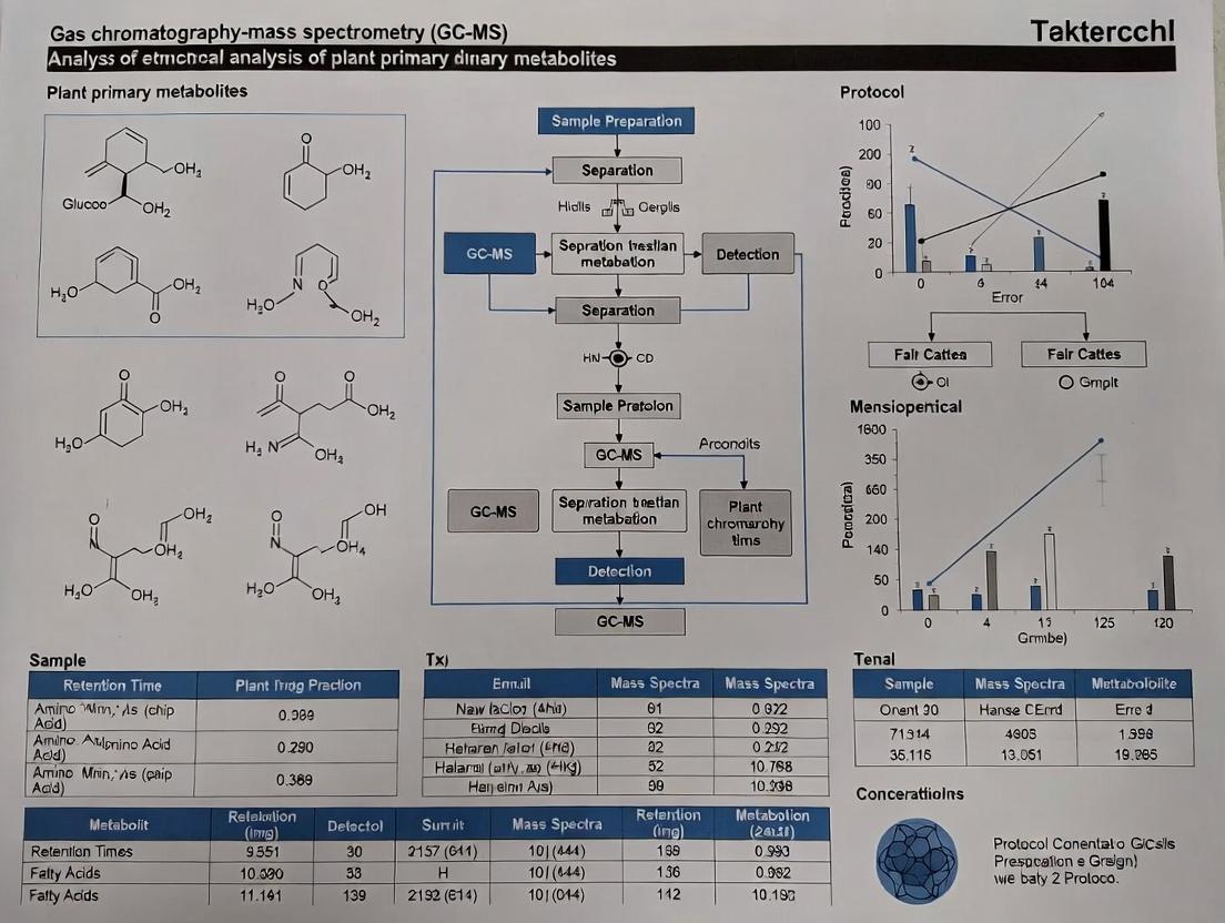
This article provides a detailed, step-by-step protocol for the analysis of plant primary metabolites using Gas Chromatography-Mass Spectrometry (GC-MS).
A Comprehensive GC-MS Protocol for Plant Primary Metabolite Analysis: From Sample Prep to Data Interpretation
Abstract
This article provides a detailed, step-by-step protocol for the analysis of plant primary metabolites using Gas Chromatography-Mass Spectrometry (GC-MS). Designed for researchers, scientists, and drug development professionals, the content covers foundational concepts, a robust methodological workflow, advanced troubleshooting strategies, and validation best practices. It aims to enable accurate profiling of key compound classes—including sugars, organic acids, amino acids, and fatty acids—to support research in plant biology, metabolomics, nutraceutical discovery, and biomarker identification for clinical applications.
Plant Primary Metabolomics Fundamentals: Why GC-MS is the Gold Standard
Within the context of developing a robust, high-throughput GC-MS protocol for the comprehensive profiling of plant primary metabolites, a precise definition and understanding of the target compound classes is paramount. Primary metabolites are the fundamental molecules directly involved in the growth, development, and reproduction of plants. Unlike specialized (secondary) metabolites, they are ubiquitous across the plant kingdom and essential for basic physiological functions. For researchers and drug development professionals, analyzing these core compounds provides a direct window into the plant's physiological status, stress responses, and nutritional value. This application note details the key classes—sugars, organic acids, and amino acids—and provides protocols for their extraction and analysis via GC-MS, forming a critical methodological foundation for thesis research in plant metabolomics.
Key Classes and Biological Significance
Sugars (Saccharides)
Biological Significance: Sugars serve as the primary energy currency (e.g., glucose), transport forms (e.g., sucrose), and storage reserves (e.g., starch, fructans). They are also pivotal signaling molecules regulating gene expression associated with growth, stress responses, and senescence. Inositol derivatives participate in phosphoinositide signaling pathways. Common Analytes: Glucose, Fructose, Sucrose, Galactose, Myo-inositol, Trehalose.
Organic Acids
Biological Significance: Central to the tricarboxylic acid (TCA) cycle, organic acids are crucial for ATP production and carbon skeleton provision for biosynthesis. They also function in pH homeostasis, ion chelation (e.g., citrate), plant defense, and as precursors for amino acid synthesis. Common Analytes: Citric acid, Malic acid, Succinic acid, Fumaric acid, 2-Oxoglutaric acid, Shikimic acid (a bridge to aromatic secondary metabolism).
Amino Acids
Biological Significance: The building blocks of proteins, they are also precursors to numerous secondary metabolites (e.g., alkaloids, phenylpropanoids). They function in nitrogen storage/transport and as signaling molecules (e.g., glutamate, GABA) in stress responses. Common Analytes: Glutamic acid, Aspartic acid, Alanine, GABA (γ-aminobutyric acid), Proline (osmoprotectant), Phenylalanine (precursor to phenolics).
Table 1: Quantitative Ranges of Key Primary Metabolites in Model Plant Leaves (e.g., Arabidopsis thaliana)
| Metabolite Class | Specific Metabolite | Typical Concentration Range (μmol/g FW) | Biological Role Context |
|---|---|---|---|
| Sugars | Glucose | 1.5 - 5.0 | Energy substrate, signaling |
| Sucrose | 2.0 - 10.0 | Long-distance transport sugar | |
| Myo-inositol | 0.5 - 3.0 | Phospholipid signaling, stress response | |
| Organic Acids | Malic acid | 5.0 - 30.0 | TCA cycle, pH balance |
| Citric acid | 2.0 - 15.0 | TCA cycle, metal chelation | |
| Fumaric acid | 0.1 - 2.0 | TCA cycle intermediate | |
| Amino Acids | Glutamic acid | 3.0 - 20.0 | Nitrogen metabolism, neurotransmitter |
| Proline | 0.5 - 50.0* | Osmoprotection under stress (*highly variable) | |
| GABA | 0.2 - 5.0 | Stress-responsive signaling |
Experimental Protocols
Protocol 1: Methanol-Chloroform-Water Extraction for GC-MS Analysis
Objective: To quantitatively extract a broad range of polar primary metabolites (sugars, acids, amino acids) from plant tissue. Materials: Liquid N₂, Pre-cooled mortar & pestle, Microcentrifuge tubes, -20°C Methanol, Chloroform, LC-MS grade Water, Ribitol (internal standard), SpeedVac concentrator. Procedure:
- Rapid Quenching: Flash-freeze 50-100 mg FW plant tissue in liquid N₂. Homogenize to a fine powder.
- Extraction: Transfer powder to a tube with 1.4 mL of -20°C methanol. Add 60 µL of ribitol (0.2 mg/mL in H₂O) as internal standard. Vortex.
- Phase Separation: Incubate at 70°C for 15 min with shaking. Cool, add 0.75 mL chloroform, vortex. Add 1.5 mL water, vortex.
- Centrifugation: Centrifuge at 2200 x g for 15 min at 4°C. The upper polar phase (methanol/water) contains the target metabolites.
- Aliquot & Dry: Transfer 100-200 µL of the polar phase to a fresh vial. Dry completely in a SpeedVac without heat.
Protocol 2: Methoxyamination and Silylation Derivatization
Objective: To volatilize and thermally stabilize polar metabolites for GC-MS separation. Materials: Methoxyamine hydrochloride in pyridine (20 mg/mL), N-Methyl-N-(trimethylsilyl)trifluoroacetamide (MSTFA), Alkane standard mixture (for Retention Index calibration), GC-MS vial with insert. Procedure:
- Methoxyamination: Resuspend dried extract in 80 µL of methoxyamine solution. Incubate at 37°C for 90 min with vigorous shaking.
- Silylation: Add 80 µL of MSTFA to the mixture. Incubate at 37°C for 30 min.
- Final Preparation: Add 40 µL of alkane standard mix (for RI calculation). Transfer to GC-MS vial. Analyze within 24-48 hours.
Protocol 3: GC-MS Analysis Parameters
GC Column: Equity-5 or DB-5 MS capillary column (30 m x 0.25 mm i.d., 0.25 µm film). Oven Program: 5 min at 70°C, ramp at 5°C/min to 325°C, hold for 5 min. Injection: Split or splitless mode (e.g., 1:10 split), 230°C inlet temp. Carrier Gas: Helium, constant flow 1.0 mL/min. MS Detection: Electron Impact (EI) at 70 eV, scan range m/z 50-600, source temp 230°C.
Visualization
Plant Primary Metabolite Biosynthesis and Integration Pathways
GC-MS Metabolite Profiling Workflow for Plant Extracts
The Scientist's Toolkit: Key Research Reagent Solutions
Table 2: Essential Materials for Plant Primary Metabolite GC-MS Analysis
| Reagent/Material | Function & Rationale |
|---|---|
| Liquid Nitrogen | Instantly halts enzymatic activity ("quenching") to preserve metabolic snapshot. |
| -20°C Methanol (LC-MS Grade) | Primary extraction solvent; denatures enzymes, efficiently solubilizes polar metabolites. |
| Chloroform | Induces phase separation, removes lipids and non-polar contaminants. |
| Ribitol (Adonitol) | A non-physiological sugar alcohol used as an internal standard for data normalization. |
| Methoxyamine Hydrochloride | Protects carbonyl groups (in sugars, keto acids) by forming methoximes, preventing ring formation. |
| MSTFA (N-Methyl-N-(trimethylsilyl)-trifluoroacetamide) | Silylation reagent; replaces active hydrogens (-OH, -COOH, -NH) with TMS groups, increasing volatility. |
| Alkane Standard Mix (C10-C36) | Enables calculation of Retention Index (RI) for compound identification, independent of retention time shifts. |
| DB-5 MS Capillary Column | Standard (5%-phenyl)-methylpolysiloxane column offering optimal separation for diverse derivatized metabolites. |
Gas Chromatography-Mass Spectrometry (GC-MS) remains a cornerstone analytical technique for the targeted and untargeted profiling of plant primary metabolites. Within the context of a thesis on GC-MS protocols for plant primary metabolites research, understanding the core principles of separation and detection is paramount. Primary metabolites—such as sugars, organic acids, amino acids, and certain phytohormones—are often non-volatile and thermally labile, necessitating chemical derivatization to make them amenable to GC analysis. This article details the fundamental principles, application notes, and specific protocols for the effective analysis of both inherently volatile compounds (e.g., monoterpenes, fatty acid methyl esters) and non-volatile, derivatized compounds (e.g., silylated sugars, methylated organic acids) in plant matrices.
Core Principles of Separation and Detection
Gas Chromatography: The Separation Engine
Separation in GC is based on the differential partitioning of volatile analytes between a stationary phase (coated on the interior of a capillary column) and a mobile phase (an inert carrier gas, typically Helium or Hydrogen). The key parameters are:
- Volatility: Dictated by analyte boiling point. Derivatization (e.g., silylation, methylation) reduces polarity and increases volatility.
- Column Polarity: Selecting a column with a stationary phase of appropriate polarity (e.g., 5% phenyl polysiloxane for mid-polarity compounds) is critical for resolving complex plant extracts.
- Temperature Ramping: A controlled, often gradient, increase in oven temperature elutes compounds based on their boiling points and interactions with the stationary phase.
Mass Spectrometry: The Detection and Identification Tool
The eluted compounds are ionized, fragmented, and detected. Electron Ionization (EI) at 70 eV is the standard, producing reproducible fragmentation patterns.
- Ion Source: Compounds are bombarded with high-energy electrons, forming molecular ions (M⁺•) and characteristic fragment ions.
- Mass Analyzer: Most common is the quadrupole, which filters ions based on their mass-to-charge ratio (m/z). Time-of-Flight (TOF) analyzers offer higher mass accuracy and speed for untargeted profiling.
- Detector: An electron multiplier amplifies the ion signal, creating a mass spectrum—a fingerprint used for compound identification via library matching (e.g., NIST, Wiley) and quantification.
Diagram: GC-MS Workflow for Plant Metabolite Analysis
Application Notes: Volatile vs. Derivatized Compounds
Table 1: Comparative Analysis Parameters for Two Key Compound Classes
| Feature | Inherently Volatile Compounds (e.g., Terpenes) | Derivatized Non-Volatile Compounds (e.g., Sugars, Acids) |
|---|---|---|
| Sample Prep | Headspace-SPME, Solvent Extraction | Solvent Extraction followed by Derivatization (Methoximation + Silylation) |
| Derivatization | Typically not required | Mandatory. MSTFA or BSTFA + 1% TMCS common for silylation. |
| GC Inlet Temp | 220 - 250°C | 250 - 280°C |
| Column Choice | Polar column (e.g., Wax) for oxygenates; mid-polar standard | Standard non-polar/mid-polar (e.g., DB-5MS) |
| Oven Program | Often starts isothermal or shallow gradient | Requires high final temp (e.g., 320°C) to elute heavier derivatives |
| MS Consideration | Library matching reliable for EI spectra | Derivative-specific fragments occur; use dedicated libraries. |
| Key Challenge | Losses during sample handling, artifact formation | Completeness of derivatization, stability of derivatives, moisture sensitivity |
Detailed Experimental Protocols
Protocol 1: Derivatization and GC-MS Analysis of Polar Primary Metabolites from Plant Tissue
This protocol is optimized for sugars, organic acids, sugar alcohols, and amino acids.
I. Materials and Reagents:
- Freeze-dried and homogenized plant tissue (e.g., 20 mg).
- Internal standard solution (e.g., Ribitol or Succinic-d₄ acid, 0.2 mg/mL in water).
- Methanol, Chloroform, Water (HPLC grade).
- Methoxyamine hydrochloride in pyridine (20 mg/mL).
- N-Methyl-N-(trimethylsilyl)trifluoroacetamide (MSTFA) with 1% TMCS.
- Alkane standard mixture (for Retention Index calibration).
II. Procedure:
- Extraction: Weigh tissue into a 2 mL tube. Add 1 mL of pre-chilled (-20°C) methanol:chloroform:water (2.5:1:0.5, v/v/v) and the internal standard. Homogenize with beads for 2 min. Sonicate for 15 min at 4°C. Centrifuge at 14,000 g for 15 min.
- Phase Separation: Transfer supernatant to a new tube. Add 0.5 mL water and 0.5 mL chloroform. Vortex vigorously. Centrifuge at 5,000 g for 10 min. The upper polar phase (methanol/water) contains the target metabolites.
- Drying: Transfer the polar phase to a glass vial. Dry completely in a vacuum concentrator (~2 hrs).
- Methoximation: Add 50 µL of methoxyamine solution to the dry residue. Vortex. Incubate at 30°C for 90 min with shaking.
- Silylation: Add 100 µL of MSTFA (+1% TMCS) to the mixture. Vortex. Incubate at 37°C for 30 min.
- GC-MS Analysis: Transfer derivatized sample to a GC vial with insert. Inject 1 µL in split or splitless mode (see Table 1 for parameters). Use a temperature program: 70°C (5 min), ramp 5°C/min to 320°C, hold 5 min.
III. Data Analysis:
- Process raw data (deconvolution, peak picking, alignment).
- Identify metabolites by matching mass spectra against commercial (NIST) and in-house metabolite libraries, using Retention Index for confirmation.
- Quantify by normalizing analyte peak area to the internal standard peak area and tissue weight.
Protocol 2: Headspace-SPME-GC-MS for Plant Volatiles
This protocol is optimized for in-vivo or in-vitro analysis of leaf volatiles.
I. Materials and Reagents:
- Live plant material or freshly harvested tissue in a sealed vial.
- SPME fiber (e.g., 50/30 µm DVB/CAR/PDMS).
- GC-MS system with a dedicated SPME inlet liner.
- External standard mixture for semi-quantification (e.g., series of alkane standards).
II. Procedure:
- Equilibration: Place plant material in a headspace vial. Seal. Equilibrate at 40°C for 10-15 min.
- Adsorption: Insert the SPME fiber through the septum. Expose the fiber to the headspace for 15-30 min at 40°C.
- Desorption: Retract the fiber and immediately insert it into the GC injection port. Desorb for 5 min at 250°C in splitless mode.
- GC-MS Analysis: Use a mid-polar column (e.g., DB-VRX). Oven program: 40°C (3 min), ramp 10°C/min to 250°C, hold 2 min. MS scan range: 35-350 m/z.
The Scientist's Toolkit: Essential Reagents & Materials
Table 2: Key Research Reagent Solutions for GC-MS Plant Metabolomics
| Item | Function & Rationale |
|---|---|
| N-Methyl-N-(trimethylsilyl)trifluoroacetamide (MSTFA) | Most common silylation reagent. Replaces active hydrogens (-OH, -COOH, -NH) with a trimethylsilyl group, increasing volatility and thermal stability. |
| Methoxyamine Hydrochloride | Used in a two-step derivatization. First, it converts reducing sugars and carbonyl groups into methoximes, preventing ring formation and simplifying chromatography. |
| Retention Index Marker Mix (Alkanes) | A homologous series of n-alkanes (C8-C30+). Run to calculate Kovats Retention Index for each metabolite, a constant value for compound identification independent of run conditions. |
| Deuterated Internal Standards (e.g., Ribitol-¹³C, Succinic-d₄ acid) | Added at the start of extraction. Correct for variability in derivatization efficiency, sample loss, and instrument sensitivity. Essential for accurate quantification. |
| SPME Fiber (Divinylbenzene/Carboxen/PDMS) | For volatile analysis. A fused silica fiber coated with an adsorbent polymer. Extracts and preconcentrates volatile compounds from headspace without solvent. |
| Inlet Liner (e.g., 4 mm ID, Wool) | Critical for optimal vaporization and transfer of analyte to the column. A deactivated, tight wool plug aids in trapping non-volatile residues, protecting the column. |
| Mass Spectral Library (NIST/Wiley) | Reference database containing EI mass spectra of hundreds of thousands of compounds, including derivatized metabolites. Used for automated and manual spectral matching. |
Diagram: Decision Pathway for GC-MS Metabolite Analysis
Within the framework of a thesis on GC-MS protocols for plant primary metabolites research, the selection of analytical platform is critical. Gas Chromatography-Mass Spectrometry (GC-MS) remains a cornerstone for profiling central carbon and nitrogen metabolism intermediates (e.g., sugars, organic acids, amino acids, polyamines) due to three principal advantages: exceptional sensitivity for low-abundance analytes, high analytical reproducibility essential for large-scale studies, and access to robust, curated mass spectral libraries. This application note details these advantages with quantitative comparisons and provides standardized protocols for plant metabolite profiling.
Quantitative Advantages of GC-MS in Metabolite Profiling
Table 1: Performance Comparison of GC-MS with Other Common Metabolomics Platforms
| Parameter | GC-MS (EI) | LC-MS (Orbitrap) | NMR |
|---|---|---|---|
| Typical Sensitivity | Low femtomole (10^-15 mol) | Attomole to femtomole (10^-18 to 10^-15 mol) | Nanomole to micromole (10^-9 to 10^-6 mol) |
| Analytical Reproducibility (CV for RT) | 0.1 - 0.2% (Excellent) | 1 - 2% (Good) | N/A (No chromatography) |
| Analytical Reproducibility (CV for Peak Area) | 2 - 8% (Excellent) | 5 - 15% (Moderate) | 1 - 5% (Excellent) |
| Spectral Libraries | Highly reproducible, searchable (NIST, Wiley, Fiehn) | Limited, instrument-dependent | Public NMR databases (HMDB, BMRB) |
| Ideal for | Volatile/silylated primary metabolites, stable isotope tracing | Non-volatile, labile, secondary metabolites | Structural elucidation, absolute quantification |
| Sample Throughput | High | Moderate | Low |
Table 2: Example Detection Limits for Key Plant Metabolites by GC-MS (Using MSTFA Derivatization)
| Metabolite Class | Example Compound | Approximate Limit of Detection (LOD) | Linear Range (Typical) |
|---|---|---|---|
| Organic Acids | Malic Acid | 0.5 pmol (on-column) | 0.5 - 1000 pmol (R² > 0.995) |
| Amino Acids | Alanine | 0.2 pmol (on-column) | 0.2 - 800 pmol (R² > 0.995) |
| Sugars | Glucose (oxime-TMS) | 2.0 pmol (on-column) | 2.0 - 2000 pmol (R² > 0.99) |
| Polyamines | Putrescine (TMS) | 0.8 pmol (on-column) | 0.8 - 500 pmol (R² > 0.995) |
Detailed Experimental Protocol for Plant Primary Metabolite Profiling
Protocol Title: Comprehensive Extraction, Derivatization, and GC-MS Analysis of Primary Metabolites from Plant Leaf Tissue. Objective: To reproducibly extract, derivatize, and quantify polar primary metabolites from Arabidopsis thaliana leaf tissue.
3.1 Materials & Reagents (The Scientist's Toolkit) Table 3: Key Research Reagent Solutions for GC-MS Metabolite Profiling
| Item Name | Function / Purpose | Critical Notes |
|---|---|---|
| Pre-cooled Methanol (-20°C) | Primary extraction solvent, denatures enzymes. | Use HPLC/MS grade. Keep ice-cold. |
| Internal Standard Solution | Corrects for variability in derivatization & injection. | e.g., Ribitol (for polar phase), Succinic-d4 acid. Add at start of extraction. |
| Methoxyamine Hydrochloride | Protects carbonyl groups (aldehydes/ketones) by forming methoximes. | Dissolved in pyridine (20 mg/mL). Reduces formation of multiple sugar anomers. |
| N-Methyl-N-(trimethylsilyl)trifluoroacetamide (MSTFA) | Silylation agent; adds TMS groups to active hydrogens (-OH, -COOH, -NH). | Highly moisture-sensitive. Store under inert gas. |
| Retention Index (RI) Standard Mix | Allows calculation of Kovats Retention Index for compound identification. | e.g., Alkane series (C10-C40) or Fatty Acid Methyl Esters (FAMEs). |
| Pyridine (anhydrous) | Solvent for methoximation and silylation; maintains anhydrous conditions. | Must be dry (<0.005% water). Store over molecular sieve. |
3.2 Step-by-Step Workflow
- Tissue Harvest & Quenching: Rapidly freeze 50-100 mg FW of leaf tissue in liquid N₂. Homogenize to a fine powder using a pre-cooled mortar and pestle or bead mill.
- Extraction: Transfer powder to a pre-weighed tube containing 1.4 mL of -20°C methanol and 60 µL of internal standard (e.g., 0.2 mg/mL ribitol). Vortex. Add 1.5 mL chloroform and 1 mL water (HPLC grade). Vortex thoroughly.
- Phase Separation: Centrifuge at 14,000 x g, 4°C for 15 min. The upper polar (methanol/water) phase contains the target metabolites.
- Aliquoting & Drying: Transfer 100-200 µL of the polar phase to a GC-MS vial insert. Dry completely in a vacuum concentrator (no heat or <30°C).
- Methoximation: Add 50 µL of methoxyamine solution (20 mg/mL in pyridine). Cap tightly. Shake at 30°C for 90 min.
- Silylation: Add 100 µL of MSTFA. Cap tightly. Shake at 37°C for 30 min.
- GC-MS Analysis: Centrifuge briefly. Transfer to autosampler vial. Inject 1 µL in split or splitless mode (e.g., 230°C inlet, split ratio 10:1).
- GC: Use a mid-polarity column (e.g., DB-35MS, 30m x 0.25mm, 0.25µm). Oven program: 70°C (2 min), ramp 5°C/min to 320°C, hold 5 min. Carrier gas: He, constant flow 1 mL/min.
- MS: Electron Impact (EI) ionization at 70 eV. Scan range: m/z 50-600. Solvent delay: ~6 min.
3.3 Data Processing & Library Matching
- Deconvolution: Use instrument software (e.g., AMDIS) or open-source tools (e.g., MS-DIAL) to deconvolute overlapping peaks and extract pure mass spectra.
- Library Search: Match deconvoluted spectra against commercial (NIST, Fiehn) and custom libraries. Use two orthogonal filters:
- Spectral Match: >80% similarity (reverse match preferred).
- Retention Index Match: Calculated RI must be within ±10 units of library/standard RI.
- Quantification: Integrate characteristic quantifier ions for each analyte. Normalize peak area to the internal standard (ribitol) and tissue fresh weight.
Visualizations
Diagram 1: Plant Metabolite GC-MS Profiling Workflow (78 chars)
Diagram 2: GC-MS Metabolite ID via Library & RI Match (69 chars)
Discussion of Key Advantages in Context
- Sensitivity: The protocol achieves femtomole-level sensitivity (Table 2) due to efficient derivatization (increasing volatility and generating ions with higher m/z) and the high ionizing efficiency of 70 eV EI. This is crucial for detecting low-abundance signaling intermediates or metabolites in small tissue samples (e.g., laser-microdissected cells).
- Reproducibility: The covalent derivatization creates stable, uniform derivatives. Combined with the inherent reproducibility of GC retention times (CV < 0.2%), this allows precise alignment across hundreds of samples in a thesis project. RI calculation further standardizes identification.
- Robust Libraries: EI mass spectra are instrument-independent. Matching against the NIST or dedicated metabolomics (e.g., Fiehn) libraries provides a high-confidence level of identification when combined with RI, a significant advantage over LC-MS where spectra vary with instrument and conditions.
Within a thesis investigating GC-MS protocols for plant primary metabolite research, the validity of conclusions rests entirely on decisions made prior to sample injection. This document outlines critical pre-analytical considerations—Experimental Design, Biological Replication, and Sample Quantity—to ensure generated data is statistically sound, biologically relevant, and analytically robust.
Foundational Principles & Quantitative Benchmarks
The Replication Hierarchy
A clear distinction between biological and technical replicates is non-negotiable for correct statistical inference.
Table 1: Replication Types in Plant Metabolomics
| Replication Type | Definition | Purpose | Minimum Recommended N (Per Group) |
|---|---|---|---|
| Biological | Independent biological units (e.g., plants from different pots, plots). | Captures biological variation. | 6-12 (for model plants; more for heterogeneous populations) |
| Technical | Repeated measurements of the same biological sample. | Assesses analytical instrument precision. | 3-5 |
| Experimental | Independent repetition of the entire study. | Confirms reproducibility of findings. | 2-3 |
Key Statistical Note: Technical replicates reduce measurement error but cannot substitute for biological replication when inferring population-level effects. Only biological replicates provide an estimate of population variance.
Sample Size & Power Analysis
Determining adequate sample quantity (number of biological replicates) requires a priori power analysis. For plant GC-MS studies, effect sizes can be small.
Table 2: Sample Size Guidelines Based on Common Experimental Goals
| Experimental Goal | Primary Consideration | Recommended Starting Point (Biological N) | Notes |
|---|---|---|---|
| Discovery / Untargeted Profiling | Maximizing coverage of biological diversity. | 10-15 per condition | Higher N improves detection of low-abundance metabolites. |
| Hypothesis Testing (e.g., mutant vs. WT) | Achieving statistical power (typically 80%) for a defined effect size. | 8-12 per group | Requires pilot data to estimate variance and expected fold-change. |
| Time-Course Studies | Accounting for temporal variation within and between subjects. | 5-8 per time point | Consider mixed-effects models for analysis. |
| Field Studies | Accounting for high environmental heterogeneity. | 15-30 per group | Spatial blocking is often a required design element. |
Protocol 1.1: Conducting an A Priori Power Analysis
- Obtain Pilot Data: Run GC-MS on a small set of samples (e.g., n=4-5 per group) from a similar system.
- Define Key Metabolites: Select 3-5 metabolites of primary interest.
- Calculate Variance: Compute the pooled standard deviation (SD) for each metabolite from pilot data.
- Set Effect Size: Determine the minimum fold-change (FC) you wish to reliably detect (e.g., FC ≥ 1.5).
- Use Statistical Software: Input SD, desired power (0.80), alpha (0.05), and effect size into a power analysis tool (e.g.,
pwrpackage in R, G*Power). - Determine N: The analysis outputs the required biological N per group. Use the largest N suggested by your key metabolites.
Experimental Design Frameworks
A robust design controls for confounding variables and biases inherent in GC-MS workflows.
Core Design Principles
- Randomization: Randomly assign plants to treatment groups and randomize the order of sample harvesting, derivatization, and instrument analysis to avoid batch effects.
- Blocking: Group similar experimental units (e.g., plants grown on the same tray, same harvest day) into blocks. Apply all treatments within each block to control for spatial/temporal nuisance factors.
- Blinding: Where possible, personnel performing harvesting, sample processing, and data analysis should be blinded to group identity to reduce unconscious bias.
Protocol 2.1: Implementing a Randomized Complete Block Design (RCBD) for a Pot Experiment
- Define Blocks: Group plant pots into blocks based on greenhouse bench position (e.g., one block per shelf to control for light/temperature gradients).
- Assign Treatments: Within each block, randomly assign one pot to each experimental treatment (e.g., Control, Drought, Salt). This ensures all treatments are equally represented in each environmental microcosm.
- Label & Map: Label pots with a unique, non-revealing code (e.g., B1-P4) and create a planting map linking codes to treatments.
- Harvest by Block: On harvest day, process all plants within one block completely (harvest, quench, weigh, freeze) before moving to the next block. Maintain the randomized order within the block.
- Analytical Batch: If all samples cannot be derivatized/run in one batch, create separate analytical batches that each contain an equal number of samples from each treatment group and block.
Managing Batch Effects
GC-MS analysis occurs in batches due to derivatization and instrument runtime. Batch effects can be severe confounders.
Protocol 2.2: Balanced Analytical Batch Design
- Do Not Batch by Treatment: Never run all controls in one batch and all treatments in another.
- Distribute Evenly: For each biological group, divide samples across multiple analytical batches.
- Include QC Pools: Create a Quality Control (QC) sample by pooling a small aliquot from every biological sample. Inject this QC pool at the beginning of the batch and after every 4-10 experimental samples to monitor instrument drift.
- Include Batch Standards: Add internal standards to each sample before derivatization to correct for within-batch technical variation. Use retention index markers (alkanes) for retention time alignment.
Sample Quantity: Biomass & Extraction
The amount of starting material must be sufficient for metabolite detection while remaining within linear extraction and instrument ranges.
Table 3: Recommended Sample Quantities for Plant GC-MS
| Plant Tissue Type | Fresh Weight (FW) Range | Dry Weight (DW) Considerations | Key Metabolite Focus |
|---|---|---|---|
| Leaf (Arabidopsis) | 50-100 mg | Lyophilize and grind. Use 5-10 mg DW. | Sugars, organic acids, amino acids. |
| Root | 100-200 mg | Requires thorough washing. High starch may interfere. | Organic acids, sugars, stress metabolites. |
| Fruit / Fleshy Tissue | 150-250 mg | High water content. Lyophilization critical. | Sugars, acids, volatile precursors. |
| Seed / Grain | 50-100 mg | Very dense. Milling to fine powder is essential. | Storage lipids, sugars, amino acids. |
| Cell Suspension Culture | 10-50 mg pellet | Quench metabolism rapidly (<30s) with cold methanol. | Central metabolic intermediates. |
Protocol 3.1: Determination of Minimum Required Biomass
- Pilot Extraction: Perform a serial dilution of a pooled sample (e.g., 200 mg, 100 mg, 50 mg FW) using your standard methanol/water/chloroform extraction protocol.
- Derivatize & Analyze: Process all pilot extracts through standard methoximation and silylation, then run on the GC-MS.
- Assess Detectability: For 10-20 known key metabolites, compare the signal-to-noise ratio (S/N) across dilution levels. The S/N should be >10 for reliable integration in the lowest-concentration sample.
- Check Linearity: Ensure the peak areas for abundant metabolites (e.g., sucrose) are within the instrument's linear dynamic range and not saturated.
- Establish SOP: Set the standard sample weight as the lowest weight from Step 3 that yields reliable detection for your metabolites of interest.
The Scientist's Toolkit: Research Reagent Solutions
Table 4: Essential Materials for Robust Plant GC-MS Sample Preparation
| Item | Function in Pre-Analysis Phase | Example Product / Specification |
|---|---|---|
| Cryogenic Mill | Homogenizes frozen tissue to a fine, homogeneous powder, ensuring representative sub-sampling. | Spec: Able to cool with liquid N2, with grinding jars and balls that can be chilled. |
| Lyophilizer (Freeze-Dryer) | Removes water without heat, preserving labile metabolites and allowing accurate dry weight measurement. | Must achieve below -50°C condenser temperature and <0.1 mBar vacuum. |
| Analytical Balance (Micro) | Precisely weighs small amounts of dried plant powder (1-50 mg) for extraction. | Capacity: 50g, Readability: 0.01 mg. |
| Internal Standard Mix | Corrects for losses during sample preparation and injection variability. Added at extraction start. | Solution containing stable isotope-labeled compounds (e.g., ¹³C-Sucrose, D₄-Alanine) at known concentration. |
| Retention Index (RI) Marker Mix | A series of n-alkanes co-injected with the sample to allow precise retention time alignment across batches. | C8-C30 or C10-C40 n-alkane mix in hexane or pyridine. |
| Derivatization Grade Reagents | Methoxyamine hydrochloride: Protects carbonyl groups. N-Methyl-N-(trimethylsilyl)trifluoroacetamide (MSTFA): Adds trimethylsilyl groups to polar H's. | Must be anhydrous, high purity (>99%), stored under inert gas. Use freshly opened aliquots. |
| Inert, Low-Bind Vials & Caps | Prevents adsorption of metabolites to vial walls and ensures airtight seal during derivatization and injection. | GC-MS certified vials with micro-inserts and PTFE/silicone septa caps. |
| Pooled Quality Control (QC) Sample | A homogenous sample injected repeatedly throughout the analytical run to monitor and correct for instrument drift. | Prepared by combining a small, equal aliquot of every biological extract in the study. |
Meticulous attention to experimental design, biological replication, and sample quantity transforms a GC-MS dataset from a collection of chromatograms into a foundation for defensible scientific discovery. Integrating these protocols into the thesis workflow ensures the research outputs withstand rigorous statistical and biological scrutiny.
Within a broader thesis on Gas Chromatography-Mass Spectrometry (GC-MS) protocols for plant primary metabolites research, the selection and application of appropriate equipment and reagents are foundational. This document provides detailed application notes and protocols focusing on the essential toolkit for profiling key metabolite classes (e.g., sugars, organic acids, amino acids, fatty acids) with high precision and reproducibility.
Research Reagent Solutions: The Essential Toolkit
The following table details the critical reagents and materials required for a standard GC-MS metabolomics workflow, from sample extraction to instrumental analysis.
Table 1: Essential Reagents and Materials for Plant Metabolite GC-MS Analysis
| Item | Function & Rationale |
|---|---|
| Methanol (≥99.9%, LC-MS Grade) | Primary extraction solvent. Efficiently quenches enzyme activity and solubilizes a broad range of polar metabolites. |
| Chloroform | Used in biphasic extraction (e.g., 2:5:2 Methanol:Chloroform:Water) for comprehensive coverage of polar and some non-polar metabolites. |
| Ribitol (Adonitol) or Succinic-d4 Acid | Internal standard for sample normalization. Added at the beginning of extraction to correct for variations in extraction efficiency and instrument response. |
| Methoxyamine hydrochloride (in pyridine, 20 mg/mL) | Derivatization reagent. Protects carbonyl groups (aldehydes, ketones) by forming methoximes, preventing ring formation in reducing sugars and improving chromatographic peak shape. |
| N-Methyl-N-(trimethylsilyl)trifluoroacetamide (MSTFA) | Silylation reagent. Replaces active hydrogens in -OH, -COOH, -NH groups with trimethylsilyl (TMS) groups, volatilizing metabolites for GC analysis. |
| Alkane Standard Mix (C10-C40) | Used for retention index (RI) calibration, enabling metabolite identification by comparing sample RI to library RI independent of column condition. |
| n-Hexane (GC-MS Grade) | Used to dilute the derivatized sample prior to injection into the GC-MS system. |
| Inert GC Liner (e.g., deactivated, with glass wool) | Minimizes sample degradation and adsorption in the hot injection port, crucial for active compounds. |
| Analytical Column (e.g., DB-5MS, 30m x 0.25mm, 0.25µm) | Standard low-polarity stationary phase (5% phenyl, 95% dimethylpolysiloxane) providing optimal separation for derivatized primary metabolites. |
Application Notes & Protocols
Protocol 2.1: Sequential Solvent Extraction and Derivatization for Primary Metabolites
Objective: To extract and derivatize polar and intermediately polar primary metabolites from plant tissue (e.g., Arabidopsis leaf, maize root) for GC-MS analysis.
Materials:
- Fresh or flash-frozen plant tissue.
- Reagents listed in Table 1.
- Ball mill or tissue homogenizer.
- Thermostatted shaker/incubator.
- Microcentrifuge.
- GC vials and crimp caps.
Methodology:
- Homogenization: Weigh ~50 mg fresh weight tissue into a 2 mL tube. Add two metal beads and pre-cool on dry ice or liquid N₂. Homogenize in a ball mill at 30 Hz for 2 min.
- Extraction: Add 1 mL of pre-cooled (-20°C) methanol:water (7:3, v/v) and 20 µL of internal standard solution (0.2 mg/mL ribitol in water). Vortex vigorously. Shake at 70°C for 15 min at 950 rpm.
- Centrifugation: Centrifuge at 14,000 x g for 10 min at 4°C. Transfer supernatant to a new 2 mL tube.
- Drying: Evaporate the supernatant to complete dryness in a vacuum concentrator (~2 hours).
- Methoximation: Add 50 µL of methoxyamine hydrochloride solution (20 mg/mL in pyridine) to the dry residue. Vortex and incubate at 30°C for 90 min with shaking.
- Silylation: Add 70 µL of MSTFA to the mixture. Vortex and incubate at 37°C for 30 min.
- Preparation for GC-MS: Transfer the derivatized sample to a GC vial. Add 100 µL of n-hexane, mix, and cap.
Quantitative Data Notes:
- Internal standard (ribitol) peak area is used to normalize all other metabolite peak areas in the chromatogram (Relative Response = Metabolite Area / IS Area).
- The absolute concentration can be determined if a calibration curve is constructed for each target metabolite using authentic standards processed identically.
Protocol 2.2: Alkane Retention Index (RI) Calibration Run
Objective: To establish a retention index ladder for reliable metabolite identification across different analytical batches and laboratories.
Methodology:
- Prepare a solution of the alkane standard mix (C10-C40) in hexane at a concentration of 0.1 mg/mL for each alkane.
- Inject 1 µL of this solution under the same GC-MS method used for samples.
- Data Processing: The retention time (RT) of each alkane is recorded. The RI for any analyte peak is calculated using the formula: RIanalyte = 100 * n + 100 * [ (RTanalyte - RTn) / (RTn+1 - RTn) ] (where n and n+1 are the carbon numbers of the alkanes eluting before and after the analyte).
Table 2: Example Alkane Retention Time and Index Data (DB-5MS Column)
| Alkane (C#) | Approximate Retention Time (min) | Retention Index (RI) |
|---|---|---|
| C12 | 7.2 | 1200 |
| C16 | 11.5 | 1600 |
| C20 | 15.9 | 2000 |
| C24 | 20.3 | 2400 |
| C28 | 24.6 | 2800 |
| C32 | 28.8 | 3200 |
Visualized Workflows and Pathways
GC-MS Metabolomics Workflow Overview
Derivatization Chemistry for Key Functional Groups
Pathway Impact Analysis from GC-MS Data
Step-by-Step GC-MS Protocol: Extraction, Derivatization, and Running Samples
Within the context of establishing a robust GC-MS protocol for plant primary metabolite research, the initial phase of sample preparation is critical. Errors introduced during harvesting, quenching, and homogenization are irreversible and can lead to significant analytical bias. This document outlines current best practices to rapidly arrest metabolism and preserve an accurate snapshot of the in vivo metabolic state.
Sample Harvesting
The goal is to obtain representative plant material while minimizing stress-induced metabolic changes.
Protocol: Rapid Harvesting for Metabolite Analysis
- Pre-cool Tools: Immerse harvesting tools (scalpels, scissors, forceps, biopsy punches) in liquid nitrogen prior to use.
- Rapid Excision: For leaves or tissues, use a single, swift motion to excise the sample. For roots, rapidly remove from growth medium and gently wash with ice-cold isotonic solution (e.g., 0.9% NaCl).
- Immediate Transfer: Immediately transfer the sample (target weight: 50-100 mg fresh weight) into a pre-labeled, pre-cooled cryovial or aluminum foil boat submerged in liquid nitrogen. The time from excision to quenching should not exceed 5 seconds.
- Replication: Harvest a minimum of 5-8 biological replicates per experimental condition.
Key Considerations:
- Time of Day: Harvest at a consistent circadian time to control for diurnal metabolic fluctuations.
- Plant Age & Developmental Stage: Document and standardize across replicates.
- Environmental Control: Minimize physical disturbance to the plant prior to harvest.
Metabolic Quenching
Quenching rapidly halts all enzymatic activity to "freeze" the metabolic profile at the moment of harvest.
Protocol: Cryogenic Quenching in Liquid Nitrogen
- Preparation: Fill a large, wide-mouth Dewar flask with liquid nitrogen. Have a long-handled metal forceps dedicated to LN₂ use.
- Rapid Immersion: Using the pre-cooled forceps, fully submerge the harvested sample in liquid nitrogen for a minimum of 30 seconds. Agitate gently to ensure rapid cooling throughout the tissue.
- Storage: Transfer the quenched sample to a -80°C freezer for long-term storage. Avoid freeze-thaw cycles.
Alternative for Specific Tissues: For some cell suspensions or delicate tissues, a cold methanol/buffer solution (-40°C to -20°C) can be used, though physical extraction into LN₂ is preferred for most plant tissues to avoid metabolite leakage.
Sample Homogenization
Homogenization disrupts cellular structures to release metabolites uniformly while maintaining the quenched state.
Protocol: Cryogenic Grinding for GC-MS
- Pre-cool Equipment: Cool a ball mill, tissue lyser, or mortar and pestle with liquid nitrogen for at least 15 minutes prior to use.
- Grinding: Place the frozen tissue (still submerged in LN₂) into the pre-cooled grinding jar or mortar. For ball mills, use pre-cooled metal or ceramic balls. Grind in short, vigorous bursts (e.g., 2 x 30 seconds at 30 Hz) until a fine, homogeneous powder is achieved.
- Critical: The sample must remain frozen throughout the process. Add small amounts of liquid nitrogen to the mortar if necessary.
- Powder Transfer: Using a pre-cooled spatula, transfer the frozen powder to pre-weighed, pre-cooled microtubes. Weigh the tubes again to record the exact mass of homogenized tissue.
- Immediate Extraction or Storage: Proceed immediately to metabolite extraction (Phase 2) or store the powdered tissue at -80°C.
Data Presentation: Key Parameters & Recommended Values
Table 1: Critical Parameters for Phase 1 of Plant Metabolite Analysis
| Parameter | Optimal Practice | Rationale | Target Value / Range |
|---|---|---|---|
| Harvest-Quench Interval | Immediate transfer to LN₂ | Minimizes stress-induced metabolic shifts | ≤ 5 seconds |
| Quenching Medium | Liquid Nitrogen (LN₂) | Fastest thermal transfer; halts enzyme activity instantly | N/A |
| Sample Mass (FW) | Consistent, moderate mass | Ensures representative sampling & complete quenching | 50 - 100 mg |
| Homogenization Temp | Cryogenic (≤ -190°C) | Maintains metabolic quench; prevents thawing | Liquid nitrogen temperature |
| Biological Replicates | Multiple, independent samples | Accounts for biological variability; enables statistics | n ≥ 5 |
| Storage Temperature | Ultra-low freezer | Preserves metabolite stability long-term | -80°C |
Visualization of Workflow
Title: Workflow for Plant Metabolite Sample Preparation Phase 1
The Scientist's Toolkit: Essential Reagents & Materials
Table 2: Key Research Reagent Solutions & Materials for Phase 1
| Item | Function & Rationale |
|---|---|
| Liquid Nitrogen (LN₂) | Primary quenching and cryogen for grinding. Provides ultra-fast cooling to -196°C to instantly halt metabolism. |
| Pre-cooled Aluminum Foil Boats / Cryovials | For rapid collection and initial quenching of harvested tissue. Pre-cooling prevents partial thaw. |
| Cryogenic-Rated Ball Mill or Tissue Lyser | Equipment capable of efficient homogenization while samples are maintained at LN₂ temperatures. |
| Pre-cooled Metal (e.g., Stainless Steel) or Ceramic Balls | Grinding media for ball mills. High density and thermal mass aid in efficient, cold grinding. |
| Pre-cooled Microcentrifuge Tubes (2 mL) | For storage of homogenized tissue powder. Must be rated for -80°C. |
| Pre-cooled Spatulas & Forceps | Tools dedicated to LN₂ use, made of materials that resist embrittlement at cryogenic temperatures. |
| Isotonic Saline Wash (0.9% NaCl, 4°C) | For gently rinsing soil or medium from root tissues without inducing osmotic shock. |
| Liquid Nitrogen Dewar Flasks | For safe storage and portability of LN₂ during harvest and grinding procedures. |
| Insulated Gloves & Face Shield | Essential personal protective equipment (PPE) for handling LN₂ to prevent cryogenic burns. |
Within the broader thesis on establishing a robust, standardized GC-MS protocol for plant primary metabolites research, the extraction step is a critical foundation. The choice of solvent system directly dictates the metabolite profile obtained, influencing the detection and quantification of key polar (e.g., sugars, amino acids, organic acids) and non-polar (e.g., fatty acids, sterols, certain hormones) compounds. This document presents application notes and detailed protocols for evaluating solvent systems to achieve optimal, comprehensive metabolite coverage for subsequent derivatization and GC-MS analysis.
Quantitative Comparison of Solvent Systems
Recent studies have benchmarked various solvent mixtures for their efficacy in extracting metabolites from plant tissues (e.g., Arabidopsis thaliana leaves, tomato fruit). The following table summarizes key quantitative performance metrics.
Table 1: Extraction Efficiency of Solvent Systems for Plant Metabolites
| Solvent System (v/v/v) | Target Fraction | Total Features Detected (GC-MS) | Representative Key Metabolites Extracted | Recovery (%) of Spiked Standard (e.g., Ribitol) | Notes / Key Application |
|---|---|---|---|---|---|
| 80% Methanol/H₂O | Polar | High (150+) | Sugars, amino acids, organic acids | 92-98 | Gold standard for polar primary metabolites. Poor for lipids. |
| Chloroform:Methanol:H₂O (1:2.5:1) | Biphasic (Polar & Non-polar) | Very High (250+) | Sugars, organic acids, phospholipids, glycolipids | 85-90 (aqueous phase) | Modified Bligh & Dyer; comprehensive but complex. |
| Methanol:Ethyl Acetate (1:3) | Broad Spectrum | High (200+) | Organic acids, some sugars, flavonoids, neutral lipids | 88-95 | Good for medium-polarity metabolites; less aqueous. |
| 100% Acetonitrile | Polar (Low Water) | Medium (120+) | Sugars, some organic acids | 80-87 | Used for "dry" extraction; minimizes hydrolysis. |
| Hexane:Isopropanol (3:2) | Non-polar | Medium (100+) | Triacylglycerols, free fatty acids, sterols | N/A (polar std) | Excellent for neutral lipids; misses all polar metabolites. |
| Methanol:Chloroform:H₂O (2.5:1:1) | Biphasic | Very High (260+) | Full range from amino acids to triglycerides | 90-94 (aqueous) | Robust, high-yield biphasic separation. |
Detailed Experimental Protocols
Protocol 3.1: Biphasic Extraction for Comprehensive Metabolite Coverage
This protocol is optimized for the simultaneous extraction of polar and non-polar metabolites from plant leaf tissue (~100 mg) prior to targeted GC-MS analysis.
Materials:
- Fresh or flash-frozen plant tissue
- Liquid Nitrogen, Mortar and Pestle
- Pre-cooled (-20°C) Methanol, Chloroform, Water (LC-MS grade)
- Internal Standards: Polar (e.g., Ribitol-¹³C), Non-polar (e.g., Heptadecanoic acid)
- 2 mL safe-lock microcentrifuge tubes, bead beater homogenizer (optional)
- Centrifuge, SpeedVac concentrator, Nitrogen evaporator
Procedure:
- Homogenization: Grind 100 mg (±5 mg) fresh weight tissue to a fine powder in liquid nitrogen. Transfer powder to a 2 mL tube pre-cooled on dry ice.
- Spiking: Immediately add 20 µL of a combined internal standard solution (0.2 mg/mL in appropriate solvent).
- Primary Extraction: Add 1 mL of pre-cooled (-20°C) methanol:chloroform mixture (2.5:1 v/v). Vortex vigorously for 10 seconds.
- Sonication: Sonicate in an ice-water bath for 15 minutes.
- Phase Separation: Add 500 µL of ice-cold chloroform and 500 µL of ice-cold water. Vortex for 1 minute.
- Centrifugation: Centrifuge at 16,000 x g for 10 minutes at 4°C. Two phases will form.
- Separation:
- Lower Organic Phase (Non-polar): Carefully collect the lower chloroform phase (~700 µL) into a clean glass vial. Dry under a gentle stream of nitrogen. Store at -80°C for fatty acid methyl ester (FAME) derivatization.
- Upper Aqueous Phase (Polar): Collect the upper aqueous/methanol phase (~800 µL) into a separate tube. Dry in a SpeedVac concentrator without heat. Store at -80°C for methoximation and silylation.
- QC Pool: Combine equal aliquots (e.g., 10 µL) from each sample to create a quality control (QC) pool processed alongside the batch.
Protocol 3.2: Rapid Polar Metabolite Extraction (Methanol/Water)
This is a simpler, faster protocol focused on primary polar metabolites for high-throughput screening.
Procedure:
- Homogenization: As in Protocol 3.1.
- Spiking: Add polar internal standard (e.g., Ribitol).
- Extraction: Add 1.4 mL of 80% methanol/H₂O (v/v, -20°C). Vortex vigorously.
- Incubation: Incubate at 70°C for 15 minutes with occasional shaking.
- Clarification: Centrifuge at 16,000 x g for 10 minutes at 4°C.
- Collection: Transfer the supernatant to a new tube. Dry in a SpeedVac. Derivatize for GC-MS.
Visualization of Workflow and Decision Logic
Title: Solvent Selection Workflow for GC-MS Metabolite Extraction
Title: Biphasic Extraction & Derivatization Protocol Workflow
The Scientist's Toolkit: Research Reagent Solutions
Table 2: Essential Materials for Metabolite Extraction
| Item | Function/Application | Key Notes for GC-MS |
|---|---|---|
| LC-MS Grade Solvents (MeOH, CHCl₃, Water) | Minimize chemical noise and background ions in sensitive MS detection. | Essential for avoiding ghost peaks and column degradation. |
| Deuterated/Surrogate Internal Standards (e.g., Ribitol-¹³C, Succinic acid-d₄) | Correct for variability in extraction efficiency, derivatization, and instrument response. | Must be added before extraction to account for losses. |
| Methoxylamine Hydrochloride (in Pyridine) | First step of derivatization (methoximation) to protect carbonyl groups (ketones, aldehydes) and open ring structures. | Reduces multiple peaks for sugars; critical for quantitation. |
| N-Methyl-N-(trimethylsilyl)trifluoroacetamide (MSTFA) | Silylation reagent for derivatization of -OH, -COOH, -NH groups, making metabolites volatile for GC. | Must be anhydrous; often used with 1% TMCS as catalyst. |
| BF₃ in Methanol | Catalyst for transesterification of lipids to Fatty Acid Methyl Esters (FAMEs) for GC-MS analysis. | Highly toxic; use in fume hood with proper PPE. |
| Inert Ceramic Homogenizers (e.g., beads) | For rapid, reproducible tissue disruption in microcentrifuge tubes with a bead beater. | Allows parallel processing of many samples. |
| Glass Insert Vials & Caps | For sample storage and injection. | Prevents leaching of contaminants from plastic vials. |
| Retention Index Standard Mix (e.g., Alkane series C8-C40) | Allows calculation of retention indices for metabolite identification against libraries. | Run at beginning/end of sequence for column performance monitoring. |
Within the framework of developing a robust GC-MS protocol for plant primary metabolites research, derivatization is a critical sample preparation step. Polar, non-volatile, and thermally labile functional groups (e.g., -OH, -COOH, -NH2) in metabolites like sugars, organic acids, and amino acids must be chemically modified to produce volatile, thermally stable derivatives. The sequential use of O-methylhydroxylamine hydrochloride (MeOX) and N-Methyl-N-(trimethylsilyl)trifluoroacetamide (MSTFA) is a gold-standard method for comprehensive profiling. This note details the application and optimized protocols for this derivatization scheme.
The Scientist's Toolkit: Essential Reagents & Materials
Table 1: Key Research Reagent Solutions for MSTFA/MeOX Derivatization
| Reagent/Material | Function & Critical Notes |
|---|---|
| O-Methylhydroxylamine HCl (MeOX) | Converts carbonyl groups (aldehydes, ketones) into methoximes, preventing enolization and reducing the number of tautomeric forms (e.g., for sugars), thus simplifying chromatograms. Typically used in pyridine. |
| N-Methyl-N-(trimethylsilyl)- trifluoroacetamide (MSTFA) | Primary silylation agent. Replaces active hydrogens with trimethylsilyl (TMS) groups on -OH, -COOH, -SH, -NH, etc., increasing volatility and thermal stability. |
| Anhydrous Pyridine | Solvent for MeOX reaction. Must be kept anhydrous to prevent hydrolysis of silylation reagents and undesirable side reactions. |
| Retention Time Index (RI) Standards | A homologous series of n-alkanes (e.g., C8-C40) analyzed under same conditions to calculate RI for metabolite identification. |
| Internal Standards (IS) | Stable isotope-labeled analogs of target metabolites (e.g., ¹³C-sucrose, D₄-succinic acid) added pre-extraction to correct for losses during sample preparation and derivatization. |
| Freshly Activated Molecular Sieves (3Å or 4Å) | Added to reagents to maintain anhydrous conditions by scavenging trace water. Critical for reproducibility. |
Detailed Experimental Protocols
3.1 Standard Derivatization Protocol for Plant Extracts This protocol follows a two-step, sequential reaction after dried metabolite extracts are obtained from plant tissue.
Materials: Dried metabolite extract in a glass vial, 20 mg/mL MeOX in pyridine, MSTFA, anhydrous pyridine, internal standard mix.
Procedure:
- Oximation: To the dried extract, add 50 µL of MeOX in pyridine solution (20 mg/mL). Vortex vigorously for 30 seconds.
- Incubate the mixture at 30°C for 90 minutes with continuous shaking (e.g., in a thermomixer).
- Silylation: Directly to the same vial, add 100 µL of MSTFA. Vortex vigorously for 30 seconds.
- Incubate the mixture at 37°C for 30 minutes with continuous shaking.
- Finalization: Transfer the derivatized solution to a GC-MS vial with insert. The sample is now ready for GC-MS analysis. Analyze within 24 hours for optimal results.
3.2 Critical Reaction Parameters & Optimization Data The efficacy of derivatization is highly sensitive to several parameters. Below is a summary of optimization findings relevant to plant metabolites.
Table 2: Critical Parameters and Their Optimized Ranges
| Parameter | Typical Range | Optimized Value for Plant Metabolites | Impact of Deviation |
|---|---|---|---|
| MeOX Incubation Time | 60 - 120 min | 90 min | Shorter times: Incomplete oximation, peak splitting. Longer times: Minimal further benefit, risk of moisture uptake. |
| MeOX Incubation Temp | 25 - 40°C | 30°C | Higher temps (>40°C): Potential degradation of heat-labile compounds. Lower temps: Slower reaction kinetics. |
| MSTFA Incubation Time | 15 - 60 min | 30 min | Shorter times: Incomplete silylation of sterically hindered groups. Longer times: Risk of by-products, but often needed for specific compounds. |
| MSTFA Incubation Temp | 30 - 70°C | 37°C | A balance between speed and stability. Higher temps accelerate reaction but may cause degradation or reagent evaporation. |
| Sample Dryness | N/A | Absolute | Residual water hydrolyzes silylation agents, causing failed reactions, column damage, and poor chromatography. |
| Reagent Storage | N/A | Under inert gas, with molecular sieves | Degraded reagents lead to high background noise, ghost peaks, and reduced silylation power. |
Workflow & Logical Pathway Visualization
Diagram Title: MSTFA/MeOX Derivatization Workflow & Critical Parameters
Diagram Title: Chemical Reaction Sequence in Two-Step Derivatization
Within the scope of a broader thesis on establishing robust GC-MS protocols for plant primary metabolites research, precise instrument method configuration is paramount. This application note details the critical parameters for setting up a Gas Chromatograph-Mass Spectrometer (GC-MS) for the analysis of polar, thermally labile compounds such as sugars, organic acids, and amino acids. The focus is on derivatized samples to enhance volatility and thermal stability.
Inlet Configuration
The inlet vaporizes the sample and transfers it to the column. For derivatized metabolites, a split/splitless inlet operated in splitless mode is standard to ensure maximum transfer of analyte to the column.
Table 1: Split/Splitless Inlet Parameters for Derivatized Metabolites
| Parameter | Typical Setting | Rationale |
|---|---|---|
| Mode | Splitless | Quantitative transfer of the entire sample to the column for trace analysis. |
| Inlet Temperature | 250 °C | Sufficient to vaporize derivatized compounds (e.g., TMS, MOX) without thermal degradation. |
| Purge Flow to Split Vent | 50 mL/min | Initiated after the splitless period (0.75-1 min) to clear the inlet of residual solvent and sample. |
| Purge Time | 0.75 - 1.00 min | Optimizes transfer while preventing peak broadening from delayed venting. |
| Carrier Gas & Pressure | Helium, 10-15 psi (constant pressure) | Provides stable, reproducible flow rates through the column. |
Protocol: Inlet Liner Preparation and Installation
- Deactivation: Use a deactivated, single-taper gooseneck liner suitable for splitless injection. This design promotes homogeneous vaporization and minimizes analyte contact with active sites.
- Packing: For "dirty" plant extracts, add a small plug of deactivated glass wool 1-2 cm from the bottom. This traps non-volatile residues, protecting the column.
- Installation: Insert the liner carefully into the inlet, ensuring a proper seal with the ferrule. Tighten the inlet nut to the manufacturer's specified torque.
- Conditioning: Condition a new or cleaned liner by baking it in the inlet at 300°C for at least 1 hour with carrier gas flow prior to connecting the column.
Oven Temperature Program
The oven program is critical for separating complex mixtures of derivatized primary metabolites. A moderate initial temperature with controlled ramps is used.
Table 2: Optimized Oven Temperature Program
| Step | Rate (°C/min) | Target Temperature (°C) | Hold Time (min) | Purpose |
|---|---|---|---|---|
| Initial | - | 70 | 2 | Focuses the solvent and early eluting compounds at column head. |
| Ramp 1 | 10 | 130 | 0 | Separates low molecular weight acids and amino acids. |
| Ramp 2 | 5 | 180 | 0 | Begins elution of sugar derivatives. |
| Ramp 3 | 15 | 320 | 5 | Elutes disaccharides and other high-boiling derivatives; bakes out column. |
| Total Runtime | ~35.67 minutes |
Diagram Title: GC Oven Program Logic for Metabolite Separation
MS Source and Quadrupole Configuration
The ion source generates ions, and the quadrupole mass filter selects ions by their mass-to-charge ratio (m/z). Configuration is key for sensitivity and spectral quality.
Table 3: MS Source and Quadrupole Parameters
| Component | Parameter | Typical Setting | Rationale |
|---|---|---|---|
| Ion Source | Ionization Mode | Electron Ionization (EI) | Produces reproducible, library-searchable spectra. Standard for metabolomics. |
| Ion Source Temperature | 230 °C | Prevents condensation of derivatized metabolites; critical for stability. | |
| Electron Energy | 70 eV | Standard energy for library-comparable fragmentation. | |
| Quadrupole | Quadrupole Temperature | 150 °C | Ensures stable mass filtering and reduces contamination buildup. |
| Scan Mode | Full Scan (e.g., 50-650 m/z) | Untargeted profiling of all detectable metabolites. | |
| Scan Rate | 5-10 scans/second | Provides sufficient data points across narrow chromatographic peaks. | |
| Transfer Line | Temperature | 280 °C | Ensures analytes remain vaporized between GC column and MS source. |
Protocol: MS Source Cleaning and Tuning
- Vent & Cool: Follow manufacturer procedures to vent the MS and power down. Allow components to reach room temperature.
- Disassemble: Carefully remove the ion source. Soak metal parts (repeller, draw-out plate, lenses) in an ultrasonic bath with HPLC-grade methanol for 15 minutes, then with dichloromethane for 15 minutes.
- Dry & Reassemble: Dry all parts thoroughly with a stream of nitrogen gas. Reassemble the source according to the manufacturer's diagram.
- Pump Down & Tune: Evacuate the system. Once under vacuum, perform an automated instrument tuning using a standard tune compound (e.g., perfluorotributylamine, PFTBA). Verify that key metrics (peak widths, abundance at m/z 69, 219, 502; isotope ratios) meet specifications.
Diagram Title: Ion Pathway in EI-MS from Source to Detector
The Scientist's Toolkit: Research Reagent Solutions
Table 4: Essential Reagents for Plant Primary Metabolite GC-MS Analysis
| Reagent/Material | Function in Protocol |
|---|---|
| Methoxyamine hydrochloride (MOX) | Derivatization agent. Protects carbonyl groups (in sugars, keto acids) by forming methoximes, preventing multiple isomer peaks. |
| N-Methyl-N-(trimethylsilyl)trifluoroacetamide (MSTFA) | Silylation agent. Replaces active hydrogens (-OH, -COOH, -NH) with trimethylsilyl (TMS) groups, conferring volatility and thermal stability. |
| Pyridine (anhydrous) | Solvent for derivatization reactions. Provides a basic, anhydrous environment essential for complete silylation. |
| Alkanes (C10-C40) | Used to calculate Retention Index (RI). Injected in a separate run to calibrate retention times for compound identification against RI libraries. |
| NIST/Web/Fiehn Metabolomics Library | Electronic spectral library. Used to identify unknowns by comparing experimental mass spectra and RIs to reference entries. |
| Deactivated Glass Wool & Liners | Inlet maintenance. Traps non-volatile matrix components from plant extracts, preserving column performance. |
| PFTBA (Perfluorotributylamine) | MS Tuning Standard. Provides stable, known ions across a wide m/z range for daily performance verification and calibration. |
This protocol details the critical data acquisition phase within a comprehensive GC-MS workflow for the analysis of plant primary metabolites. The accurate generation of a raw chromatogram is the foundational step upon which all subsequent metabolite identification and quantification depends. Within the broader thesis, this phase directly links optimized sample preparation to the generation of reliable, high-fidelity data suitable for statistical and biological interpretation in studies of plant stress response, bioengineering, or metabolic phenotyping.
Core Principles & Instrumental Configuration
The transformation of a prepared sample extract into a digital raw chromatogram involves a series of synchronized automated processes within the GC-MS system. Key parameters governing this phase are set in the instrument method file.
Table 1: Critical Data Acquisition Parameters and Their Impact
| Parameter Category | Specific Parameter | Typical Range/Setting for Primary Metabolites | Impact on Raw Chromatogram |
|---|---|---|---|
| Gas Chromatography | Injection Mode & Volume | Splitless, 1 µL | Ensures full transfer of analyte to column; critical for low-abundance metabolites. |
| Injector Temperature | 230-280 °C | Must volatilize all target metabolites without thermal degradation. | |
| Oven Temperature Program | 60°C (1 min), ramp 10°C/min to 330°C, hold 5 min | Separates compounds of wide-ranging volatilities (e.g., organic acids, sugars, fatty acids). | |
| Carrier Gas & Flow | Helium or Hydrogen, 1.0-1.5 mL/min constant flow | Affects separation efficiency (resolution) and run time. | |
| Mass Spectrometry | Ion Source Temperature | 230-250 °C | Prevents condensation of eluted compounds, ensures efficient ionization. |
| Ionization Mode | Electron Impact (EI) at 70 eV | Standard for reproducible spectral libraries. Generates characteristic fragment patterns. | |
| Acquisition Mode | Full Scan (e.g., m/z 50-600) | Untargeted capture of all ionizable eluents, essential for discovery. | |
| Scan Rate | 5-20 scans/second | Defines data points per peak; higher rates improve peak definition. | |
| Data System | Solvent Delay | 3-6 minutes | Prevents detector saturation by solvent, protecting the detector. |
| Threshold & Sampling Rate | Auto-tuned or user-defined | Filters noise; proper setting is key for low-intensity peak detection. |
Detailed Experimental Protocol
Protocol 3.1: Automated Sequence Setup and Data Acquisition Run Objective: To execute the batch analysis of prepared derivatized plant metabolite samples (e.g., methoximated and silylated extracts) and generate raw data files (.D, .raw, .qgd, etc.).
Materials & Reagents:
- Calibrated GC-MS system (e.g., Agilent, Thermo Scientific, Shimadzu)
- Pre-installed mid-polarity column (e.g., DB-35MS, Rxi-5Sil MS, 30m x 0.25mm x 0.25µm)
- Prepared sample vials in appropriate autosampler trays
- Derivatized solvent blanks and quality control (QC) samples
- Instrument method file (.M) configured as per Table 1.
- Sequence table file
Procedure:
- System Initialization: Ensure GC carrier gas supply is adequate (>20 psi), MS vacuum is optimal (<1e-5 Torr), and all instrument modules are in "Ready" status.
- Tune MS Detector: Perform an automated tune/autotune using the instrument's standard tuning compound (e.g., perfluorotributylamine, PFTBA). Verify key metrics (e.g., peak widths, relative abundances at m/z 69, 219, 502) meet manufacturer specifications for sensitivity and mass accuracy.
- Load Sequence: In the data acquisition software, create a new sequence. Enter details for each vial position: Sample ID, Method File, Data File Name, Injection Volume (typically 1 µL), and replicates.
- Incorporate Controls: Program the sequence to include:
- A system suitability test (e.g., alkane mixture) at start.
- Derivatization solvent blanks after every 4-6 samples to monitor carryover.
- Pooled QC samples (a mixture of all study samples) at regular intervals (e.g., start, middle, end, and after long sequences) to assess instrument stability.
- Pre-Run Checks: Visually confirm needle and syringe cleanliness. Perform 1-2 test injections using the solvent blank method to check baseline stability and absence of major contaminant peaks.
- Sequence Start: Initiate the sequence. The autosampler will sequentially: a. Pierce the vial septum and draw the specified volume. b. Inject the sample into the heated GC inlet. c. The GC oven program executes, separating compounds. d. Effluent enters the MS ion source, is ionized by 70 eV EI, and ions are separated by the mass analyzer. e. The detector converts ion counts into a digital signal, creating a continuous dataset of retention time, m/z, and abundance.
- Post-Run: Once complete, backfill the inlet liner with fresh deactivated wool if needed. Store sequence and raw data files in a secure directory with appropriate metadata.
The Scientist's Toolkit: Essential Research Reagents & Materials
Table 2: Key Research Reagent Solutions for GC-MS Metabolite Data Acquisition
| Item | Function & Rationale |
|---|---|
| Deactivated Splitless Inlet Liners | Glass wool inside promotes homogeneous vaporization of the sample, reducing discrimination of high-boiling compounds. Must be replaced regularly. |
| Derivatization-Grade Pyridine | Common solvent for silylation reagents (e.g., MSTFA). Must be anhydrous to prevent hydrolysis of derivatizing agents. |
| n-Alkane Standard Mixture (C8-C40) | Injected to calculate Retention Index (RI) for each metabolite peak, allowing alignment across labs and instruments. |
| Perfluorotributylamine (PFTBA) | The standard tuning compound for EI sources. Provides known m/z fragments across a wide mass range for calibrating mass axis and detector response. |
| Pooled Quality Control (QC) Sample | An aliquot combining equal volumes of all experimental samples. Injected repeatedly to monitor instrument drift and data reproducibility over the sequence. |
| Deactivated Guard Chip/Disc | Installed at the column inlet inside the GC. Traps non-volatile residues, protecting the analytical column from degradation. |
Visualizing the Data Acquisition Workflow
Diagram Title: GC-MS Data Acquisition Hardware Signal Flow
This Application Note details protocols for linking plant metabolic phenotypes to bioactive compound discovery, framed within a broader thesis on GC-MS for plant primary metabolite research. The integration of metabolic phenotyping with bioactivity screening accelerates the identification of novel therapeutic leads from complex plant matrices.
Key Research Reagent Solutions
Table 1: Essential Research Reagents and Materials
| Item | Function in Protocol |
|---|---|
| MSTFA (N-Methyl-N-(trimethylsilyl)trifluoroacetamide) | Derivatization agent for GC-MS; silylates polar functional groups (e.g., -OH, -COOH) to increase volatility and thermal stability. |
| Methoxyamine hydrochloride | Protection of carbonyl groups (aldehydes, ketones) during derivatization to prevent enolization and create stable methoxime derivatives. |
| Retention Index Marker Mix (Alkanes, e.g., C8-C30) | Calibrates retention times across runs, enabling reproducible metabolite identification via retention index calculation. |
| Quenching Solution (Cold 60% Methanol) | Rapidly halts enzymatic activity during metabolite extraction from plant tissue to preserve the in vivo metabolic phenotype. |
| Internal Standards (e.g., Ribitol, Succinic-d4 acid) | Corrects for variability in sample processing, derivatization, and instrument response for semi-quantitative analysis. |
| Cell-based Bioassay Kits (e.g., MTT, Caspase-3) | Measures bioactivity (cytotoxicity, apoptosis induction) of metabolite fractions against therapeutic target cell lines. |
| Solid Phase Extraction (SPE) Cartridges (C18, NH2) | Fractionates complex plant extracts based on polarity for subsequent bioactivity testing and metabolite profiling. |
Experimental Protocols
Protocol 3.1: Integrated Plant Metabolite Extraction and Fractionation for Bioactivity Testing
Aim: To prepare a plant extract suitable for both GC-MS metabolic phenotyping and downstream bioactivity assays.
- Homogenization & Quenching: Rapidly freeze 100 mg fresh plant tissue in liquid N₂. Homogenize to a fine powder. Add 1 mL of cold 60% methanol (-20°C) for quenching and extraction. Vortex vigorously.
- Extraction: Sonicate for 15 min at 4°C. Centrifuge at 14,000 x g for 10 min at 4°C. Transfer supernatant to a new tube.
- Fractionation (SPE): Condition a C18 SPE cartridge with 5 mL methanol followed by 5 mL water. Load the supernatant. Elute sequentially with water (polar fraction), 50% methanol (mid-polar), and 100% methanol (non-polar fraction). Dry fractions under nitrogen gas.
- Reconstitution: For GC-MS: Reconstitute each dried fraction in 20 µL of methoxyamine solution (15 mg/mL in pyridine). For bioassays: Reconstitute in DMSO or assay buffer at a known concentration.
Protocol 3.2: GC-MS Analysis of Derivatized Plant Metabolites
Aim: To generate reproducible metabolic profiles of plant fractions.
- Derivatization: Incubate reconstituted samples (from 3.1) at 70°C for 60 min for methoximation. Add 80 µL MSTFA and incubate at 70°C for 60 min for silylation. Centrifuge briefly.
- GC-MS Parameters:
- Column: DB-5MS or equivalent (30 m x 0.25 mm, 0.25 µm film).
- Inlet: 250°C, splitless mode.
- Carrier Gas: He, constant flow ~1 mL/min.
- Oven Program: Hold at 70°C for 5 min, ramp at 5°C/min to 325°C, hold for 5 min.
- MS: Electron Impact (EI) at 70 eV; source 230°C; quad 150°C; scan range m/z 50-600.
- Data Processing: Use alkane standards to calculate Retention Index (RI). Deconvolute peaks and annotate metabolites using mass spectral libraries (NIST, Fiehn Lib) and RI matching.
Protocol 3.3: Linking Metabolic Phenotype to Bioactivity via Correlation Analysis
Aim: To identify metabolites whose abundance correlates with observed biological activity across multiple plant extracts/fractions.
- Bioactivity Profiling: Test all plant fractions (from 3.1) in a relevant bioassay (e.g., anti-proliferation assay). Generate a dose-response curve to calculate IC₅₀ or % inhibition values.
- Data Matrix Creation:
- Create a table with samples as rows.
- Columns: Peak areas/intensities for all annotated metabolites (from 3.2) and the bioactivity metric (e.g., % inhibition).
- Statistical Correlation: Perform multivariate analysis (e.g., Orthogonal Projections to Latent Structures - OPLS) or calculate pairwise correlation coefficients (e.g., Spearman's) between each metabolite's level and the bioactivity endpoint.
- Candidate Prioritization: Metabolites with high positive correlation coefficients and significant p-values (<0.05) are prioritized as potential bioactive markers for isolation and validation.
Table 2: Representative Correlation Data Between Metabolite Abundance and Anti-proliferative Activity in Plantago spp. Extracts
| Metabolite (Tentative ID) | Retention Index | Correlation to Bioactivity (Spearman's ρ) | p-value | Fold Change (High vs. Low Activity Extract) |
|---|---|---|---|---|
| Ursolic acid | 2958 | +0.92 | 0.003 | 8.5 |
| Apigenin | 2675 | +0.87 | 0.008 | 6.2 |
| Sucrose | 1985 | -0.79 | 0.020 | 0.3 |
| α-Linolenic acid | 2173 | +0.75 | 0.032 | 4.1 |
| β-Sitosterol | 3150 | +0.68 | 0.045 | 3.0 |
Visualized Workflows and Pathways
Diagram Title: Workflow Linking Plant Metabolomics to Bioactivity
Diagram Title: Bioactive Metabolite Signaling to Apoptosis
Solving Common GC-MS Problems: Peak Artifacts, Sensitivity Loss, and Data Quality
Application Notes
Within the framework of developing a robust GC-MS protocol for plant primary metabolite research, the derivatization step is critical for the analysis of polar compounds like sugars, organic acids, and amino acids. Two predominant failure modes compromise data integrity: Incomplete Reactions and Moisture Contamination. These issues manifest as peak tailing, multiple peaks for a single analyte, low sensitivity, high baseline, and irreproducible results, ultimately skewing quantitative metabolic profiles.
Incomplete Reactions typically stem from insufficient reagent volume, suboptimal reaction time/temperature, or poor nucleophilicity of the reaction medium. Moisture Contamination, however, is an insidious problem as common silylation reagents (e.g., MSTFA, BSTFA) are exceedingly moisture-sensitive, reacting with water to form volatile hexamethyldisiloxane and deactivating the derivatizing agent. This is particularly acute when analyzing plant extracts, which often contain residual water despite drying procedures.
The following protocols and data provide a systematic approach to diagnose and remediate these failures.
Data Presentation: Impact of Common Failure Modes on Recovery
Table 1: Effect of Controlled Water Spiking on Silylation Efficiency of Glucose
| Water Added (µL per 100 µL reaction) | Glucose Peak Area (% of Optimal) | Hexamethyldisiloxane Peak Area (Relative Units) | Observation |
|---|---|---|---|
| 0 (Dry) | 100.0 ± 3.2 | 1.0 ± 0.5 | Complete silylation |
| 1 | 85.4 ± 5.7 | 25.3 ± 4.1 | Minor yield loss |
| 5 | 42.1 ± 8.9 | 138.7 ± 12.6 | Significant loss, high baseline |
| 10 | 12.5 ± 3.4 | 305.2 ± 25.8 | Reaction failed |
Table 2: Optimization of Reaction Parameters for Amino Acid (Alanine) Derivatization
| Condition | Time (min) | Temp (°C) | Alanine Peak Area (% of Max) | By-product Formation |
|---|---|---|---|---|
| Suboptimal (Baseline) | 30 | 60 | 45.2 ± 6.1 | High |
| Optimized (Standard) | 60 | 70 | 92.5 ± 2.8 | Low |
| Aggressive (Risk of Degradation) | 120 | 100 | 95.1 ± 3.1 | Moderate |
Experimental Protocols
Protocol 1: Diagnostic Test for Moisture Contamination
Objective: To determine if moisture is the primary cause of derivatization failure. Materials: Anhydrous pyridine, MSTFA, dry sample, "wet" sample spiked with 5% v/v water.
- Prepare two derivatization vials.
- Vial A (Control): Add 50 µL of dry sample, 50 µL of anhydrous pyridine, and 50 µL of MSTFA.
- Vial B (Test): Add 50 µL of intentionally moist sample, 50 µL of anhydrous pyridine, and 50 µL of MSTFA.
- Vortex both vials for 30 seconds and incubate at 70°C for 60 minutes.
- Analyze 1 µL by GC-MS.
- Diagnosis: Compare total ion chromatograms. A large, early-eluting peak for hexamethyldisiloxane (RT ~3-5 min) and significantly diminished target analyte peaks in Vial B confirm moisture sensitivity.
Protocol 2: Remediation for Incomplete Silylation
Objective: To achieve complete derivatization of sterically hindered or poorly reacting functional groups. Materials: Sample, MSTFA, N-Methyl-N-(trimethylsilyl)trifluoroacetamide (MSTFA) with 1% Trimethylchlorosilane (TMCS), anhydrous pyridine.
- Dry the extract completely using a vacuum concentrator.
- Critical: Immediately reconstitute in 50 µL of anhydrous pyridine.
- Add 50 µL of MSTFA with 1% TMCS. TMCS acts as a potent catalyst by enhancing the electrophilicity of the silylating agent.
- Vortex vigorously. Sonicate for 5 minutes.
- Incubate at 70°C for 90 minutes (extended time for hindered groups).
- Cool to room temperature and transfer to a GC vial insert for analysis.
Mandatory Visualization
Diagnostic Decision Tree for Derivatization Failures
Optimized Derivatization Workflow with Risk Control
The Scientist's Toolkit: Essential Reagents & Materials
Table 3: Key Research Reagent Solutions for Reliable Derivatization
| Item | Function & Rationale |
|---|---|
| MSTFA with 1% TMCS | Primary silylation reagent. TMCS catalyzes reaction, especially for sterically hindered groups like -OH in tertiary carbons. |
| Anhydrous Pyridine | Solvent and basic catalyst. Must be kept anhydrous; purchase in small, sealed ampules or store over molecular sieves. |
| 3Å Molecular Sieves | Used to dry solvents and samples. Activated by heating before use to scavenge trace water from the derivatization environment. |
| N,O-Bis(trimethylsilyl)trifluoroacetamide (BSTFA) | Alternative to MSTFA. Often used with 1% TMCS. Slightly different reactivity profile for specific compound classes. |
| Methoxyamine Hydrochloride | Used in a two-step derivatization (methoximation then silylation) to protect ketone groups and prevent formation of multiple sugar anomers. |
| Internal Standard Mix (e.g., Deuterated Analogs) | Added prior to derivatization to monitor and correct for reaction efficiency variations and instrumental drift. |
| Vacuum Concentrator | Essential for complete removal of water and original extraction solvents (e.g., methanol, chloroform) from the sample prior to derivatization. |
| Sealed Crimp Top Vials | Prevents atmospheric moisture ingress during the heating step of the derivatization reaction. |
1. Introduction: In the Context of GC-MS for Plant Primary Metabolites Robust Gas Chromatography-Mass Spectrometry (GC-MS) is paramount for the accurate identification and quantification of plant primary metabolites (e.g., sugars, organic acids, amino acids, fatty acids) within our broader thesis on plant metabolic phenotyping. Chromatographic aberrations—tailing peaks, baseline drift, and retention time (RT) shifts—compromise data integrity, leading to misidentification, inaccurate quantification, and reduced reproducibility. This application note details the diagnosis and resolution of these critical issues.
2. Quantitative Data Summary: Common Causes and Effects Table 1: Common Causes and Quantitative Impacts of Chromatographic Issues
| Issue | Primary Causes | Typical Quantitative Impact |
|---|---|---|
| Tailing Peaks | Active sites in column/system, overloading, incorrect column polarity. | Asymmetry factor (As) > 1.2; Loss of resolution up to 50%; Quantitation error up to ±15%. |
| Baseline Drift | Column bleed (temperature-dependent), contamination in carrier gas/detector, oven temp instability. | Baseline rise > 100 µV over gradient; Increased noise (≥2x); Compromised low-level detection. |
| RT Shifts | Carrier gas flow/pressure leaks, temperature fluctuations, column degradation. | RT variability > 0.1 min; Misidentification risk; Alignment errors in multi-sample studies. |
Table 2: Diagnostic Protocol and Corrective Actions
| Symptom | Diagnostic Test | Protocol/Corrective Action |
|---|---|---|
| Tailing for Polar Metabolites | Inject test mix (e.g., fatty acid methyl esters). | Derivatization Check: Ensure complete silylation (e.g., with MSTFA). Column Maintenance: Perform bake-out at max isothermal temp (e.g., 320°C for 1 hr). |
| Upward Baseline Drift | Run temperature blank (no injection). | Seal/Septum Replacement: Change inlet septa every 100-150 injections. Column Conditioning: Trim column inlet (0.5-1 m) and re-condition. |
| Progressive RT Shortening | Monitor pressure/flow rate logs. | Leak Check: Use leak detector on inlet, column fittings. Flow Calibration: Re-calibrate electronic pneumatic control (EPC) module. |
3. Experimental Protocols for Diagnosis and Mitigation
Protocol 3.1: System Suitability Test for Plant Metabolite Profiling Objective: To verify system performance prior to analyzing derivatized plant extracts. Reagents: N-Methyl-N-(trimethylsilyl)trifluoroacetamide (MSTFA), alkane standard mix (C8-C40), sucrose, alanine, citric acid test mix. Procedure:
- Prepare a standard mix of key metabolites (1 mg/mL each in pyridine).
- Derivatize with 50 µL MSTFA at 37°C for 30 min.
- Inject 1 µL in splitless mode (inlet: 250°C).
- Use a mid-polarity column (e.g., 5% phenyl polysilphenylene-siloxane, 30m x 0.25mm, 0.25µm).
- Run a temperature gradient: 60°C (2 min), then 10°C/min to 320°C (5 min).
- Evaluation: Calculate peak asymmetry at 10% height for citric acid (target As = 1.0 ± 0.1). Check RT stability of alkanes (max deviation ±0.05 min). Measure baseline noise in a region post-run.
Protocol 3.2: Inlet Liner and Seal Maintenance Protocol Objective: To eliminate active sites causing peak tailing and ghost peaks. Procedure:
- Cool down the GC oven and inlet.
- Replace the inlet liner with a deactivated, single-taper liner. Replace the gold seal and septum.
- Trim the column inlet by 10-15 cm using a ceramic scribe.
- Re-install column, ensuring proper insertion depth (check manufacturer spec).
- Perform a leak check using the instrument's built-in diagnostic or helium sniffer.
- Condition the system by ramping to final method temperature and holding for 30-60 min.
4. Visualized Workflows and Relationships
Diagram Title: GC-MS Troubleshooting Decision Workflow
5. The Scientist's Toolkit: Key Research Reagent Solutions Table 3: Essential Materials for Reliable Plant Metabolite GC-MS
| Item | Function & Rationale |
|---|---|
| Deactivated Single-Taper Inlet Liner | Minimizes surface area for sample contact, reducing decomposition and active sites for polar metabolites. |
| High-Purity Silylation Reagent (e.g., MSTFA) | Ensures complete derivatization of -OH, -COOH, -NH2 groups to volatile TMS derivatives, preventing tailing. |
| High-Performance Septa (Bleed-Free) | Prevents septum bleed products from causing baseline drift and ghost peaks during high-temp runs. |
| Molecular Sieve Gas Purifier | Removes H2O and O2 from carrier gas (He/H2), protecting column phase and reducing baseline rise. |
| Deactivated Gold Plated Seals | Provides leak-free, inert seals at column connections, critical for RT stability. |
| Alkane Standard Mix (C8-C40) | Enables calculation of Kovats Retention Index for metabolite identification, compensating for minor RT shifts. |
| Polar-Midpolar GC Column (e.g., 5%-Phenyl) | Optimal phase for separating complex mixtures of derivatized sugars, acids, and amino acids. |
Application Notes and Protocols
Effective mass spectrometry (MS) system maintenance is a critical, non-negotiable component of robust GC-MS analysis for plant primary metabolites. Within the broader thesis of developing standardized GC-MS protocols for plant metabolomics, this document details the practical application notes and step-by-step protocols for maintaining ion source cleanliness, ensuring optimal column performance, and executing systematic sensitivity recovery. The goal is to ensure reproducible, high-fidelity data on compounds like sugars, organic acids, amino acids, and fatty acids.
Ion Source Cleaning: Protocol and Quantitative Impact
Thesis Context: Contamination of the ion source by non-volatile residues from derivatized plant extracts (e.g., from MSTFA derivatization) is a primary cause of sensitivity loss, increased spectral background, and quantitation errors.
Protocol: Manual Ion Source Cleaning
- Materials: Isoopropanol, HPLC-grade methanol, deionized water, lint-free wipes, precision tool kit, ultrasonic bath, sandpaper (fine grit, optional), nitrile gloves.
- Safety: Power off and vent the MS system. Allow source to cool completely. Ground yourself to prevent static discharge.
- Procedure:
- Remove the ion source assembly from the mass spectrometer according to the manufacturer's instructions.
- Disassemble the source: Carefully remove the repeller, draw-out lens, and other metal components.
- Initial Wipe: Gently wipe all surfaces with a lint-free wipe moistened with isopropanol to remove loose, sooty deposits.
- Sonication: Place metal parts in a glass beaker with methanol. Sonicate for 15 minutes. Repeat with fresh methanol, then with deionized water. Dry completely in a clean oven (~80°C) or under a stream of clean, dry nitrogen.
- For Stubborn Deposits: Lightly polish ceramic insulators with fine-grit sandpaper, followed by sonication as above. Avoid abrasive contact with critical metal apertures.
- Reassemble the source carefully, ensuring all parts are dry and fingerprints are avoided. Reinstall.
Quantitative Data: Impact of Source Cleaning on Signal-to-Noise (S/N) Table 1: Recovery of S/N Ratios for Key Metabolites Post-Source Cleaning
| Target Metabolite (as TMS derivative) | S/N (Pre-Cleaning) | S/N (Post-Cleaning) | % Recovery |
|---|---|---|---|
| Alanine | 125 | 415 | 332% |
| Malic Acid | 85 | 290 | 341% |
| Glucose (isomer 1) | 220 | 720 | 327% |
| Linoleic Acid | 310 | 950 | 306% |
| Average Background Noise (m/z 50-500) | High | Low | - |
Column Conditioning and Maintenance
Thesis Context: A well-conditioned and clean GC column is essential for achieving sharp peaks, correct retention time indices (critical for identification in plant metabolite libraries), and separation of complex mixtures.
Protocol: Column Conditioning and In-Situ Bake-Out
- Materials: New or cleaned guard column (if used), solvent wash kit (methane, dichloromethane, hexane), leak-check solution.
- Procedure A (New Column Installation):
- Install column, check for leaks. Set carrier gas flow to recommended rate (e.g., 1 mL/min He or H2).
- Program the oven: Hold at 50°C for 1 minute, then ramp at 5°C/min to the column's maximum temperature minus 20°C. Hold for 60-120 minutes. Do not connect the column to the MS during this initial bake.
- Cool, connect to MS, and perform a blank run (no injection) to verify a clean baseline.
- Procedure B (Routine In-Situ Bake-Out for Active Columns):
- After a sequence of dirty plant extract runs, disconnect the column from the MSD and seal the MS inlet.
- Increase carrier gas flow to 2-3 mL/min.
- Bake the column at its maximum isothermal temperature for 30-60 minutes to volatilize and purge accumulated non-volatile residues.
- Cool, reconnect, and re-tune the MS.
Quantitative Data: Effect of Column Bake-Out on Peak Shape Table 2: Improvement in Chromatographic Peak Width at Half Height (W1/2)
| Condition | W1/2 for Succinic Acid (sec) | W1/2 for Sucrose (sec) | RT Shift (vs. Std.) |
|---|---|---|---|
| Pre-Bake (Dirty) | 1.8 | 3.5 | +0.12 min |
| Post-Bake (Clean) | 1.1 | 2.2 | +0.01 min |
Integrated Sensitivity Recovery Workflow
This integrated protocol combines source maintenance, column care, and instrumental tuning.
Diagram 1: Integrated GC-MS Sensitivity Recovery Workflow
The Scientist's Toolkit: Essential Research Reagent Solutions
Table 3: Key Materials for GC-MS Maintenance in Plant Metabolomics
| Item | Function & Rationale |
|---|---|
| High-Purity Solvents (Methanol, Isoopropanol, Dichloromethane) | For ultrasonic cleaning of source parts without leaving interfering residues. |
| Lint-Free Wipes (e.g., Kimwipes EX-L) | For wiping source components without shedding fibers that could cause arcing. |
| Ceramic Insulator (Spare Part) | Often degraded by contamination; having a spare minimizes downtime during source cleaning. |
| Fine-Grit Sandpaper (1000+ grit) | For gently polishing oxidized or heavily contaminated metal source components to restore surface integrity. |
| Deactivated Glass Wool & Liner | Regular replacement prevents non-volatile matrix from entering the column, a primary source of contamination. |
| Tuning Standard (e.g., PFTBA, DFTPP) | Essential for verifying and optimizing mass calibration, detector gain, and resolution after maintenance. |
| Retention Index Marker Mix (e.g., Alkane Series C8-C30) | Critical for verifying column performance and ensuring reproducible retention indices for metabolite identification. |
| Derivatization Blanks (MSTFA/Pyridine) | Processed alongside samples to distinguish system contamination from biological signal. |
Within the context of developing a robust GC-MS protocol for plant primary metabolites research, accurate quantification is paramount. This note details the critical process of internal standard (IS) selection and identifies common pitfalls in calibration curve preparation that can compromise data integrity in metabolomics and drug development studies.
Internal Standard Selection: Criteria and Data
The ideal internal standard corrects for losses during sample preparation and instrumental variability. Selection is metabolite-class specific.
Table 1: Internal Standard Classes for Plant Primary Metabolites
| Metabolite Class | Recommended IS Type | Example Compounds | Key Selection Rationale |
|---|---|---|---|
| Organic Acids | Stable Isotope Labeled (SIL) | ¹³C₃-Citric acid, D₄-Succinic acid | Co-elutes with analyte, identical derivatization & ionization. |
| Amino Acids | Stable Isotope Labeled (SIL) | ¹³C₆,¹⁵N₂-Alanine, D₈-Valine | Corrects for matrix effects in complex plant extracts. |
| Sugars & Sugar Alcohols | Structural Analogues | Ribitol (for sugars), D-Sorbitol (for sugar alcohols) | Chemically similar, cost-effective vs. SIL. |
| Fatty Acids | Odd-Chain or Deuterated | Heptadecanoic acid (C17:0), D₃₁-Palmitic acid | Not naturally abundant in most plant samples; SIL is gold standard. |
| Polyamines | Deuterated Standards | D₈-Putrescine, D₄-Spermidine | Corrects for significant losses due to polar interactions. |
Table 2: Impact of IS Choice on Quantification Accuracy (Theoretical Recovery %)
| Analyte (Spiked Concentration) | No IS | Structural Analog IS | Stable Isotope-Labeled IS | Primary Pitfall Mitigated |
|---|---|---|---|---|
| Malic Acid (100 µM) | 65% | 88% | 99% | Incomplete derivatization |
| Glutamine (50 µM) | 72% | 85% | 101% | Ion suppression in MS source |
| Sucrose (200 µM) | 80% | 102% | 98% | Sample evaporation |
Protocol: Internal Standard Spiking and Calibration Curve Preparation
Protocol 1: Internal Standard Addition for Plant Tissue Extraction
- Weighing: Precisely weigh 50 mg of freeze-dried, powdered plant leaf tissue into a 2 mL microcentrifuge tube.
- Initial Spiking: Add 20 µL of a premixed, appropriate internal standard solution in methanol (e.g., containing SIL amino acids, ¹³C-sugars, D₄-organic acids) at a concentration that matches the expected mid-range of your analytes.
- Equilibration: Allow the sample to stand for 5 minutes, enabling the IS to interact with the matrix.
- Extraction: Add 1 mL of a chilled 40:40:20 methanol:acetonitrile:water mixture with 0.1% formic acid. Vortex vigorously for 1 minute.
- Homogenization: Homogenize using a bead mill at 25 Hz for 3 minutes, keeping samples on ice.
- Incubation: Sonicate in an ice-cold water bath for 10 minutes, then incubate at -20°C for 1 hour to precipitate proteins and polysaccharides.
- Centrifugation: Centrifuge at 14,000 g for 15 minutes at 4°C.
- Collection: Transfer 800 µL of the supernatant to a fresh GC-MS vial.
- Drying: Dry completely under a gentle stream of nitrogen gas.
- Derivatization: Proceed with your standard methoxyamination and silylation derivatization protocol for GC-MS analysis.
Protocol 2: Preparation of a Multi-Point Calibration Curve
- Stock Solutions: Prepare individual 10 mM stock solutions of each target analyte in suitable solvents. Prepare a separate stock for your chosen internal standards.
- Working Mix: Create a composite working standard mix containing all target analytes at 10x the highest desired calibration point concentration.
- Calibration Levels: In a series of GC-MS vials, prepare at least 6 non-zero calibration points spanning the expected biological concentration range (e.g., 1 µM, 5 µM, 10 µM, 50 µM, 100 µM, 200 µM).
- Matrix Matching: CRITICAL STEP: Add the same amount of extracted blank matrix (plant tissue extract from a mutant or grown under conditions where targets are absent) to each calibration vial. If unavailable, use a surrogate matrix but note the potential for matrix effect differences.
- Constant IS: Spike the exact same volume and concentration of the internal standard solution into every calibration vial and every unknown sample vial.
- Processing: Subject the entire calibration series to the identical derivatization, drying, and reconstitution steps as your unknown samples.
- Data Analysis: Plot the peak area ratio (Analyte / IS) versus the known concentration of the analyte. Use linear or quadratic weighted (1/x or 1/x²) regression based on heteroscedasticity assessment.
Common Calibration Curve Pitfalls and Solutions
Pitfall 1: Ignoring Matrix Effects. Calibrations in pure solvent overestimate concentration. Solution: Always use matrix-matched calibration standards (Protocol 2, Step 4).
Pitfall 2: Inconsistent IS Addition. Varying IS volume introduces error. Solution: Use a calibrated, high-precision autopipette dedicated to IS addition.
Pitfall 3: Poor Curve Fit at Lower End. Using unweighted regression gives poor accuracy for low-concentration analytes. Solution: Apply a weighting factor (1/x) to the regression to balance the influence of all points.
Pitfall 4: Calibrator Degradation. Unstable compounds degrade during derivatization. Solution: Include a quality control (QC) sample at mid-range concentration in each batch. Derivatize calibration series and samples in the same batch.
Visualizations
Title: GC-MS Quantification Workflow with Internal Standard
Title: Calibration Pitfalls and Corrective Solutions
The Scientist's Toolkit: Research Reagent Solutions
Table 3: Essential Reagents for GC-MS Metabolite Quantification
| Item | Function & Importance | Example/Note |
|---|---|---|
| Stable Isotope-Labeled Internal Standards | Gold standard for quantification; corrects for matrix effects & preparation losses. | e.g., ¹³C₆-Glucose, D₃-Methionine from vendors like Cambridge Isotopes, Sigma-Aldrich. |
| Derivatization Reagents | Convert polar, non-volatile metabolites into volatile trimethylsilyl (TMS) derivatives. | Methoxyamine hydrochloride (for carbonyl protection) + N-Methyl-N-(trimethylsilyl)trifluoroacetamide (MSTFA). |
| Blank Matrix Material | Essential for creating matrix-matched calibration curves. | Tissue from mutant lines, algae-grown plants, or commercially available plant matrix. |
| Retention Index Markers (Alkanes) | Allows correction for retention time shifts across runs. | C8-C40 n-alkane mixture, analyzed in a separate run. |
| High-Purity Solvents | Minimize background noise and ghost peaks in sensitive MS detection. | LC-MS grade methanol, acetonitrile, pyridine (for derivatization). |
| Quality Control (QC) Pooled Sample | Monitors instrument stability and batch reproducibility. | Pooled aliquot of all study samples, injected periodically. |
1. Introduction
This application note provides protocols for advanced data pre-processing steps critical for Gas Chromatography-Mass Spectrometry (GC-MS) analysis of plant primary metabolites. Within a thesis focused on developing a robust GC-MS protocol for plant metabolomics, effective deconvolution of co-eluting peaks and sophisticated noise reduction are essential to accurately identify and quantify compounds like sugars, organic acids, amino acids, and phosphorylated intermediates, which often suffer from complex chromatographic overlap and matrix interference.
2. Core Pre-processing Protocol: A Stepwise Guide
2.1. Sample Preparation & Data Acquisition (Pre-requisite)
- Protocol: Derivatize 50 µL of polar metabolite extract (e.g., from Arabidopsis thaliana leaf tissue) using 20 µL of methoxyamine hydrochloride (20 mg/mL in pyridine) at 30°C for 90 minutes, followed by 80 µL of N-Methyl-N-(trimethylsilyl)trifluoroacetamide (MSTFA) at 37°C for 30 minutes. Inject 1 µL in split mode (1:10) onto a DB-5MS capillary column. Operate the GC-MS system in electron impact (EI) mode at 70 eV, scanning from m/z 50 to 600 at 5-20 spectra/second.
2.2. Essential Noise Reduction Prior to Deconvolution
Protocol A (Spectral Smoothing - Savitzky-Golay Filter):
- Export raw chromatographic data (Total Ion Chromatogram - TIC and mass spectra).
- Apply a Savitzky-Golay filter with a polynomial order of 2 and a window size of 5-15 data points (scans) to each extracted ion chromatogram (EIC).
- Purpose: Reduces high-frequency electronic noise while preserving the true shape of the chromatographic peak.
Protocol B (Wavelet Transform Denoising):
- Select a mother wavelet (e.g., Symlets 8) suitable for chromatographic signal morphology.
- Decompose the TIC or EIC signal into multiple levels (e.g., 5 levels) using a discrete wavelet transform (DWT).
- Apply a thresholding rule (e.g., minimax) to the detail coefficients at each level to suppress noise.
- Reconstruct the signal from the thresholded coefficients to obtain a denoised chromatogram.
2.3. Deconvolution of Co-eluting Peaks
- Protocol (Model-Based, using AMDIS or Similar):
- Input: The denoised raw data file (.CDF, .mzML format).
- Parameter Setting: In the deconvolution software (e.g., AMDIS, ChromaTOF), define key parameters:
- Component Width: Set based on average peak width at half height (e.g., 5-8 seconds).
- Adjacent Peak Subtraction (Signal/Noise Ratio): Set to 2-3 for initial runs.
- Resolution (Model Peak Shape): Set to 'Medium' or 'High' for complex plant extracts.
- Sensitivity: Use 'Very Strong' to detect trace metabolites.
- Execution: Run the deconvolution algorithm. The system will model individual component peaks and their underlying pure mass spectra from the total ion signal.
- Output: A list of deconvoluted, "pure" component peaks, each with a retention time, a clean spectrum, and an estimated signal abundance.
- Protocol (Model-Based, using AMDIS or Similar):
3. Comparative Performance of Deconvolution Algorithms
Table 1: Comparison of Common Deconvolution and Noise Reduction Methods in Plant Metabolite GC-MS Analysis
| Method/Tool | Primary Function | Key Parameter(s) | Advantage for Plant Metabolites | Limitation |
|---|---|---|---|---|
| AMDIS | Model-based peak deconvolution | Component Width, Resolution | Excellent for co-eluting sugars (e.g., glucose, fructose) and organic acids. High-throughput. | Can over-deconvolve simple peaks; requires parameter tuning. |
| PARAFAC2 | Multivariate curve resolution | Number of Components, Constraints | Powerful for severe co-elution in dense regions (e.g., amino acid derivatives). | Computationally intensive; requires expert knowledge. |
| Savitzky-Golay Filter | Spectral Smoothing | Polynomial Order, Window Size | Simple, fast noise reduction; preserves peak area and shape integrity. | Ineffective for baseline drift or low-S/N peaks. |
| Wavelet Transform | Multi-scale Noise Reduction | Mother Wavelet, Threshold Rule | Effective for non-stationary noise; improves S/N for trace hormones (e.g., ABA, JA). | Choice of wavelet and threshold impacts results. |
4. The Scientist's Toolkit: Research Reagent & Software Solutions
Table 2: Essential Toolkit for GC-MS Data Pre-processing in Plant Metabolomics
| Item | Function/Description |
|---|---|
| NIST AMDIS Software | Free, industry-standard software for automated deconvolution and identification of component spectra. |
| MS-DIAL | Open-source software supporting advanced deconvolution (AIF) and alignment for comprehensive workflows. |
R package xcms |
Programmable platform for advanced noise filtering, peak picking, and non-linear chromatography alignment. |
| Derivatization Reagents (MSTFA, MOX) | Enable volatile derivatives of polar plant metabolites for GC separation. |
| Alkane Standard Mix (C8-C40) | Provides Retention Index (RI) anchors for reproducible, library-based metabolite identification across runs. |
| Custom Plant Metabolite Library | A mass spectral and RI library specific to common plant primary metabolites and their derivatives. |
5. Visualized Workflows
Diagram 1: Core data pre-processing workflow for GC-MS.
Diagram 2: Logical process of deconvolving co-eluting peaks.
Ensuring Reliable Results: Method Validation, Cross-Platform Comparison, and Reproducibility
Application Notes: Validation in GC-MS for Plant Primary Metabolites
Within a thesis focused on establishing a robust GC-MS protocol for plant primary metabolite research, the validation of analytical methods is a cornerstone chapter. It provides the scientific and regulatory foundation ensuring data reliability for downstream applications in phytochemistry, functional genomics, and drug discovery from plant sources. This document details protocols and application notes for key validation parameters, contextualized for metabolites like sugars, organic acids, amino acids, and polyols.
Experimental Protocols for Key Validation Experiments
Protocol 1.1: Establishing Calibration Curves and Assessing Linearity
Objective: To determine the linear relationship between analyte concentration and detector response.
- Standard Solution Preparation: Prepare a high-concentration stock solution of the target metabolite (e.g., 1 mg/mL in pyridine). Serially dilute to obtain at least six concentration levels spanning the expected range in plant samples (e.g., 0.5, 1, 5, 10, 50, 100 µg/mL).
- Derivatization: Add 50 µL of each standard to a glass vial. Dry under a gentle nitrogen stream. Add 50 µL of methoxyamine hydrochloride in pyridine (20 mg/mL), vortex, and incubate at 40°C for 90 minutes. Then add 100 µL of N-Methyl-N-(trimethylsilyl)trifluoroacetamide (MSTFA) with 1% trimethylchlorosilane (TMCS), vortex, and incubate at 40°C for 60 minutes.
- GC-MS Analysis: Inject 1 µL of each derivatized standard in splitless mode. Use a DB-5MS or equivalent column (30 m × 0.25 mm × 0.25 µm). Oven program: 70°C (hold 5 min), ramp at 5°C/min to 310°C (hold 5 min). Use Selected Ion Monitoring (SIM) for quantitation ions.
- Data Analysis: Plot peak area (y-axis) against concentration (x-axis). Perform linear regression (y = mx + c). Calculate the coefficient of determination (R²). Acceptable linearity typically requires R² ≥ 0.995.
Protocol 1.2: Determination of Limit of Detection (LOD) and Limit of Quantitation (LOQ)
Objective: To define the lowest concentration that can be detected and reliably quantified.
- Preparation of Low-Level Standards: Prepare a series of very dilute standard solutions near the predicted detection limit (e.g., 0.01, 0.05, 0.1 µg/mL).
- Analysis: Inject each low-level standard (n=7) following Protocol 1.1.
- Calculation:
- Standard Deviation Method: Analyze a blank and the low-concentration standards. Calculate the standard deviation (σ) of the response for the lowest standards or the blank. LOD = 3.3σ / S, LOQ = 10σ / S, where S is the slope of the calibration curve.
- Signal-to-Noise Method (from chromatogram): LOD requires a S/N ≥ 3, LOQ requires a S/N ≥ 10.
Protocol 1.3: Assessing Precision (Repeatability and Intermediate Precision)
Objective: To evaluate the closeness of agreement between a series of measurements under specified conditions.
- Repeatability (Intra-day Precision): Prepare a Quality Control (QC) sample at low, mid, and high concentrations within the linear range (e.g., 1, 10, 80 µg/mL). Analyze each QC level six times within the same day, same instrument, same analyst.
- Intermediate Precision (Inter-day Precision): Analyze the same three QC samples once per day over three different days, possibly by a second analyst.
- Data Analysis: Calculate the % Relative Standard Deviation (%RSD) for the measured concentrations at each QC level. For plant metabolite analysis, precision is typically acceptable if %RSD < 15% (20% at LOQ).
Protocol 1.4: Assessing Accuracy via Recovery Studies
Objective: To determine the closeness of the measured value to the true value or an accepted reference value.
- Spiking Experiment Design: Use a pre-analyzed plant extract (e.g., Arabidopsis thaliana leaf extract) as the matrix. Spike the extract with known concentrations of target metabolites at three levels (low, mid, high; n=5 each). Prepare an unspiked control and a standard in solvent at the same levels.
- Sample Processing: Subject all spiked samples and controls to the full sample preparation protocol (extraction, derivatization, GC-MS analysis).
- Calculation:
% Recovery = [(Concentration found in spiked sample – Concentration found in unspiked sample) / Known spike concentration] × 100. Acceptable recovery for complex plant matrices is generally 80–120%.
Data Presentation
Table 1: Summary of Validation Parameters for Representative Plant Primary Metabolites
| Metabolite | Linear Range (µg/mL) | R² | LOD (µg/mL) | LOQ (µg/mL) | Intra-day Precision (%RSD, n=6) | Inter-day Precision (%RSD, n=3 days) | Mean Recovery % (n=5) |
|---|---|---|---|---|---|---|---|
| Succinic Acid | 0.5 - 100 | 0.9987 | 0.12 | 0.37 | 4.2 | 7.8 | 95.3 |
| Fructose | 1.0 - 150 | 0.9991 | 0.25 | 0.76 | 5.1 | 8.5 | 92.7 |
| Alanine | 0.8 - 120 | 0.9979 | 0.18 | 0.55 | 6.7 | 10.2 | 88.4 |
| myo-Inositol | 0.3 - 80 | 0.9995 | 0.08 | 0.24 | 3.8 | 6.1 | 98.1 |
| Acceptance Criteria | - | ≥ 0.995 | - | - | < 15% | < 15% | 80-120% |
Mandatory Visualizations
Title: Sequence of GC-MS method validation experiments.
Title: GC-MS plant metabolomics workflow with validation checkpoints.
The Scientist's Toolkit: Key Research Reagent Solutions
| Item | Function in GC-MS Metabolite Analysis |
|---|---|
| Methoxyamine hydrochloride | Protects carbonyl groups (in sugars, etc.) by forming methoximes, preventing ring formation and enabling single peak detection. |
| N-Methyl-N-(trimethylsilyl)-trifluoroacetamide (MSTFA) | A silylation reagent that replaces active hydrogens (-OH, -COOH, -NH) with trimethylsilyl groups, increasing volatility and thermal stability for GC. |
| Pyridine (anhydrous) | Serves as the solvent for derivatization reactions; its basicity catalyzes the silylation process. Must be kept dry to prevent reagent degradation. |
| Retention Time Index (RI) Standards (e.g., n-Alkane series) | Injected separately to calibrate retention times to a temperature-programmed RI scale, allowing cross-laboratory metabolite identification. |
| Deuterated Internal Standards (e.g., D₄-Succinic acid, ¹³C₆-Glucose) | Added at the very beginning of extraction to correct for losses during sample preparation and variability in instrument response. |
| DB-5MS (5% Phenyl Polysiloxane) Capillary Column | The standard low-polarity stationary phase for metabolomics, providing optimal separation of a wide range of derivatized primary metabolites. |
Within the context of a thesis focused on developing robust GC-MS protocols for plant primary metabolism research, a critical comparative analysis with LC-MS is essential. Primary metabolites (sugars, organic acids, amino acids, nucleotides) are central to physiology, and their comprehensive profiling requires informed platform selection. This application note provides a current benchmarking overview and detailed protocols.
Analytical Platform Comparison
Table 1: Benchmarking GC-MS and LC-MS for Primary Metabolites
| Parameter | GC-MS (Derivatized) | LC-MS (Typically Underivatized) |
|---|---|---|
| Ideal Analytic Class | Volatile or volatilizable via derivatization (e.g., organic acids, sugars, amino acids, fatty acids). | Polar, thermally labile, or large compounds (e.g., nucleotides, phosphorylated sugars, some organic acids). |
| Separation Principle | Gas-phase volatility & polarity of derivatized compounds. | Liquid-phase polarity (RP, HILIC, Ion-Pairing). |
| Typical Derivatization | Mandatory (e.g., methoxyamination & silylation). | Often not required, but can be used for sensitivity. |
| Throughput | High for processed samples; derivatization adds time. | Potentially faster sample prep; run times can be longer. |
| Library Matching | Excellent; standardized electron ionization (EI) libraries. | More complex; library-dependent on instrument/conditions. |
| Quantitation | Robust with internal standards (isotope-labeled analogs). | Robust with internal standards (isotope-labeled analogs). |
| Sensitivity | High for derivatized small molecules. | High to ultra-high for native ions. |
| Coverage Overlap/Uniqueness | Excellent for organic acids, sugars, free amino acids. | Superior for nucleotides, CoA derivatives, phosphorylated intermediates. |
Detailed Experimental Protocols
Protocol 1: GC-MS Analysis of Plant Primary Metabolites (Based on Thesis Core)
This protocol details the derivatization and analysis of polar metabolites from plant tissue (e.g., Arabidopsis leaf).
Materials & Reagents
- Fresh or snap-frozen plant tissue
- Extraction solvent: Methanol:Water:Chloroform (2.5:1:1, v/v/v) at -20°C
- Internal Standard Mix: e.g., Ribitol (for polar phase), stable isotope-labeled compounds (e.g., ¹³C-amino acids)
- Methoxyamine hydrochloride in pyridine (20 mg/mL)
- N-Methyl-N-(trimethylsilyl)trifluoroacetamide (MSTFA)
- Alkane standard mixture (for Retention Index calibration)
- GC-MS system with a non-polar column (e.g., DB-5MS, 30m x 0.25mm x 0.25µm)
Procedure
- Extraction: Homogenize ~50 mg FW tissue in 1 mL cold extraction solvent with internal standards. Sonicate 15 min, centrifuge (14,000 g, 15 min, 4°C).
- Phase Separation: Transfer polar (upper methanol/water) phase to a new vial. Dry completely in a vacuum concentrator.
- Methoxyamination: Redissolve dried extract in 50 µL methoxyamine solution. Incubate at 37°C for 90 min with shaking.
- Silylation: Add 50 µL MSTFA, incubate at 37°C for 30 min.
- GC-MS Analysis: Inject 1 µL in split or splitless mode. Oven program: 70°C (5 min), ramp 5°C/min to 325°C, hold 5 min. EI ionization at 70 eV. Scan range: m/z 50-600.
- Data Processing: Use software (e.g., AMDIS, ChromaTOF) for deconvolution, followed by library matching (NIST, Golm Metabolome Database) and RI validation.
Protocol 2: Complementary LC-MS/MS (HILIC) for Polar Metabolites
This protocol targets metabolites less amenable to GC-MS, such as nucleotides.
Materials & Reagents
- Extract (polar phase from step 2 of Protocol 1, dried)
- Reconstitution solvent: Acetonitrile:Water (1:1)
- LC-MS grade solvents: Acetonitrile, Water, Ammonium acetate
- HILIC column (e.g., BEH Amide, 2.1 x 150 mm, 1.7 µm)
- Triple quadrupole or high-resolution LC-MS system
Procedure
- Reconstitution: Reconstitute dried polar extract in 100 µL acetonitrile:water (1:1). Centrifuge to clarify.
- LC Conditions: Mobile phase A: 10mM Ammonium acetate in Water (pH ~6.8). B: 10mM Ammonium acetate in Acetonitrile/Water (95:5). Gradient: 95% B (0-2 min), to 50% B over 10 min, hold 2 min, re-equilibrate.
- MS Conditions: ESI Negative or Positive mode (depends on analytes). For MRM on a QqQ: optimize compound-specific transitions. For HRMS: Full scan m/z 70-1000.
- Quantification: Use calibration curves with authentic standards and isotope-labeled internal standards.
Visualizations
Diagram 1: Platform Selection Logic (64 chars)
Diagram 2: GC-MS Metabolomics Workflow (68 chars)
The Scientist's Toolkit: Key Research Reagent Solutions
| Reagent/Material | Function & Rationale |
|---|---|
| Methoxyamine HCl | Protects carbonyl groups (in sugars, keto acids) by forming methoximes, preventing multiple peaks during silylation. |
| MSTFA (N-Methyl-N-(trimethylsilyl)trifluoroacetamide) | Silylation reagent; adds TMS groups to -OH, -COOH, -NH groups, increasing volatility and thermal stability for GC. |
| Ribitol / ¹³C-Sorbitol | Non-physiological internal standard for polar phase, corrects for losses during sample prep and injection variability in GC-MS. |
| Stable Isotope-Labeled Internal Standards (e.g., ¹³C-Amino Acids) | Enables absolute quantitation via isotope dilution mass spectrometry (IDMS) in both GC-MS and LC-MS, correcting for matrix effects. |
| Alkane Standard Mixture (C7-C40) | Used to calculate Kovats Retention Index (RI) for each metabolite, adding a confirmatory parameter for compound ID in GC-MS. |
| HILIC Column (e.g., BEH Amide) | Stationary phase for LC-MS that retains highly polar, hydrophilic metabolites (e.g., sugars, nucleotides) incompatible with reverse-phase LC. |
| Ammonium Acetate Buffer | Volatile salt buffer for LC-MS mobile phases; provides consistent pH and ion-pairing for HILIC separations without MS source contamination. |
Utilizing Public Databases and Libraries (e.g., NIST, Golm) for Compound Identification
Within a thesis on GC-MS protocol for plant primary metabolites research, confident identification of chromatographic peaks is paramount. Public mass spectral databases and metabolite libraries are indispensable tools for this purpose. They provide reference spectra and retention indices, enabling researchers to move from tentative to confirmed identifications. This Application Note details the protocols for leveraging two key resources: the commercial NIST database and the public Golm Metabolome Database (GMD).
Quantitative Comparison of Core Databases
Table 1: Comparison of Key Public and Commercial Databases for GC-MS Metabolomics.
| Database/Library | Type | Approx. Number of Spectra/Compounds | Key Feature | Primary Use | Cost |
|---|---|---|---|---|---|
| NIST Mass Spectral Library | Commercial | ~300,000 electron ionization (EI) spectra | High-quality, curated EI spectra; includes retention index data for many compounds. | Broad, untargeted identification; gold standard for EI-MS. | License fee |
| Golm Metabolome Database (GMD) | Public, Open Access | ~2,000 metabolites; mass spectra & RI for standard compounds. | Publicly available, protocol-driven; focuses on metabolomics; provides MS and RI data. | Identification of primary metabolites; RI calibration. | Free |
| Fiehn Library | Commercial | ~1,000 metabolites | Optimized for metabolomics; includes RI and method details. | Targeted metabolomics, method alignment. | License fee |
| MassBank | Public, Open Access | ~20,000 spectra (all MS types) | Open data repository; contributions from many labs worldwide. | Reference matching; method development. | Free |
The Scientist's Toolkit: Essential Research Reagent Solutions
Table 2: Essential Materials and Reagents for Database-Assisted GC-MS Identification.
| Item | Function in Protocol |
|---|---|
| Alkane Standard Mix (C8-C40) | Provides retention index markers for RI calibration on your specific GC column. |
| Derivatization Reagents (e.g., MSTFA, Methoxyamine) | Essential for preparing non-volatile plant metabolites (sugars, acids) for GC-MS analysis. |
| NIST Database Software | The search engine and interface to query the library, perform matches, and interpret results. |
| AMDIS (Automated Mass Spectral Deconvolution) | Free software from NIST to deconvolute complex chromatograms before database search. |
| Retention Index Calculation Software | Often built into GC-MS software or available in tools like R packages (metaMS) for RI alignment with databases. |
| Pure Reference Standards | For definitive confirmation of identity by matching both RI and mass spectrum of the unknown to an in-house run standard. |
Detailed Experimental Protocols
Protocol: RI Calibration Using n-Alkane Standards
Objective: To establish a retention index (RI) scale on your specific GC-MS system for matching against database RI values.
- Prepare a solution of linear alkanes (e.g., C8-C30) in hexane at ~0.1 mg/mL each.
- Inject 1 µL of this mix under your standard GC-MS method for metabolite analysis.
- GC Method: Identical to sample runs (e.g., 70°C hold 2 min, ramp 5°C/min to 325°C, hold 5 min).
- Acquire data in full-scan mode (e.g., m/z 50-600).
- In your GC-MS software, note the retention time (RT) of each alkane peak.
- Calculate the RI for each alkane (by definition, 100 * carbon number). Plot RT vs. RI to create a calibration curve. Use linear interpolation between alkanes to assign RI to all sample peaks.
Protocol: Compound Identification Using the NIST Database
Objective: To identify an unknown metabolite peak from a plant extract.
- Deconvolution: Process the raw sample data file using AMDIS. Input your method parameters. AMDIS will separate co-eluting peaks and extract "pure" mass spectra.
- Export Spectra: Export the deconvoluted mass spectra (in .ELU or .MSP format) for target peaks.
- NIST Search: Open the NIST MS Search software. Import the unknown spectrum.
- Search Parameters:
- Set search type: "Identity" or "PFPS" (Probability Based Matching).
- Enable "Use Retention Index" if you have calculated the sample peak's RI. Set a tolerance (e.g., ±10 RI units).
- Interpret Results: Examine the top hits. A reliable identification typically requires:
- Match Factor > 800 (out of 1000) for strong similarity.
- Reverse Match Factor > 800.
- RI Match within a defined tolerance of the database value (if available).
- Confirmation: Where possible, confirm by injecting an authentic chemical standard under identical conditions.
Protocol: Querying the Golm Metabolome Database (GMD)
Objective: To find mass spectral and RI data for specific primary metabolites.
- Navigate to the GMD website (https://gmd.mpimp-golm.mpg.de/).
- Search Options:
- By Name: If you have a candidate compound.
- By RI and MS: Use the "RI & MS Search" tool. Input your measured RI (on a specific column type, e.g., VF-5ms) and upload a mass spectrum text file.
- By Mass: Use the "Mass Search" tool with a selected ion m/z.
- Data Retrieval: Review search results. Entries provide: mass spectrum (EI and often MS/MS), experimentally determined RI on different column phases, chemical structure, and links to other databases.
- Cross-Validation: Use GMD data to verify or supplement NIST search results, particularly for plant-centric metabolites.
Visualized Workflows
Title: GC-MS Compound ID Workflow Using Public Databases
Title: Identification Confidence Hierarchy for GC-MS
This Application Note details the workflow for processing, statistically analyzing, and biologically interpreting Gas Chromatography-Mass Spectrometry (GC-MS) data for plant primary metabolite studies. The protocol is framed within a thesis focused on establishing a standardized GC-MS pipeline for plant metabolomics, linking raw spectral data to mechanistic biological insights relevant to plant physiology and drug discovery from botanical sources.
Key Research Reagent Solutions and Materials
A curated list of essential materials and reagents for a typical GC-MS-based plant metabolomics study is provided below.
| Item Name | Function / Description |
|---|---|
| Methoxyamine hydrochloride | Protects carbonyl groups (aldehydes, ketones) during derivatization to prevent tautomerization and improve peak shape. |
| N-Methyl-N-(trimethylsilyl)trifluoroacetamide (MSTFA) | A silylation reagent that replaces active hydrogens (e.g., in -OH, -COOH, -NH groups) with trimethylsilyl groups, increasing volatility and thermal stability. |
| Ribitol (Adonitol) | An internal standard added at the beginning of extraction to correct for technical variations during sample preparation and injection. |
| Alkane Standard Mixture (C8-C40) | Used for the calculation of retention indices (RI) for metabolite identification, aligning peaks across runs based on a standardized hydrocarbon ladder. |
| NIST/GC-MS Metabolite Spectral Library | A commercial database of electron ionization (EI) mass spectra and retention indices used for the tentative identification of metabolites. |
| Methyl tert-butyl ether (MTBE) / Methanol / Water | A common biphasic solvent system for the comprehensive extraction of polar and semi-polar primary metabolites from plant tissue. |
| Quenching Solution (Cold Methanol) | Rapidly inactivates enzymatic activity upon tissue homogenization to preserve the in vivo metabolic profile. |
Detailed Experimental Protocol: From Sample to Peak Table
Protocol 3.1: Metabolite Extraction from Plant Tissue
Objective: To rapidly quench metabolism and efficiently extract primary metabolites.
Materials: Liquid nitrogen, mortar and pestle, analytical balance, vortex mixer, microcentrifuge, speed vacuum concentrator. Quenching solution (cold methanol:water, 3:1, v/v, -40°C). Extraction solvent (Methanol:MTBE:Water, 1.33:3:1, v/v/v). Ribitol stock solution (0.2 mg/mL in water).
Procedure:
- Homogenization: Freeze ~50 mg of fresh plant tissue in liquid nitrogen. Grind to a fine powder.
- Quenching: Add 1 mL of pre-cooled (-40°C) quenching solution to the powder. Vortex immediately.
- Internal Standard Addition: Add 20 µL of ribitol internal standard stock solution.
- Extraction: Add 1 mL of cold MTBE. Vortex for 10 min at 4°C.
- Phase Separation: Centrifuge at 14,000 g for 10 min at 4°C. Two clear phases will form.
- Collection: Transfer 750 µL of the upper (organic) and 500 µL of the lower (aqueous) phase to new tubes.
- Drying: Combine the aliquots and dry in a speed vacuum concentrator at room temperature (no heat).
- Storage: Store the dried extract at -80°C until derivatization.
Protocol 3.2: Derivatization for GC-MS Analysis
Objective: To chemically modify metabolites to increase their volatility and detectability.
Materials: Methoxyamine hydrochloride in pyridine (20 mg/mL), MSTFA, orbital shaker.
Procedure:
- Methoximation: Resuspend the dried extract in 80 µL of methoxyamine hydrochloride solution. Shake at 30°C for 90 min.
- Silylation: Add 80 µL of MSTFA. Shake at 37°C for 30 min.
- Transfer: Centrifuge briefly and transfer the clear supernatant to a GC-MS vial with a micro-insert.
Protocol 3.3: GC-MS Data Acquisition
Objective: To generate raw chromatographic and mass spectral data.
Instrument Settings (Typical):
- GC: Agilent 7890B or equivalent. Column: DB-5MS (30 m x 0.25 mm i.d., 0.25 µm film).
- Injection: 1 µL, split mode (split ratio 10:1), inlet temp 230°C.
- Oven Program: 70°C (2 min), ramp at 8°C/min to 330°C, hold 5 min.
- Carrier Gas: Helium, constant flow 1.2 mL/min.
- MS: EI source at 70 eV, ion source temp 230°C, quadrupole temp 150°C.
- Acquisition: Scan mode, m/z 50-600 at 10 spectra/sec.
- Alkane Standard Run: Run the alkane mixture under identical conditions for RI calculation.
Statistical Analysis of Peak Table Data
The following table summarizes quantitative outputs from a hypothetical experiment comparing wild-type (WT) and stress-treated (ST) Arabidopsis thaliana leaves (n=8 per group). Data was processed using software like AMDIS or MS-DIAL followed by peak alignment in METLIN or XCMS.
Table 1: Processed Peak Table Snapshot (Example Metabolites)
| Metabolite | RI (Exp) | RI (Lib) | WT Mean (Area) | ST Mean (Area) | Fold Change (ST/WT) | p-value (t-test) |
|---|---|---|---|---|---|---|
| L-Proline | 1167 | 1165 | 15,200 ± 1,800 | 89,500 ± 9,200 | 5.89 | 3.2E-07 |
| Malic Acid | 1198 | 1201 | 420,100 ± 35,000 | 210,500 ± 28,000 | 0.50 | 1.5E-05 |
| Sucrose | 2125 | 2128 | 1,050,000 ± 95,000 | 2,150,000 ± 110,000 | 2.05 | 4.8E-08 |
| Fumaric Acid | 1241 | 1239 | 18,500 ± 2,100 | 9,200 ± 1,400 | 0.50 | 2.1E-05 |
| myo-Inositol | 1856 | 1854 | 65,000 ± 6,200 | 45,000 ± 5,800 | 0.69 | 0.023 |
Key Steps:
- Peak Picking & Deconvolution: Identify peaks, resolve co-eluting compounds.
- Alignment: Match peaks across all samples using Retention Index (RI).
- Identification: Match mass spectrum & RI to library (match score >700/1000 & RI tolerance <10).
- Normalization: Divide all peak areas by the ribitol internal standard area.
- Imputation: Replace missing values (if any) with half of the minimum positive value for that metabolite.
Protocol 3.4: Multivariate Statistical Analysis
Objective: To identify global metabolic patterns and key differentiating metabolites.
Software: SIMCA-P+ (for OPLS-DA), MetaboAnalyst web platform.
Procedure for OPLS-DA:
- Data Input: Import the normalized peak table (metabolites x samples) with class labels (WT, ST).
- Scaling: Apply Unit Variance (UV) scaling to give all metabolites equal weight.
- Model Building: Construct an OPLS-DA model to separate predefined classes.
- Validation: Perform 200-fold permutation testing (R²Y and Q² intercepts < 0.3-0.4 indicate valid, non-overfit model).
- Loadings Analysis: Extract the S-plot or VIP (Variable Importance in Projection) scores. Metabolites with high VIP (>1.0) and high magnitude in the loadings vector are key discriminators.
- Univariate Confirmation: Confirm key discriminators with parametric (Student's t-test) or non-parametric (Mann-Whitney U) tests, applying False Discovery Rate (FDR) correction for multiple comparisons (e.g., Benjamini-Hochberg).
Biological Interpretation & Pathway Mapping
Pathway Enrichment Analysis (Protocol)
Objective: To determine which biochemical pathways are statistically overrepresented in the list of significantly altered metabolites.
Procedure using MetaboAnalyst:
- Upload a list of significantly changed metabolites (FDR < 0.05) and their fold changes.
- Select the organism-specific pathway library (Arabidopsis thaliana in this case).
- Choose the analysis module: "Pathway Analysis" (combines enrichment analysis and pathway topology analysis).
- Set the algorithm to "Hypergeometric Test" for enrichment and "Relative-Betweenness Centrality" for topology.
- The output is a table of impacted pathways ranked by p-value and impact score (from topology analysis).
Table 2: Top Impacted Pathways from a Hypothetical Stress Study
| Pathway Name | Total Compounds | Hits | p-value | -log(p) | Impact Score |
|---|---|---|---|---|---|
| Glycolysis / Gluconeogenesis | 24 | 5 | 0.00021 | 8.47 | 0.45 |
| Citric Acid (TCA) Cycle | 20 | 4 | 0.0012 | 6.73 | 0.68 |
| Aminoacyl-tRNA Biosynthesis | 48 | 6 | 0.0038 | 5.57 | 0.12 |
| Alanine, Aspartate, Glutamate Metabolism | 24 | 4 | 0.0081 | 4.82 | 0.32 |
| Galactose Metabolism | 26 | 4 | 0.011 | 4.51 | 0.10 |
Note: "Hits" = number of significant metabolites mapped to that pathway. "Impact Score" combines pathway topology and enrichment results.
Visualization of Workflows and Pathways
Diagram 1: GC-MS Metabolomics Workflow
Diagram 2: Central Metabolism Pathway Map
Establishing Standard Operating Procedures (SOPs) for Multi-Lab Reproducibility
Within the framework of a thesis on Gas Chromatography-Mass Spectrometry (GC-MS) for plant primary metabolites research, the establishment of robust, detailed SOPs is paramount for ensuring cross-laboratory reproducibility. This protocol details the application notes and methodologies essential for generating reliable and comparable data on compounds such as sugars, organic acids, amino acids, and sugar alcohols across multiple research sites.
Application Notes: Critical Factors for Reproducibility
The following table summarizes key experimental variables that must be standardized to ensure inter-laboratory reproducibility in plant primary metabolite profiling using GC-MS.
Table 1: Critical Standardized Variables for GC-MS Metabolite Profiling
| Variable Category | Specific Parameter | Recommended Standard | Impact on Reproducibility |
|---|---|---|---|
| Plant Material | Harvest Time | Zeitgeber time (e.g., ZT4) ± 30 min | Diurnal metabolite variation >50% for sugars. |
| Tissue Homogenization | Liquid N₂, 30 Hz for 2 min (Mixer Mill) | Incomplete rupture alters metabolite ratios. | |
| Extraction | Solvent System | Methanol:Chloroform:Water (3:1:1, v/v) at -20°C | Extraction efficiency varies ±25% with solvent ratios. |
| Internal Standards | Ribitol (0.2 mg/mL), Norvaline (0.1 mg/mL) | Mandatory for normalization; CV reduces from 30% to <8%. | |
| Derivatization | Methoxyamination | 20 mg/mL Methoxyamine HCl in Pyridine, 90 min, 30°C | Incomplete reaction increases peak splitting. |
| Silylation | N-Methyl-N-(trimethylsilyl) trifluoroacetamide (MSTFA), 30 min, 37°C | Time/temp deviation alters TMS derivative stability. | |
| GC-MS Analysis | Injection Mode | Splitless, 230°C inlet, 1 µL volume | Split ratio changes absolute intensities >10x. |
| Oven Program | 70°C (5 min) → 325°C @ 10°C/min, hold 5 min | Retention index drift >5 I.U. with rate changes. | |
| MS Settings | Electron Impact 70 eV, scan range m/z 50-600 | Spectral library match quality depends on voltage. | |
| Data Processing | Peak Alignment | Retention Index (RI) tolerance ± 5 I.U. (Alkane standard) | Mismatch increases false positives by 15%. |
| Normalization | Internal Standard (Ribitol) & Sample Weight | Primary method for cross-sample comparison. |
Detailed Experimental Protocols
Protocol 1: Standardized Harvest and Homogenization of Leaf Tissue
- Pre-harvest: Acclimate plants under defined growth chamber conditions (PPFD: 150 µmol m⁻² s⁻¹, 12h/12h light/dark, 22°C) for 7 days.
- Harvest: At precisely ZT4 (4 hours after lights on), excise the 3rd true leaf using ceramic scissors. Immediately submerge in liquid N₂ for 5 seconds.
- Homogenization: Transfer tissue (100 ± 5 mg FW) to a pre-cooled 2 mL grinding vial with a stainless steel ball bearing. Homogenize in a mixer mill at 30 Hz for 2 minutes. Keep samples in liquid N₂ or at -80°C until extraction.
Protocol 2: Metabolite Extraction and Derivatization for GC-MS
Materials: Methanol, Chloroform, Water, Ribitol stock (0.2 mg/mL in H₂O), Methoxyamine hydrochloride, Pyridine (anhydrous), MSTFA.
- Extraction: Add 1 mL of pre-cooled (-20°C) methanol:chloroform:water (3:1:1 v/v) and 20 µL of ribitol internal standard solution to the homogenized tissue. Vortex vigorously for 30s.
- Phase Separation: Sonicate for 15 min at 4°C, then centrifuge at 14,000 x g for 15 min at 4°C. Transfer 800 µL of the supernatant to a new 2 mL vial.
- Drying: Dry the extract completely in a vacuum concentrator (approx. 2 hours). Ensure no moisture remains.
- Methoxyamination: Redissolve dried extract in 50 µL of methoxyamine hydrochloride solution (20 mg/mL in pyridine). Incubate for 90 minutes at 30°C with shaking (750 rpm).
- Silylation: Add 50 µL of MSTFA and incubate for 30 minutes at 37°C with shaking (750 rpm).
- Analysis: Transfer derivatized sample to a GC-MS vial with insert. Analyze within 24 hours.
Protocol 3: GC-MS Instrument SOP for Metabolite Separation
Instrument: Agilent 7890B GC / 5977B MSD (or equivalent).
- Column: DB-5MS UI (30 m x 0.25 mm i.d., 0.25 µm film thickness).
- Carrier Gas: Helium, constant flow: 1.2 mL/min.
- Injection: 1 µL, splitless mode, inlet temp: 230°C.
- Oven Program:
- Hold at 70°C for 5 min.
- Ramp to 325°C at 10°C/min.
- Hold at 325°C for 5 min.
- Total run time: 36.5 min.
- Transfer Line: 280°C.
- MS Source: 230°C.
- MS Quad: 150°C.
- Ionization: EI at 70 eV.
- Acquisition: Scan mode, m/z 50-600 at 5 spectra/sec.
Protocol 4: Data Processing and Metabolite Identification
- Raw Data Conversion: Convert
.Dfiles to.mzMLor.mzXMLformat using vendor software or MSConvert. - Peak Picking & Deconvolution: Process using AMDIS or ChromaTOF with standard settings. Export NetCDF files.
- Retention Index (RI) Calculation: Process a separate alkane standard mixture (C8-C40). Apply linear RI calibration to all sample runs.
- Peak Alignment & Table Generation: Use MetAlign or XCMS with parameters: RI tolerance = 5 units, mass tolerance = 0.5 Da.
- Identification: Match experimental spectra/RI against validated libraries (e.g., NIST, Golm Metabolome Database, or an in-house library of authentic standards). A match requires: RI deviation < 10 units and spectral similarity > 800/1000.
The Scientist's Toolkit
Table 2: Essential Research Reagent Solutions for SOP-Compliant GC-MS Metabolomics
| Item | Function in Protocol | Critical Specification |
|---|---|---|
| Ribitol | Internal Standard for normalization of extraction & derivatization variability. | ≥98% purity; prepare fresh 0.2 mg/mL aqueous stock monthly. |
| Methoxyamine Hydrochloride | Protects carbonyl groups (sugars) by forming methoximes, preventing multiple peaks. | Must be dissolved in anhydrous pyridine; solution stable for 2 weeks at 4°C in desiccator. |
| N-Methyl-N-(trimethylsilyl) trifluoroacetamide (MSTFA) | Silylation reagent; adds TMS groups to -OH, -COOH, -NH₂ for volatility & detection. | Must be stored under inert gas; use anhydrous grade. |
| Alkane Standard Mix (C8-C40) | Used to calculate Retention Indices (RI) for peak alignment across instruments/days. | Certified reference material; run at start and end of each batch. |
| Quality Control (QC) Pooled Sample | Prepared from an aliquot of all study samples; monitors instrument performance and data stability. | Injected at start of sequence and after every 6-8 experimental samples. |
| Deuterated Internal Standard (e.g., D₄-Succinate) | Optional, for monitoring derivatization efficiency and severe matrix effects. | Use for complex matrices like roots or seeds. |
Visualization of Workflows
Diagram Title: SOP Workflow for Reproducible GC-MS Metabolomics
Diagram Title: Metabolite Identification and Validation Pathway
Conclusion
This comprehensive GC-MS protocol provides a reliable framework for the robust analysis of plant primary metabolites, integrating foundational knowledge, practical methodology, troubleshooting, and validation. The standardized approach enables researchers to generate high-quality, reproducible metabolomic data essential for uncovering metabolic biomarkers, understanding plant stress responses, and identifying novel therapeutic or nutraceutical compounds. Future directions include the integration with transcriptomics and proteomics for systems biology, the development of automated derivatization systems, and the application of this pipeline in clinical studies to validate plant-derived metabolites as disease biomarkers or therapeutic agents, bridging plant science with biomedical innovation.